The ribosome, biology’s universal translation apparatus, is one of the deepest molecular imprints of life’s early evolution. Among its structural motifs, the RNA A-helix is the most abundant and fundamental architectural element. Here, we investigate how A-helices collectively shape the three-dimensional organization of the large ribosomal subunit (LSU) across bacteria, archaea, eukaryotes, and mitochondria. By mapping each A-helix onto a centroid-based geometric framework, we identify a conserved peak-centered radial distribution of A-helices that characterizes the spatial organization of LSUs. Applying this approach across diverse ribosomes reveals reproducible architectural states while accommodating lineage-specific structural variation. Notably, mitochondrial LSUs exhibit remodeled radial distributions while retaining the core architectural pattern, consistent with extensive structural adaptation accompanying mitochondrial ribosome evolution. Together, these findings establish the A-helix as a fundamental architectural element of the LSU and provide a generalizable framework for describing ribosomal structure across evolutionary diversity.
The emergence of molecular oxygen on early Earth is conventionally attributed to the evolution of oxygenic photosynthesis. A persistent challenge for early life, however, was the management of reactive oxygen species such as hydrogen peroxide (H2O2), which could arise through a variety of abiotic processes. Here we report that some RNA molecules, when coordinated with ferrous iron (Fe2+), catalyze the oxidation of H2O2 into O2 and H2O under anoxic conditions that mimic the early Earth environment. This previously unrecognized RNA-based redox activity suggests that ancient RNA-metal complexes may have contributed to the detoxification of H2O2 and the management of oxidative stress prior to the evolution of protein enzymes. Such RNA-Fe complexes provide a plausible molecular mechanism linking early geochemical oxidants to primitive biological redox chemistry.
This study sought to clone and sequence the interferon-gamma (IFN-gamma) gene of the Fischer's lovebird parrot (Agapornis fischeri). Raw264.7 cells treated with the expressed IFN-gamma protein exhibited an upregulation in inducible nitric oxide synthase protein expression and nitric oxide (NO) production coupled with increases in phagocytosis and pinocytosis, as well as an induction of interferon-stimulated genes through the activation of the NF-kappa B factor, all of which are indicators of the innate immune responses of the activated macrophages. Similar to the IFN-gamma protein of other species, the NO production activity of the parrot IFN-gamma protein decreased by 80% after exposure at 60 degrees C for 4 min. Additionally, only half of the NO production activity of the parrot IFN-gamma protein remained upon exposure to HCl for 30 min. These findings suggested that the parrot IFN-gamma protein was heat-labile and sensitive to acidic conditions. Therefore, all of these effects contributed to the blockage of the uptake of BFDV virus-like particles (VLPs) by cells, the nuclear entry of the Cap protein of BFDV VLPs, and the clearance of the virus from BFDV-infected parrots by the IFN-gamma protein of Agapornis fischeri. This study is the first to describe the cloning of the IFN-gamma gene of Agapornis fischeri and characterize the anti-beak and feather disease virus activity of the IFN-gamma protein of Agapornis fischeri.
Beak and feather disease virus (BFDV) is a single-stranded circular DNA icosahedral virus that belongs to the Circoviridae family. This virus is the causative pathogen of beak and feather disease, which leads to feather loss, malformed claws, and immunosuppression of psittacine birds. Our study produced BFDV virus-like particles (VLPs) including capsid proteins, mutant Cap proteins (Cap Delta NLS54, Cap Delta NLS62, Cap C228S, and Cap Delta NES) and chimeric Cap proteins carrying the epitope (amino acid residues 64-70) of the replication-associated protein (R-Cap, Cap-R, R-Cap Delta NLS54, and Cap Delta NLS54-R). All of the aforementioned VLPs were observed via transmission electron microscopy and verified through immunogold labeling. The nuclear localization sequence (NLS) of the Cap protein was identified between amino acid residues 55-62. Nuclear export of the Cap protein depended on the nuclear export sequence (NES). All VLPs except Cap Delta NLS62 and Cap Delta NES entered the cells 2 h post-infection (hpi) and were shuttled into the nucleus at 8 hpi. Wheat germ agglutinin (WGA) blocked the nuclear entry of Cap proteins at 8 hpi and the nuclear export of Cap proteins at 16 hpi was inhibited by leptomycin B. The nuclear entry of Cap protein was inhibited by importin a and importin p inhibitors, as well as NLS peptides. Moreover, the interactions of Cap proteins and Cap VLPs with both importin alpha and importin beta were characterized via the GST pull-down and immunofluorescence assays. These interactions were blocked by the presence of importin alpha and importin beta inhibitors, as well as NLS peptides. Therefore, our study is the first to describe the precise position of the NLS of the BFDV Cap protein and the interaction of Cap protein with importin alpha and importin beta in vitro.
Life does not sustain without water. For water, there is a natural abundance of stable isotope hydrogen and oxygen. Water molecules get across cell membranes through a plasma membrane protein, named aquaporin. Moreover, the kidney is the main organ to maintain water homeostasis. Here, we study the stable isotopic ratios of hydrogen and oxygen in human blood plasma and erythrocyte corresponding to kidney functions. We extract waters from human plasma and erythrocyte, collected from 110 participants, including 51 clinically stable outpatients with end-stage renal disease (ESRD) and 59 subjects with normal renal function (NRF). We observed that (i) both extracellular (blood plasma) and intracellular (erythrocyte) biology waters are isotopic differences between the ESRD and NRF participants, (ii) the natural abundance of isotopic waters of ESRD is hypo-isotopic, and (iii) the isotopic enrichment of water between erythrocyte and blood plasma are distinct. In addition, we introduce an empirical formula using entropy transformation to describe isotopic water enrichment for biology. Accordingly, the natural abundance of stable isotope water of blood plasma and erythrocyte may be possibly put in practice a new sign for assessments of kidney dysfunctions.
The ribosome is responsible for protein synthesis in all living organisms. It is best known to exist around 3.5-3.7 Ga whereat life on Earth inhabited anoxic environment with abundant soluble irons. The RNAs and proteins are the two biopolymers that constitute the ribosome. However, both proteins and RNAs require metal cations to fold and to function. There are four Mg-microcluster (Mg2+-lc) structures conserved in core of large subunit, and the 23S ribosomal RNA (rRNA) was shown to catalyze electron transfer in an anoxic environment in the presence of Fe2+. The Mg2+-lc features two idiosyncratic Mg(2+)ions that are chelated and bridged by a common phosphate group and along with that, the adjacent residues of RNA backbone together forming ten-membered chelation ring(s). Here, we utilized four rRNA fragments of the large subunit 23S rRNA of Haloarcula marismortui, that includes the residues that form the four Mg(2+)lc's. These four rRNA fragments are shown competent to assemble with Mg2+. Our results show that when these rRNA fragments fold or assembly in the presence of Fe(2+)under anoxic conditions, each Fe2+-microcluster can catalyze electron transfer. We propose that Fe2+-microclusters of the ribosome, which use Fe(2+)as a cofactor to regulate electron transfer, are pivotal and primordial and may be an origin in evolution of the ribosome.
Beak and feather disease virus (BFDV) belongs to the family Circoviridae. A rolling-circle replication strategy based on a replication-associated protein (Rep) has been proposed for BFDV. The Rep gene of BFDV was expressed and purified, and it was shown to cleave short oligonucleotides containing the conserved nonanucleotide sequence found in the replication origin of circoviruses. This endonuclease activity was most efficient in the presence of the divalent metal ions Mg2+ and Mn2+. Rep proteins containing mutation in the ATPase/GTPase motifs and the 14FTLNN18, 61KKRLS65, 89YCSK92, and 170GKS172 motifs lacked endonuclease activity. The endonuclease activity was not affected by ATPase inhibitors, with the exception of N-ethylmaleimide (NEM), or by GTPase inhibitors, but it was decreased by treatment with the endonuclease inhibitor L-742001. Both the ATPase and GTPase activities were decreased by site-directed mutagenesis and deletion of the ATPase/GTPase and endonuclease motifs. The Rep protein was able to bind a double-stranded DNA fragment of P36 (dsP36) containing the stem-loop structure of the replication origin of BFDV. All of the Rep mutant proteins showed reduced ability to bind this fragment, suggesting that all the ATPase/GTPase and endonuclease motifs are involved in the binding. Other than NEM, all ATPase, GTPase, and endonuclease inhibitors inhibited the binding of the Rep protein to the dsP36 fragment. This is the first report describing the endonuclease activity of the Rep protein of BFDV.
Virus Like Particles (VLPs) are devices for RNA packaging, protection and delivery, with utility in fundamental research, drug discovery, and disease treatment. Using E. coli for combined expression and packaging of non-viral RNAs into Qβ VLPs, we investigated the extent of chemical protection conferred by packaging of RNA in VLPs. We also probed relationships between packaging efficiency and RNA size, sequence and intrinsic compaction. We observe that VLP packaging protects RNA against assault by small diffusible damaging agents such as hydroxyl radicals and divalent cations. By contrast, the extent of unmediated cleavage, in the absence of reactive species, is the same for RNA that is free or packaged within VLPs, and is very slow. In vivo packaging of RNA within VLPs appears to be more efficient for intrinsically compact RNAs, such as rRNA, and less efficient for unstructured, elongated RNA such as mRNA. Packaging efficiency is reduced by addition of the ribosome binding site to a target RNA. The Qβ hairpin is necessary but not sufficient for efficient packaging.
We describe here a one pot RNA production, packaging and delivery system based on bacteriophage Q beta. We demonstrate a method for production of a novel RNAi scaffold, packaged within Q beta virus-like particles (VLPs). The RNAi scaffold is a general utility chimera that contains a functional RNA duplex with paired silencing and carrier sequences stabilized by amiR-30 stem-loop. The Q beta hairpin on the 5' end confers affinity for the Q beta coat protein (CP). Silencing sequences can include mature miRNAs and siRNAs, and can target essentially any desired mRNA. The VLP-RNAi assembles upon co-expression of CP and the RNAi scaffold in E. coli. The annealing of the scaffold to form functional RNAs is intramolecular and is therefore robust and concentration independent. We demonstrate dose-and time-dependent inhibition of GFP expression in human cells with VLP-RNAi. In addition, we target the Q beta UTR of oncogenic Ras mRNA and suppress Pan-Ras expression, which attenuates cell proliferation and promotes mortality of brain tumor cells. This combination of RNAi scaffold design with Q beta VLP packaging is demonstrated to be targetspecific and efficient.
Accurate secondary structures are important for understanding ribosomes, which are extremely large and highly complex. Using 3D structures of ribosomes as input, we have revised and corrected traditional secondary (2°) structures of rRNAs. We identify helices by specific geometric and molecular interaction criteria, not by co-variation. The structural approach allows us to incorporate non-canonical base pairs on parity with Watson-Crick base pairs. The resulting rRNA 2° structures are up-to-date and consistent with three-dimensional structures, and are information-rich. These 2° structures are relatively simple to understand and are amenable to reproduction and modification by end-users. The 2° structures made available here broadly sample the phylogenetic tree and are mapped with a variety of data related to molecular interactions and geometry, phylogeny and evolution. We have generated 2° structures for both large subunit (LSU) 23S/28S and small subunit (SSU) 16S/18S rRNAs of Escherichia coli, Thermus thermophilus, Haloarcula marismortui (LSU rRNA only), Saccharomyces cerevisiae, Drosophila melanogaster, and Homo sapiens. We provide high-resolution editable versions of the 2° structures in several file formats. For the SSU rRNA, the 2° structures use an intuitive representation of the central pseudoknot where base triples are presented as pairs of base pairs. Both LSU and SSU secondary maps are available (http://apollo.chemistry.gatech.edu/RibosomeGallery). Mapping of data onto 2° structures was performed on the RiboVision server (http://apollo.chemistry.gatech.edu/RiboVision).
RiboVision is a visualization and analysis tool for the simultaneous display of multiple layers of diverse information on primary (1D), secondary (2D), and three-dimensional (3D) structures of ribosomes. The ribosome is a macromolecular complex containing ribosomal RNA and ribosomal proteins and is a key component of life responsible for the synthesis of proteins in all living organisms. RiboVision is intended for rapid retrieval, analysis, filtering, and display of a variety of ribosomal data. Preloaded information includes 1D, 2D, and 3D structures augmented by base-pairing, base-stacking, and other molecular interactions. RiboVision is preloaded with rRNA secondary structures, rRNA domains and helical structures, phylogeny, crystallographic thermal factors, etc. RiboVision contains structures of ribosomal proteins and a database of their molecular interactions with rRNA. RiboVision contains preloaded structures and data for two bacterial ribosomes (Thermus thermophilus and Escherichia coli), one archaeal ribosome (Haloarcula marismortui), and three eukaryotic ribosomes (Saccharomyces cerevisiae, Drosophila melanogaster, and Homo sapiens). RiboVision revealed several major discrepancies between the 2D and 3D structures of the rRNAs of the small and large subunits (SSU and LSU). Revised structures mapped with a variety of data are available in RiboVision as well as in a public gallery (). RiboVision is designed to allow users to distill complex data quickly and to easily generate publication-quality images of data mapped onto secondary structures. Users can readily import and analyze their own data in the context of other work. This package allows users to import and map data from CSV files directly onto 1D, 2D, and 3D levels of structure. RiboVision has features in rough analogy with web-based map services capable of seamlessly switching the type of data displayed and the resolution or magnification of the display. RiboVision is available at .
The origins and evolution of the ribosome, 3-4 billion years ago, remain imprinted in the biochemistry of extant life and in the structure of the ribosome. Processes of ribosomal RNA (rRNA) expansion can be "observed" by comparing 3D rRNA structures of bacteria (small), yeast (medium), and metazoans (large). rRNA size correlates well with species complexity. Differences in ribosomes across species reveal that rRNA expansion segments have been added to rRNAs without perturbing the preexisting core. Here we show that rRNA growth occurs by a limited number of processes that include inserting a branch helix onto a preexisting trunk helix and elongation of a helix. rRNA expansions can leave distinctive atomic resolution fingerprints, which we call "insertion fingerprints." Observation of insertion fingerprints in the ribosomal common core allows identification of probable ancestral expansion segments. Conceptually reversing these expansions allows extrapolation backward in time to generate models of primordial ribosomes. The approach presented here provides insight to the structure of pre-last universal common ancestor rRNAs and the subsequent expansions that shaped the peptidyl transferase center and the conserved core. We infer distinct phases of ribosomal evolution through which ribosomal particles evolve, acquiring coding and translocation, and extending and elaborating the exit tunnel.
Mg2+ is essential for RNA folding and catalysis. However, for the first 1.5 billion years of life on Earth RNA inhabited an anoxic Earth with abundant and benign Fe2+. We hypothesize that Fe2+ was an RNA cofactor when iron was abundant, and was substantially replaced by Mg2+ during a period known as the 'great oxidation', brought on by photosynthesis. Here, we demonstrate that reversing this putative metal substitution in an anoxic environment, by removing Mg2+ and replacing it with Fe2+, expands the catalytic repertoire of RNA. Fe2+ can confer on some RNAs a previously uncharacterized ability to catalyse single-electron transfer. We propose that RNA function, in analogy with protein function, can be understood fully only in the context of association with a range of possible metals. The catalysis of electron transfer, requisite for metabolic activity, may have been attenuated in RNA by photosynthesis and the rise of O-2.
Ancient components of the ribosome, inferred from a consensus of previous work, were constructed in silico, in vitro and in vivo. The resulting model of the ancestral ribosome presented here incorporates ∼20% of the extant 23S rRNA and fragments of five ribosomal proteins. We test hypotheses that ancestral rRNA can: (i) assume canonical 23S rRNA-like secondary structure, (ii) assume canonical tertiary structure and (iii) form native complexes with ribosomal protein fragments. Footprinting experiments support formation of predicted secondary and tertiary structure. Gel shift, spectroscopic and yeast three-hybrid assays show specific interactions between ancestral rRNA and ribosomal protein fragments, independent of other, more recent, components of the ribosome. This robustness suggests that the catalytic core of the ribosome is an ancient construct that has survived billions of years of evolution without major changes in structure. Collectively, the data here support a model in which ancestors of the large and small subunits originated and evolved independently of each other, with autonomous functionalities.
We present a de novo re-determination of the secondary (2°) structure and domain architecture of the 23S and 5S rRNAs, using 3D structures, determined by X-ray diffraction, as input. In the traditional 2° structure, the center of the 23S rRNA is an extended single strand, which in 3D is seen to be compact and double helical. Accurately assigning nucleotides to helices compels a revision of the 23S rRNA 2° structure. Unlike the traditional 2° structure, the revised 2° structure of the 23S rRNA shows architectural similarity with the 16S rRNA. The revised 2° structure also reveals a clear relationship with the 3D structure and is generalizable to rRNAs of other species from all three domains of life. The 2° structure revision required us to reconsider the domain architecture. We partitioned the 23S rRNA into domains through analysis of molecular interactions, calculations of 2D folding propensities and compactness. The best domain model for the 23S rRNA contains seven domains, not six as previously ascribed. Domain 0 forms the core of the 23S rRNA, to which the other six domains are rooted. Editable 2° structures mapped with various data are provided (http://apollo.chemistry.gatech.edu/RibosomeGallery).
The three-dimensional structure of the ribosomal large subunit (LSU) reveals a single morphological element, although the 23S rRNA is contained in six secondary structure domains. Based upon maps of inter- and intra-domain interactions and proposed evolutionary pathways of development, we hypothesize that Domain III is a truly independent structural domain of the LSU. Domain III is primarily stabilized by intra-domain interactions, negligibly perturbed by inter-domain interactions, and is not penetrated by ribosomal proteins or other rRNA. We have probed the structure of Domain III rRNA alone and when contained within the intact 23S rRNA using SHAPE (selective 2'-hydroxyl acylation analyzed by primer extension), in the absence and presence of magnesium. The combined results support the hypothesis that Domain III alone folds to a near-native state with secondary structure, intra-domain tertiary interactions, and inter-domain interactions that are independent of whether or not it is embedded in the intact 23S rRNA or within the LSU. The data presented support previous suggestions that Domain III was added relatively late in ribosomal evolution.
Mg²⁺ shares a distinctive relationship with RNA, playing important and specific roles in the folding and function of essentially all large RNAs. Here we use theory and experiment to evaluate Fe²⁺ in the absence of free oxygen as a replacement for Mg²⁺ in RNA folding and catalysis. We describe both quantum mechanical calculations and experiments that suggest that the roles of Mg²⁺ in RNA folding and function can indeed be served by Fe²⁺. The results of quantum mechanical calculations show that the geometry of coordination of Fe²⁺ by RNA phosphates is similar to that of Mg²⁺. Chemical footprinting experiments suggest that the conformation of the Tetrahymena thermophila Group I intron P4-P6 domain RNA is conserved between complexes with Fe²⁺ or Mg²⁺. The catalytic activities of both the L1 ribozyme ligase, obtained previously by in vitro selection in the presence of Mg²⁺, and the hammerhead ribozyme are enhanced in the presence of Fe²⁺ compared to Mg²⁺. All chemical footprinting and ribozyme assays in the presence of Fe²⁺ were performed under anaerobic conditions. The primary motivation of this work is to understand RNA in plausible early earth conditions. Life originated during the early Archean Eon, characterized by a non-oxidative atmosphere and abundant soluble Fe²⁺. The combined biochemical and paleogeological data are consistent with a role for Fe²⁺ in an RNA World. RNA and Fe²⁺ could, in principle, support an array of RNA structures and catalytic functions more diverse than RNA with Mg²⁺ alone.
Increasingly exact measurement of single crystal X-ray diffraction data offers detailed characterization of DNA conformation, hydration and electrostatics. However, instead of providing a more clear and unambiguous image of DNA, highly accurate diffraction data reveal polymorphism of the DNA atomic positions and conformation and hydration. Here we describe an accurate X-ray structure of B-DNA, painstakingly fit to a multistate model that contains multiple competing positions of most of the backbone and of entire base pairs. Two of ten base-pairs of CCAGGCCTGG are in multiple states distinguished primarily by differences in slide. Similarly, all the surrounding ions are seen to fractionally occupy discrete competing and overlapping sites. And finally, the vast majority of water molecules show strong evidence of multiple competing sites. Conventional resolution appears to give a false sense of homogeneity in conformation and interactions of DNA. In addition, conventional resolution yields an average structure that is not accurate, in that it is different from any of the multiple discrete structures observed at high resolution. Because base pair positional heterogeneity has not always been incorporated into model-building, even some high and ultrahigh-resolution structures of DNA do not indicate the full extent of conformational polymorphism.
Some of the magnesium ions in the ribosome are coordinated by multiple rRNA phosphate groups. These magnesium ions link distal sequences of rRNA, primarily by incorporating phosphate groups into the first coordination shell. Less frequently, magnesium interacts with ribosomal proteins. Ribosomal protein L2 appears to be unique by forming specific magnesium-mediated interactions with rRNA. Using optimized models derived from X-ray structures, we subjected rRNA/magnesium/water/rProtein L2 assemblies to quantum mechanical analysis using the density functional theory and natural energy decomposition analysis. The combined results provide estimates of energies of formation of these assemblies, and allow us to decompose the energies of interaction. The results indicated that RNA immobilizes magnesium by multidentate chelation with phosphate, and that the magnesium ions in turn localize and polarize water molecules, increasing energies and specificities of interaction of these water molecules with L2 protein. Thus, magnesium plays subtle, yet important, roles in ribosomal assembly beyond neutralization of electrostatic repulsion and direct coordination of RNA functional groups.
 Loren Williams合作论文数School of Chemistry and Biochemistry, College of Sciences, Georgia Institute of Technology20
 Nicholas Hud合作论文数School of Chemistry and Biochemistry, College of Sciences, Georgia Institute of Technology10
 Steve Harvey合作论文数Georgia Institute of Technology10
 Jessica C. Bowman合作论文数9
 Roger Wartell合作论文数School of Biology;Georgia Institute of Technology;School of Biology, Georgia Institute of Technology9
 Anton S. Petrov合作论文数School of Biological Sciences, College of Sciences, Georgia Institute of Technology8
 Chad Bernier合作论文数6Athavale Shreyas S合作论文数5Waterbury Chris C合作论文数4Jui-Kai Chen合作论文数3
 Dana Marie Schneider合作论文数3Srividya Mohan合作论文数3Gossett J Jared合作论文数3
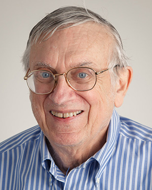 George E. Fox合作论文数Department of Biology and Biochemistry, College of Natural Sciences & Mathematics, University of Houston3Hershkovits Eli合作论文数3Fang Po-Yu合作论文数3
 Eli Hershkovitz合作论文数Chemical Physics Department, Weizmann Institute of Science3
 Chi-Young Wang合作论文数3
 Lizzette Gomez Ramos合作论文数2Ying-Chi Wang合作论文数2Eric O'Neill合作论文数2
 Thanawadee Preeprem合作论文数School of Biology, Georgia Institute of Technology Atlanta2Woods Kristen Kruger合作论文数2Watkins Derrick合作论文数2
 C. Denise Okafor合作论文数Department of Biochemistry and Molecular Biology, Eberly College of Science, The Pennsylvania State University2VanDeusen Halena合作论文数2Lie Lively合作论文数2
 Allen Tannenbaum合作论文数Department of Computer Science, Stony Brook University;Department of Applied Mathematics & Statistics, Stony Brook University2
 Eric Gaucher合作论文数Department of Biology, College of Arts & Sciences, Georgia State University2
 Martha Anne Grover (M. A. Grover)合作论文数School of Chemical and Biomolecular Engineering, College of Engineering, Georgia Institute of Technology2
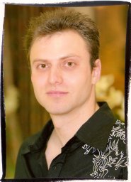 Emmanuel Tannenbaum合作论文数Department of Chemistry;Ben-Gurion University of the Negev;Department of Chemistry, Ben-Gurion University of the Negev2Benson Kalahar合作论文数2Yuzhen Xue合作论文数The University of Oklahoma2
 Victor G Stepanov合作论文数Institute of Molecular and Structural Biology, University of Aarhus2
 Thirunavukkarasu Periyasamy合作论文数1
 Stefany Y Holguin合作论文数1Ashlyn M. Norris合作论文数1Hsin-Yi Lin合作论文数1Moulaei Tinoush合作论文数1
 Maehigashi Tatsuya合作论文数School of Chemistry and Biochemistry, Georgia Institute of Technology1Larry Freil合作论文数1Kuo H-S合作论文数1Migliozzi Blacki L R合作论文数1Wang Lan合作论文数1Xiong Xiao合作论文数1Li Fengbo合作论文数1Jett James合作论文数1
 Peters Jessica K合作论文数Department of Chemistry and Biochemistry, University of California1
 Nicholas Attila Kovacs合作论文数Department of Biochemistry &Molecular Biology, Michigan State University;Department of Biochemistry & Molecular Biology, Michigan State University1




