OBJECTIVE:To explore the significance of BMP signaling in osteoarthritis (OA) etiology, and thereafter propose a disease-modifying therapy for OA.METHODS:To examine the role of the BMP signaling in pathogenesis of OA, an Anterior Cruciate Ligament Transection (ACLT) surgery was performed to incite OA in C57BL/6J mouse line at postnatal day 120 (P120). Thereafter, to investigate whether activation of BMP signaling is necessary and sufficient to induce OA, we have used conditional gain- and loss-of-function mouse lines in which BMP signaling can be activated or depleted, respectively, upon intraperitoneal injection of tamoxifen. Finally, we locally inhibited BMP signaling through intra-articular injection of LDN-193189 pre- and post-onset surgically induced OA. The majority of the investigation has been conducted using micro-CT, histological staining, and immuno histochemistry to assess the disease etiology.RESULTS:Upon induction of OA, depletion of SMURF1-an intra-cellular BMP signaling inhibitor in articular cartilage coincided with the activation of BMP signaling, as measured by pSMAD1/5/9 expression. In mouse articular cartilage, the BMP gain-of-function mutation is sufficient to induce OA even without surgery. Further, genetic, or pharmacological BMP signaling suppression also prevented pathogenesis of OA. Interestingly, inflammatory indicators were also significantly reduced upon LDN-193189 intra-articular injection which inhibited BMP signaling and slowed OA progression post onset.CONCLUSION:Our findings showed that BMP signaling is crucial to the etiology of OA and inhibiting BMP signaling locally can be a potent strategy for alleviating OA.
Objectives Osteophytes are highly prevalent in osteoarthritis (OA) and are associated with pain and functional disability. These pathological outgrowths of cartilage and bone typically form at the junction of articular cartilage, periosteum and synovium. The aim of this study was to identify the cells forming osteophytes in OA. Methods Fluorescent genetic cell-labelling and tracing mouse models were induced with tamoxifen to switch on reporter expression, as appropriate, followed by surgery to induce destabilisation of the medial meniscus. Contributions of fluorescently labelled cells to osteophytes after 2 or 8 weeks, and their molecular identity, were analysed by histology, immunofluorescence staining and RNA in situ hybridisation. Pdgfrα-H2BGFP mice and Pdgfrα-CreER mice crossed with multicolour Confetti reporter mice were used for identification and clonal tracing of mesenchymal progenitors. Mice carrying Col2-CreER, Nes-CreER, LepR-Cre, Grem1-CreER, Gdf5-Cre, Sox9-CreER or Prg4-CreER were crossed with tdTomato reporter mice to lineage-trace chondrocytes and stem/progenitor cell subpopulations. Results Articular chondrocytes, or skeletal stem cells identified by Nes, LepR or Grem1 expression, did not give rise to osteophytes. Instead, osteophytes derived from Pdgfrα-expressing stem/progenitor cells in periosteum and synovium that are descendants from the Gdf5-expressing embryonic joint interzone. Further, we show that Sox9-expressing progenitors in periosteum supplied hybrid skeletal cells to the early osteophyte, while Prg4-expressing progenitors from synovial lining contributed to cartilage capping the osteophyte, but not to bone. Conclusion Our findings reveal distinct periosteal and synovial skeletal progenitors that cooperate to form osteophytes in OA. These cell populations could be targeted in disease modification for treatment of OA.
Cartilage loss leads to osteoarthritis, the most common cause of disability for which there is no cure. Cartilage regeneration, therefore, is a priority in medicine. We report that agrin is a potent chondrogenic factor and that a single intraarticular administration of agrin induced long-lasting regeneration of critical-size osteochondral defects in mice, with restoration of tissue architecture and bone-cartilage interface. Agrin attracted joint resident progenitor cells to the site of injury and, through simultaneous activation of CREB and suppression of canonical WNT signaling downstream of β-catenin, induced expression of the chondrogenic stem cell marker GDF5 and differentiation into stable articular chondrocytes, forming stable articular cartilage. In sheep, an agrin-containing collagen gel resulted in long-lasting regeneration of bone and cartilage, which promoted increased ambulatory activity. Our findings support the therapeutic use of agrin for joint surface regeneration.
Growth and Differentiation Factor 5 (GDF5) is a key risk locus for osteoarthritis (OA). However, little is known regarding regulation of Gdf5 expression following joint tissue damage. Here, we employed Gdf5-LacZ reporter mouse lines to assess the spatiotemporal activity of Gdf5 regulatory sequences in experimental OA following destabilisation of the medial meniscus (DMM) and after acute cartilage injury and repair. Gdf5 expression was upregulated in articular cartilage post-DMM, and was increased in human OA cartilage as determined by immunohistochemistry and microarray analysis. Gdf5 expression was also upregulated during cartilage repair in mice and was switched on in injured synovium in prospective areas of cartilage formation, where it inversely correlated with expression of the transcriptional co-factor Yes-associated protein (Yap). Indeed, overexpression of Yap suppressed Gdf5 expression in chondroprogenitors in vitro. Gdf5 expression in both mouse injury models required regulatory sequence downstream of Gdf5 coding exons. Our findings suggest that Gdf5 upregulation in articular cartilage and synovium is a generic response to knee injury that is dependent on downstream regulatory sequence and in progenitors is associated with chondrogenic specification. We propose a role for Gdf5 in tissue remodelling and repair after injury, which may partly underpin its association with OA risk.
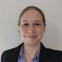 Anke J. Roelofs合作论文数Institute of Medical Sciences, University of Aberdeen4
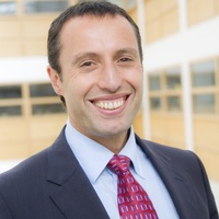 Cosimo De Bari合作论文数The Institute of Medical Sciences, School of Medicine, Medical Sciences and Nutrition, King's College, University of Aberdeen4
 Karolina Kania合作论文数2
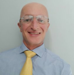 Francesco Dell'Accio合作论文数William Harvey Research Institute, Queen Mary University of London2Anna H. K. Riemen合作论文数2
 Akrit Pran Jaswal合作论文数1Ahmed Ali合作论文数1Stephanie T Kuwahara合作论文数1Fabio Colella合作论文数1Meike Sambale合作论文数1Helen Lydon合作论文数1Maxwell A. Serowoky合作论文数1Aida Barawi合作论文数1Bhupendra Kumar合作论文数1Sanap Vaibhav Nanasaheb合作论文数1Alexandra Rafipay合作论文数1
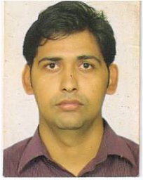 Amaresh Kumar Singh合作论文数1Bethan L Thomas合作论文数1Nitin Agnihotri合作论文数Department of Biological Sciences and Bioengineering, Indian Institute of Technology Kanpur1Joanna Sherwood合作论文数1Danielle Strachan合作论文数1
 Beatriz F Fernandez合作论文数Kings Coll London, Rayne Inst1
 Joanna Smeeton合作论文数Columbia University, Columbia University1
 Zeyu Guan合作论文数Moorfields Eye Hosp1
 Suzanne Elizabeth Eldridge合作论文数William Harvey Research Institute, Queen Mary University of London1Anne-Sophie Thorup合作论文数1Thomas Aigner合作论文数1
 Christopher Little合作论文数School of Health Sciences, Faculty of Medicine and Health, The University of Sydney1
 Costantino Pitzalis合作论文数William Harvey Research Institute, Barts and The London School of Medicine and Dentistry, Queen Mary University of London1
 Simón Méndez-Ferrer合作论文数Department of Haematology, University of Cambridge;Jeffrey Cheah Biomedical Centre, Wellcome-MRC Cambridge Stem Cell Institute, University of Cambridge1Crump J Gage合作论文数Dept Stem Cell Biol & Regenerat Med, Univ Southern Calif1
 Sadaf Ashraf合作论文数1Caxaria S合作论文数1Collins Fraser L合作论文数1Kapeni Chrysa合作论文数1
 John F. Bateman合作论文数Department of Paediatrics, University of Melbourne1Terence D. Capellini合作论文数Weill Medical College, Cornell University1
 Thomas Pap合作论文数1
 James R Whiteford合作论文数1
 Magdalena Kaneva合作论文数School of Biosciences, University of Westminster1Kenneth A. Howard合作论文数1
 Frances M. D. Henson合作论文数Department of Surgery, University of Cambridge1
 Francesca Mariani合作论文数1
 Andrew W. Mccaskie合作论文数Wellcome - MRC Cambridge Stem Cell Institute, University of Cambridge/Department of Surgery, University of Cambridge1
 Amitabha Bandyopadhyay合作论文数Department of Developmental and Molecular Biology;Albert Einstein College of Medicine, Yeshiva University;Pearl Resnick Campus;Albert Einstein College of Medicine, Yeshiva University1Lynn Rowley合作论文数1




