Glioblastoma is the most common and deadly brain cancer in adults. Although the disease disrupts the blood-brain barrier and exposes the tumor to the systemic circulation, using circulating cell-free DNA as a non-invasive biomarker for diagnosing glioblastoma remains elusive. The main obstacles are the confounding effects from artifactual variants in cell-free DNA caused by errors during next-generation sequencing and real variants in cell-free DNA originating from white blood cells that acquired somatic mutations during normal aging. Here, we developed and validated TrueR sequencing to overcome next-generation sequencing-related false positives in cell-free DNA. Subsequently, TrueR identified molecular features unique to tumor-derived cell-free DNA (i.e., circulating tumor DNA) that are detectable in blood during a tumor-naïve search. Specifically, variants associated with glioblastoma circulating tumor DNA were exclusively present in cell-free DNA (i.e., absent in white blood cell DNA) and showed gene-specific subclones. These features were uncommon in low-grade glioma, stroke, and age-related somatic mosaicism of white blood cells. We also detail a variant-agnostic method, demonstrating that copy number gains and losses in short cell-free DNA fragments (<90 bp) support the detection of glioblastoma. Finally, we present evidence that our findings extend beyond glioblastoma, supporting the broader advancement of the liquid biopsy.
Introduction The successful treatment of high-grade gliomas (HGGs) relies on advanced magnetic resonance (MR) imaging technologies to increase the resection of active tumor areas and impact patient quality of life (QoL) and overall survival (OS). The objective of this prospective pilot study was to evaluate whether preoperative MR perfusion parameters (cerebral blood volume (CBV), cerebral blood flow (CBF)) and intraoperative MRI (iMRI) can predict extent of resection (EOR), OS and progression-free survival (PFS), and postoperative QoL, with a secondary aim of determining whether QoL measures provide prognostic information earlier than conventional neurological or performance status assessments. Methods A prospective, single-center study of HGG patients, including grade 3 astrocytoma and glioblastoma, who underwent surgery guided by advanced MRI, was performed. Preoperative CBV, preoperative CBF, and EOR were compared with patient complications and QoL metrics via the MD Anderson Symptom Inventory and Functional Assessment of Cancer Therapy-Brain questionnaires. Patients were divided into good versus poor outcomes by median OS (10 months). Results Fourteen patients (mean age: 52.2, 35.7% male), including three isocitrate dehydrogenase 1/2 (IDH1/2) mutant and 11 IDH1/2 wild-type cases, were studied. For patients with good outcomes, OS and PFS were 27 ± 15 months (p = 0.002) and 21 ± 20 months (p = 0.05), respectively. Higher preoperative CBV and CBF were associated with worse OS (p < 0.05). iMRI perfusion was difficult to quantify and limited in correlating with patient outcomes. The mean EOR was 96 ± 5% during iMRIs, and no EOR difference was observed between patients with good or poor survival. Patients with worse OS and PFS showed a significant decline in three-month QoL metrics, especially in social/family well-being domains that preceded changes in Karnofsky Performance Status. Conclusion Advanced MRI and quantitative QoL measurements may help predict long-term survival for HGG patients but require further exploration and analysis.
Fabry disease is a rare X-linked deficiency of lysosomal alpha-galactosidase that causes glycolipid accumulation in tissues, including the brain. The most common neurologic sequelae of Fabry are cognitive decline and white matter lesions (WMLs) on brain magnetic resonance imaging (MRI). In the at-large population, however, WMLs are non-specific, highly prevalent, and most are clinically silent. Thus, we compared Fabry to typical brain aging to identify factors unique to Fabry-related cognitive decline. Twenty adult Fabry patients (75% female; median age 36.4 yrs, range: 19.8-63.2 yrs; 95% on enzyme replacement therapy) without a history of stroke or other neurologic diseases and 20 age/sex-matched healthy controls were enrolled in a case-control study. All participants underwent a neurocognitive assessment and a 3.0 T MRI study of the brain that used structural MRI (e.g., fluid-attenuated inversion recovery, FLAIR), semi-quantitative MRI (e.g., normalized FLAIR signal intensity), and quantitative MRI (diffusion tensor imaging, bound-pool fraction imaging). During a blinded review of structural MRIs, a neuroradiologist's categorization of case-control status did not correspond to disease status (Fisher's test, P > 0.99) but rather to age (P = 0.004), indicating qualitative changes associated with Fabry were similar to normal age-related brain alterations. Using quantitative MRI, however, we detected evidence of microstructural damage in the white matter of younger Fabry adults (<40 yrs). With age, WML severity increased and the corpus callosum atrophied in Fabry, phenomena absent in controls and consistent with progressive tissue damage. Neurocognitive assessments identified trends for lower verbal intelligence quotient and executive function in the younger Fabry participants, which became statistically significant in the older Fabry patients. Our data suggest that the early onset of microstructural damage in Fabry drives the insidious degeneration of white matter, leading to impaired cognition. Aging Fabry patients may benefit from serial cognitive assessments to identify unmet therapeutic needs.
Abstract Leptomeningeal metastatic disease (LMD) is the spread of cancer cells into the cerebrospinal fluid (CSF)-filled spaces surrounding the central nervous system (CNS). Once LMD occurs, patients endure devastating neurologic symptoms and survive for only a few weeks to months. The clinical diagnosis of LMD has risen as patient survival from non-CNS metastatic disease improves. Unfortunately, detection of LMD even in symptomatic patients is <50% using the current gold standard – CSF cytology. Therefore, a strong clinical need exists for the sensitive and early detection of LMD. Here, we sought to develop a minimally invasive liquid biopsy approach to diagnose LMD using next-generation sequencing. Because copy number variations (CNVs) are common in cancer and increase after chemoradiation, we hypothesized that CNV detection in CSF may offer a means to detect LMD since patients have already undergone treatment for the underlying disease. Specifically, we sought to develop low-pass whole genome sequencing (lpWGS) of cell-free DNA in CSF to detect CNVs associated with LMD. A novel lpWGS algorithm was developed using FASTQ files trimmed to 30 million total paired reads. The genome was partitioned into one million continuous base pair segments excluding problematic areas yielding 2,445 regions across 22 autosomes. Control samples were used to model the read depth for each region to establish the euploid state. Read depth across the genome for all samples was 0.6X. CSF samples from lung cancer, breast cancer, and melanoma patients with LMD demonstrated prominent evidence of monosomies and trisomies throughout the genome. Lung cancer exhibited frequent amplifications (>4-fold). Notably, we observed 17p monosomy in CSF regardless of cancer type which indicates TP53 haploinsufficiency may support LMD progression, a conjecture supported by a codeletion of TP53 in one patient. lpWGS detects LMD in solid tumor cancers and may also identify novel mechanisms for LMD progression.
Supplemental Table S2. Number of PCR cycles used during library preparation and panel capture-enrichment.
Supplemental Figure S15. Distribution of allele frequencies for somatic mutations discovered by True2 in tumor and peritumoral edema of adult diffuse gliomas.
Supplemental Figure S5. Effect of using the dbSNP database to remove potential contamination.
Supplemental Table S4. Truth set of single nucleotide variants in HD701 DNA and associated dilutions.
Abstract Leptomeningeal metastatic disease (LMD) is the spread of cancer cells to the cerebrospinal fluid (CSF)-filled spaces surrounding the central nervous system. After symptom onset, LMD patients survive for only weeks to months while enduring devastating neurologic symptoms. Current clinical tools to diagnose LMD, such as CSF cytology and magnetic resonance imaging, do not detect mutations susceptible to treatment, quantitatively monitor response to therapy, or identify mutation-based resistance mechanisms. Here, we sought to profile LMD using next-generation sequencing by studying CSF-derived cell-free DNA (CSF-cfDNA) from 9 breast cancer and 5 lung cancer patients with LMD and 4 LMD-negative controls. To conduct an unbiased search for somatic mutations, we used a custom-designed gene panel and our validated True2 sequencing workflow with a specificity of <1 false positive per 100kb panel positions and a 97.6% sensitivity for detecting single nucleotide variants (SNVs) with a variant allele frequency (VAF) ≥0.1%. Although CSF-cfDNA quantity ranged widely (<1 to 91 ng), genomic alterations were observed in 100% of LMD patients vs. none in the controls. In 13 patients with a SNV, an average of 4.5±3.3 variants were detected with a VAF ranging from 0.051% (HER2 p.S1151L associated with a 12-fold amplification and read depth of 25,542X) to 98.7% (PTEN p.Q733X associated with a loss of heterozygosity). Importantly, subclones in cancer driver genes were detected in 10 of 13 (76.9%) samples consistent with a high prevalence in LMD. Somatic mutations in genes common to both cancers were discovered (e.g., TP53, KMT2D, and EGFR), while other affected genes segregated based on cancer type (e.g., PIK3CA, ATM, and CDH1 in breast cancer; PTEN, MTOR and PDGFRA in lung cancer). An amplification or deletion was detected in 10 of 14 patients (71.4%), a subset of which segregated by cancer type (e.g., HER2 and ESR1 amplifications in breast cancer; CDKN2A and TP53 codeletions in lung cancer). Using serially collected CSF samples, we observed: (1) loss of subclones in response to therapy followed by resurgence at clinical recurrence showing suppression of disease but not eradication; (2) multiple subclones in the same gene supporting evidence of convergent evolution; and (3) emergence of new subclones at clinical recurrence consistent with mutation evolution in response to therapy. For example, in a breast cancer patient with a known HER2 amplification receiving intrathecal Trastuzumab, a previously absent HER2 p.V777L mutation was present at the end of therapy with a VAF of 18.6% which increased at clinical recurrence to 94.8% in association with a 2.4-fold HER2 amplification indicating the mutated HER2 subclone was already therapy-resistant. Our findings support a panel-based approach to detect and monitor LMD-derived somatic mutations using CSF-cfDNA. Moreover, the untargeted search for somatic mutations via True2 has the potential to guide personalized, adaptive therapeutics for LMD patients which may prove essential in improving patient survival. Citation Format: Yingqi Zhang, Rachna Malani, Charlie S Dean, Laura A Boatz, Brion E Harrison, Mei Wei, Wallace Akerley, Mary P Bronner, Hunter R Underhill. Molecular profiling of serially collected cerebrospinal fluid uncovers therapy-resistant subclones in recurrent leptomeningeal metastatic disease [abstract]. In: Proceedings of the AACR Special Conference: Liquid Biopsy: From Discovery to Clinical Implementation; 2024 Nov 13-16; San Diego, CA. Philadelphia (PA): AACR; Clin Cancer Res 2024;30(21_Suppl):Abstract nr B043.
Supplemental Figure S18. Copy number variation (CNV) detection demonstrates differences in CNVs between samples from the same patient.
Supplemental Figure S8. Alternative experimental and in vitro noise reduction strategies without substantive improvement in error.
Supplemental Figure S3. Effects of proper-pair-plus (PP+), alignment quality (MQ), and base quality (BQ) on read depth and false positives.
Supplemental Figure S10. Comparison of True2 sensitivity using reduced sample complexity and iterative captures.
Glutamine synthetase (GS), encoded by GLUL, catalyzes the conversion of glutamate to glutamine. GS is pivotal for the generation of the neurotransmitters glutamate and gamma-aminobutyric acid and is the primary mechanism of ammonia detoxification in the brain. GS levels are regulated post-translationally by an N-terminal degron that enables the ubiquitin-mediated degradation of GS in a glutamine-induced manner. GS deficiency in humans is known to lead to neurological defects and death in infancy, yet how dysregulation of the degron-mediated control of GS levels might affect neurodevelopment is unknown. We ascertained nine individuals with severe developmental delay, seizures, and white matter abnormalities but normal plasma and cerebrospinal fluid biochemistry with de novo variants in GLUL. Seven out of nine were start-loss variants and two out of nine disrupted 5′ UTR splicing resulting in splice exclusion of the initiation codon. Using transfection-based expression systems and mass spectrometry, these variants were shown to lead to translation initiation of GS from methionine 18, downstream of the N-terminal degron motif, resulting in a protein that is stable and enzymatically competent but insensitive to negative feedback by glutamine. Analysis of human single-cell transcriptomes demonstrated that GLUL is widely expressed in neuro- and glial-progenitor cells and mature astrocytes but not in post-mitotic neurons. One individual with a start-loss GLUL variant demonstrated periventricular nodular heterotopia, a neuronal migration disorder, yet overexpression of stabilized GS in mice using in utero electroporation demonstrated no migratory deficits. These findings underline the importance of tight regulation of glutamine metabolism during neurodevelopment in humans.
Supplemental Figure S13. Relative to the overall median read depth for each sample, the median read depth for the TERT promoter is less compared to other gene-specific regions (IDH1, IDH2, and NF1).
Supplemental Table S1. Alphabetical listing of 115 genes included on the custom-designed, 114 kb, hotspot panel.
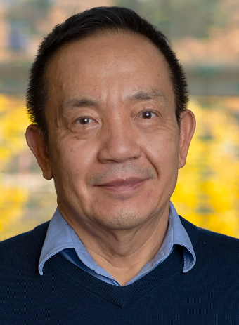 Chun Yuan合作论文数Department of Biomedical Engineering, John and Marcia Price College of Engineering, The University of Utah;Department of Radiology, The University of Utah;Department of Biomedical Informatics, The University of Utah65
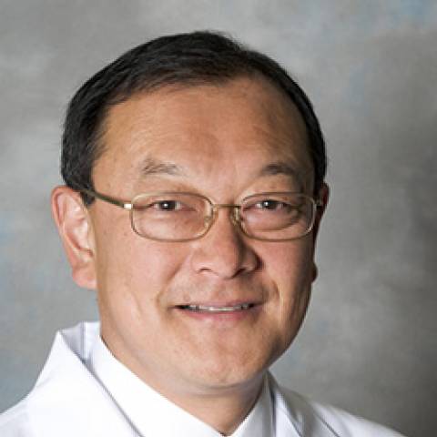 Thomas S. Hatsukami合作论文数Division of Vascular Surgery, Department of Surgery, School of Medicine, University of Washington;Vascular Imaging Laboratory, Department of Radiology, School of Medicine, University of Washington48
 Mary P. Bronner合作论文数41Sabine Hellwig合作论文数38
 Baocheng Chu合作论文数38Colman Howard合作论文数32
 Randy L. Jensen合作论文数University of Utah32Charlie Dean合作论文数31Brion E. Harrison合作论文数30Christian Davidson合作论文数30
 Michael Karsy合作论文数Department of Pathology, New York Medical College30Sydney Vincenti合作论文数28Samuel Stevenson合作论文数28Eric Goold合作论文数28
 Drew L. Sellers合作论文数Department of Bioengineering, University of Washington27Minako Oikawa合作论文数24
 William S. Kerwin合作论文数Cardiovascular Research and Training Center, University of Washington24
 Tobias Saam合作论文数Die Radiologie21Vasily L. Yarnykh合作论文数20
 Xihai Zhao (赵锡海)合作论文数Center for Biomedical Imaging Research, Department of Biomedical Engineering, School of Medicine, Tsinghua University16
 Jianming Cai (蔡建明)合作论文数School of Public Health and Management, Wenzhou Medical University15
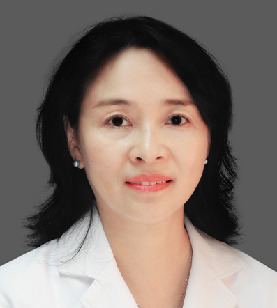 Wei Yu (于薇)合作论文数Beijng Anzhen Hospital, Capital Medical University13Norihide Takaya合作论文数13Li Dong合作论文数12
 Hideki Ota合作论文数Department of Diagnostic Radiology, Tohoku University Hospital12
 Xueqiao Zhao合作论文数Clinical Atherosclerosis Research Lab, Division of Cardiology, School of Medicine, University of Washington;Diabetes Research Center, University of Washington10Marina S. Ferguson合作论文数10Ts Hatsukami合作论文数9
 Dongxiang Xu合作论文数9Fei Liu合作论文数8David A. Nix合作论文数8
 Daniel S Hippe合作论文数Clinical Research Division, Fred Hutchinson Cancer Center8Nayak Polissar合作论文数8N Takaya合作论文数6Carrie Fuertes合作论文数6
 Preetida J Bhetariya合作论文数AstraZeneca6Gabor T. Marth合作论文数Department of Biology, Boston College6Alun Thomas合作论文数5
 Zhaoqi Zhang (张兆琪)合作论文数Beijing Anzhen Hospital, Capital Medical University5Wendy Hamar合作论文数5Kenneth R. Maravilla合作论文数5James G. Terry合作论文数Department of Internal Medicine;School of Medicine;Wake Forest University;School of Medicine, Wake Forest University5Tom Hatsukami合作论文数4N L Polissar合作论文数4Brandon A Zielinski合作论文数4Jinnan Wang合作论文数4ZiXu Yan (晏子旭)合作论文数4




