Abstract— Photocrosslinking of proteins to DNA by single‐pulse UV laser has been used only in analytical experiments, carried out with reconstituted complexes of a single DNA binding protein and a labeled target sequence. Here we propose a large‐scale technique for irradiation of nuclei, generating preparative quantities of covalently linked protein‐DNA complexes for further analysis of the partner molecules. The use of a flow cuvette allows a milligram of DNA in either nuclei or chromatin to be irradiated by a single pulse within few minutes. The efficiency of crosslinking varies from 6 to 12% of the total nuclear proteins. The presence of histones and other chromosomal proteins in the crosslinked protein‐DNA complexes was demonstrated by using specific antibodies. The irradiation procedure can be fully automated using a microcomputer.
A picosecond UV laser radiation was used to cross-link proteins to DNA in nuclei, whole cells and different chromating preparations. All histones as well as high-mobility group 1 proten were identified immunochemically in the covalently linked protein-DNA complexes. Irradiation of the nuclieohistone resulted in cross-linking 20% of bound histones to DNA as a result of two-quantum photoreaction with a maximum quantum yield 3.10 -4 for double stranded DNA. When nuclei, total bromatin Hi-depleted chromatin and core particles were irradiated and then trypsinized or treated with clostripain to cleave respectively the N-, C- and N- terminal histone tails, no histones have been found covalently linked to DNA. However whilst the yield of cross-links was similar in total and H1-depleted chromatin in core particples the efficiencey was 3-4 times lower for H2A, H2B and H4 10-12 times lower for H3. This finding we consider as a direct evidence for interaction of non structured N- tails of core histones with linker DNA. Cross-linking in core particles depends on the ionic strength. All histones were identified in the complex formed up to 0.4 N NaCl, no cross-linking was observed when irradiation was carried out at salt concentration higher than 0.4 M. The cross-linking ability was preserved both upon physiological acetylation of histones knows to be restriced to the N-terminal tails and with chemically acetylated chromatin. This finding is direct evidence that postsynthetic histone acetylation does not release the N-terminal tails from interaction with DNA.
A method is suggested for isolation of highly purified mouse centromeric heterochromatin. Treatment of mouse liver nuclei with decreasing concentrations of Ca2+ resulted in the gradual unraveling of chromatin in the nucleus and at 0.1 mM Ca2+ electron microscopy revealed several dense particles per nucleus, surrounded by decondensed chromatin. These particles, assumed to represent centromere regions of interphase chromosomes by in situ hybridization with radioactive mouse satellite DNA and by differential staining for centromere heterochromatin, were isolated in preparative amounts and their DNA and protein composition was analyzed. The preparation represented practically pure mouse centromere heterochromatin, since more than 90% of its DNA was satellite DNA.
Cation-induced folding of 10 nm chromatin filament to 30 nm fiber was studied with hyperacetylated chromatin using light scattering at 90 degrees and flow linear dichroism. Acetylated chromatin folded in a way indistinguishable from that of the control chromatin: both the compactness of chromatin and the orientation of nucleosomes relative to the fiber axis were identical at a given salt concentration.
A model of chromatin fiber structure is presented in which a repeating unit of a trinucleosome forms a 3‐dimensional zigzag. Twisting and compression of the zigzag result in a triple helix structure. The model is built mainly on the flow linear dichroism data showing that (a) nucleosomal disc faces are tilted relative to the fiber axis, (b) the orientation of nucleosomes does not change upon folding and unfolding of chromatin, and (c) the orientation of nucleosomes is maintained by the globular domain of histone HI.
FEBS LettersVolume 112, Issue 2 p. 143-146 Full-length articleFree Access Electron microscopic evidence for structurl rearrangement of H1-depleted chromatin during thermal denaturation I. Tsaneva, I. Tsaneva Institute of Molecular Biology, Bulgarian Academy of Sciences, 1113 Sofia, BulgariaSearch for more papers by this authorSt. Dimitrov, St. Dimitrov Institute of Molecular Biology, Bulgarian Academy of Sciences, 1113 Sofia, BulgariaSearch for more papers by this authorI. Pashev, I. Pashev Institute of Molecular Biology, Bulgarian Academy of Sciences, 1113 Sofia, BulgariaSearch for more papers by this authorR. Tsanev, R. Tsanev Institute of Molecular Biology, Bulgarian Academy of Sciences, 1113 Sofia, BulgariaSearch for more papers by this author I. Tsaneva, I. Tsaneva Institute of Molecular Biology, Bulgarian Academy of Sciences, 1113 Sofia, BulgariaSearch for more papers by this authorSt. Dimitrov, St. Dimitrov Institute of Molecular Biology, Bulgarian Academy of Sciences, 1113 Sofia, BulgariaSearch for more papers by this authorI. Pashev, I. Pashev Institute of Molecular Biology, Bulgarian Academy of Sciences, 1113 Sofia, BulgariaSearch for more papers by this authorR. Tsanev, R. Tsanev Institute of Molecular Biology, Bulgarian Academy of Sciences, 1113 Sofia, BulgariaSearch for more papers by this author First published: April 07, 1980 https://doi.org/10.1016/0014-5793(80)80166-9Citations: 4AboutPDF ToolsRequest permissionExport citationAdd to favoritesTrack citation ShareShare Give accessShare full text accessShare full-text accessPlease review our Terms and Conditions of Use and check box below to share full-text version of article.I have read and accept the Wiley Online Library Terms and Conditions of UseShareable LinkUse the link below to share a full-text version of this article with your friends and colleagues. Learn more.Copy URL Share a linkShare onFacebookTwitterLinked InRedditWechat Citing Literature Volume112, Issue2April 07, 1980Pages 143-146 ReferencesRelatedInformation
 Stefan Dimitrov合作论文数Univ Grenoble 1, INSERM, Inst Albert Bonniot, U823, F-38042 Grenoble 9, France7
 Dimitar Angelov合作论文数LBMC4
 Evdokia A. Pasheva合作论文数Institute of Molecular biology, Bulgarian Academy of Sciences4E Keskinova合作论文数2V. Makarov合作论文数2Victor Y. Stefanovsky合作论文数2Valya Russanova合作论文数2
 Iva Ugrinova合作论文数Institute of Molecular biology, Bulgarian Academy of Sciences2Y Chentsov合作论文数1i ugninova合作论文数1yu stefanovsky合作论文数1Mitkova Elena合作论文数1Tzveta Apostolova合作论文数1
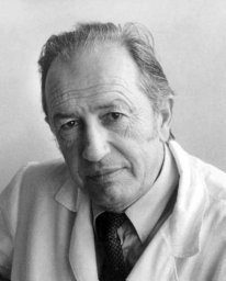 Roumen Tsanev合作论文数Institute of Molecular Biology, Bulgarian Academy of Sciences1Elena Stephanova合作论文数1Irina R. Tsaneva合作论文数Department of Biochemistry and Molecular Biology, University College1V. S. Smirnov合作论文数1
 Vesco Mutskov合作论文数Laboratory of Molecular Biology;National Institutes of Health1Cendrine Moskalenko合作论文数Laboratoire Joliot Curie, Ecole Normale Supérieure de Lyon1




