PURPOSE:Recurrent and metastatic human papillomavirus (HPV)-associated head and neck squamous cell carcinoma (R/M HPV+ HNSCC) remains largely incurable, with genetic drivers incompletely defined. We profiled the genetic landscape of R/M HPV+ HNSCC and functionally characterized genetic alterations strongly enriched in this cancer type. EXPERIMENTAL DESIGN:We identified genetic alterations uniquely enriched in 159 R/M HPV+ tumors. High-priority alterations were functionally modeled, examining proliferation, clonogenicity, migration/invasion, apoptosis, therapy response, in vivo growth, metastasis, and immune contexture. RESULTS:Compared with HPV+ primary tumors, R/M HPV+ tumors were enriched for TP53 mutations (prespecified FDR threshold met; OR, 6.23; P = 0.02) and were associated with poorer survival. Within R/M disease, cylindromatosis lysine 63 deubiquitinase (CYLD) alterations were specific to HPV+ tumors (21% vs. 0% in HPV-). TP53 mutations were predominantly clonal and associated with whole-genome duplication. Expression of TP53 gain-of-function (GOF) mutants (R175H, G245C, R273C) in HPV+ HNSCC cells increased clonogenic survival, migration/invasion, lung metastatic burden in vivo, and cisplatin IC50, without altering radiation sensitivity. CYLD knockdown accelerated cellular growth yet increased radiosensitivity. Transcriptomic analyses linked CYLD loss to NF-κB/TNFα pathway activation, a T cell-inflamed microenvironment, and checkpoint upregulation. CONCLUSIONS:R/M HPV+ HNSCC is genomically and functionally shaped by two axes with therapeutic implications: TP53 GOF mutations promote metastatic phenotypes and cisplatin resistance, whereas CYLD loss defines an HPV-specific subset with enhanced radiation sensitivity and immune activation. These data support using TP53 and CYLD as predictive biomarkers to guide investigation into precision strategies for systemic therapy choices, p53-targeted/Wee1 strategies, and radiotherapy-immunotherapy combinations in high-risk or R/M HPV+ HNSCC.
Importance:A unified salivary gland carcinoma (SGC)-specific tumor-node-metastasis (TNM) classification can enhance prognostic accuracy, support clinical decision-making, and improve the quality of patient care. Objective:To derive and validate an SGC-specific pTNM classification with improved prognostic accuracy and optimized stage distribution for version nine of the American Joint Committee on Cancer/Union for International Cancer Control staging protocol. Design, Setting, and Participants:This retrospective prognostic cohort study derived a novel pTNM classification using data from the National Cancer Database (NCDB) of patients with surgically treated major SGC (2012-2017) and validated it in an international major SGC cohort (2008-2021) and a single-institution minor SGC cohort (Memorial Sloan Kettering Cancer Center; 1985-2016). Data were analyzed from June to November 2024. Exposures:Surgery with or without postoperative radiotherapy or chemoradiotherapy. Main Outcomes and Measures:The primary end point was overall survival (OS). Cox proportional hazards multivariable analysis was used to confirm the prognostic importance of pathologically positive lymph node (LN) number and extranodal extension (pENE) and derive an optimal pTNM classification. Results:The NCDB dataset included 8409 patients with SGC: 7659 with M0 disease (5748 with pN0 disease and 1911 with pN+ disease) and 750 with M1 disease. Among the 7659 patients with M0 disease, the median (IQR) age was 60 (48-71) years, and 3861 (50.4%) were male. The median (IQR) follow-up was 88.4 (72.3-108.5) months. The 5-year OS was 87.2% (95% CI, 86.3-88.0) for N0 disease, 68.2% (95% CI, 63.9-72.8) for 1 positive LN without pENE, 60.2% (95% CI, 53.5-67.5) for 2 positive LNs without pENE, 68.4% (95% CI, 58.0-76.6) for 3 positive LNs without pENE, 47.5% (95% CI, 41.6-52.8) for more than 3 positive LNs without pENE, and 41.4% (38.1-44.8) for pENE-positive LNs. Multivariable analysis confirmed the independent prognostication of LN count compared with pN0 disease (1 positive LN: adjusted hazard ratio [aHR], 1.70; 95% CI, 1.44-2.01; 2 positive LNs: aHR, 1.61; 95% CI, 1.31-1.98; 3 positive LNs: aHR, 2.10; 95% CI, 1.65-2.68; 4 positive LNs : aHR, 2.46; 95% CI, 1.87-3.24; more than 4 positive LNs: aHR, 2.07; 95% CI, 2.08-2.91) and pENE-positive LNs compared with pENE-negative LNs (aHR, 1.27; 95% CI, 1.10-1.48). The proposed pN classification were pN1 for 1 to 3 positive LNs and pENE negativity and pN2 for more than 3 positive LNs or pENE positivity. Model fit improved with the proposed pN classification vs the current pN classification (Akaike Information Criterion, 26 442 vs 26 483). Based on the aHR model, the following stage groups were proposed: stage I: T1N0 (1 [reference]); stage II: T2N0 (aHR, 1.34; 95% CI, 1.11-1.61); stage IIIA: T1-2N1 or T3-4N0 (aHR, 2.36; 95% CI, 1.99-2.80); stage IIIB: T1-2N2 or T3-4N1-2 (aHR, 5.15; 95% CI, 4.38-6.06); and stage IV: M1 disease (aHR, 13.61; 95% CI, 11.37-16.29). The C index values were similar (proposed classification: 0.792; current classification: 0.790), while the AIC improved slightly (proposed classification: 26 441; current classification: 26 482). Stage-specific OS differences were evident in both the international major SGC cohort (n = 1015) and Memorial Sloan Kettering Cancer Center minor SGC cohort (n = 444). Conclusions and Relevance:This unified, SGC-specific staging system improved prognostic accuracy and sample size balance and was applicable to both major and minor SGCs.
Background Oral cavity squamous cell carcinoma (OSCC) is a global health burden, where negative margins are essential for reducing recurrence and improving survival. Intraoperative frozen-section analysis is limited by time, sampling error, and interpretive variability, underscoring the need for more reliable margin assessment. Reflectance confocal microscopy (RCM) enables real-time, in vivo high-resolution imaging, but accuracy depends on expert interpretation. This study evaluated the diagnostic performance of an artificial intelligence (AI)-driven model for RCM in OSCC, aiming to develop a point-of-care platform for intraoperative use. Methods Patients with biopsy-confirmed OSCC underwent in vivo RCM imaging using a handheld intraoral probe before biopsy. Histopathology was the reference standard. A deep learning model was developed with the Google Cloud Vertex AI Automated Machine Learning (AutoML) Vision platform and trained on 4,090 annotated RCM images (1,998 benign, 2,092 malignant). Performance was compared with blinded expert pathologist and RCM readers. Results The AI model achieved an area under the precision-recall curve (AUC-PR) of 0.99 and an area under the receiver operating characteristic curve (AUC-ROC) of 0.99, with sensitivity 98.09%, specificity 95.00%, accuracy 96.58%, positive predictive value (PPV) 95.35%, and negative predictive value (NPV) 97.94%. Expert readers showed sensitivity 90.00%, specificity 98.30%, accuracy 94.15%, PPV 88.20%, and NPV 96.60%. Inter-reader agreement was 95.00% for benign and 81.70% for malignant cases. Conclusions AI-driven RCM interpretation provides an accurate, rapid, noninvasive approach for OSCC diagnosis and intraoperative margin assessment. It outperformed expert readers and can reduce reliance on frozen-section analysis, streamline workflows, and improve outcomes.
Importance:Patients with papillary thyroid carcinoma (PTC) with lateral neck metastases (N1b) are usually treated with total thyroidectomy (TT), neck dissection, and adjuvant radioactive iodine (RAI). This philosophy comes with higher risks of complications and sequela than thyroid lobectomy (TL) and neck dissection alone. There are no prior studies on patients from the Western hemisphere that compare survival and recurrence outcomes between these groups. Objective:To compare recurrence and survival outcomes in propensity-matched TL vs TT + RAI patients who presented with ipsilateral N1b PTC at a tertiary cancer center in the US. Design, Setting, and Participants:This cohort propensity-matched study was conducted at a single US tertiary cancer center included 37 TL patients and 37 of 561 TT + RAI patients (after excluding patients with M1 disease), with a median (IQR) follow-up of 113 (58-241) and 90 (48-185) months, respectively. Adult patients with PTC with lateral neck node metastases (N1b) were identified from a thyroid cancer database. The study included patients undergoing surgery at Memorial Sloan Kettering Cancer Center from 1986 to 2020, inclusive, and the study was conducted from 2024 to 2025. Main Outcomes and Measures:Overall survival (OS), disease-specific survival (DSS), and recurrence-free survival (RFS). Results:Of 598 total individuals, the median (IQR) age was 41 (33-55) years, and 341 (57%) were female. The 5-year OS was 96.9% in the TL group and 96.8% in the TT + RAI group (hazard ratio [HR], 0.2; 95% CI, 0.03-1.58). The 5-year DSS was 96.7% in the TL group and 100% in the TT +RAI group. The 5-year RFS was 89.8% in the TL group and 88.9% in the TT + RAI group (HR, 1.48; 95% CI, 0.39-5.58). The survival rates did not change between 5 and 10 years. Conclusions and Relevance:This cohort study found that a select group of patients with N1b PTC treated with TL had no important difference in survival and recurrence outcomes compared with patients treated with TT + RAI. Therefore, TL is an effective and safe treatment option in carefully selected and appropriately counselled patients with N1b PTC with unilateral tumors and low-volume regional lymph node metastases without clinical extranodal extension.
Introduction:Adenoid cystic carcinoma (ACC) of the head and neck is a rare malignancy with a high risk of perineural invasion, local recurrence, and distant metastasis. Recurrent ACC poses unique challenges due to its aggressive nature, resection limited by morbidity, and prior radiation. Proton therapy, with its ability to spare normal tissues via the Bragg peak, may offer a promising and safe re-irradiation approach, especially in patients not amenable to resection requiring high definitive doses. The purpose of this study was to evaluate outcomes of patients with recurrent ACC treated with proton radiation. Methods:We reviewed the records of all patients with recurrent head and neck ACC treated with proton therapy between November 2012 and December 2023. This cohort included patients with both resected and unresected disease. Standard imaging, including MRI for perineural spread and PET for distant recurrence, was used in all cases. Descriptive statistics were used to analyze patient, tumor, and treatment characteristics, and the Kaplan-Meier method and cumulative incidence curves were utilized to estimate overall survival (OS) and loco-regional recurrence (LRR), respectively. Results:A total of 19 patients were included, with 6 (32%) receiving adjuvant re-irradiation and 13 (68%) receiving definitive proton re-irradiation. Median dose in the adjuvant group was 63.36 (59.40-70.07), and 59.20 (14.80-70.00) in the primary RT group. The median OS for all patients was 62% at 24 months and 48% at 48 months. Patients receiving adjuvant therapy had an OS of 80% at 24 and 48 months, while those not receiving surgery had an OS of 52% and 31% at 24- and 48-months. LRR at 24 months was 11% for all patients. No grade 5 toxicities were observed. Conclusion:This study demonstrates the feasibility and potential benefits of proton re-irradiation for recurrent ACC, with promising local-regional control and minimal toxicity.
BACKGROUND:Robust outcome data for anterior skull base cutaneous squamous cell carcinoma (cSCC) are limited, particularly as emerging therapies reshape management. We evaluated survival and prognostic factors following craniofacial resection. METHODS:Retrospective multi-institutional case-series (1995-2015) of patients who underwent skull base surgery for anterior face/scalp cSCC. Kaplan-Meier and Cox regression analyses assessed disease-free (DFS), disease-specific (DSS), and overall survival (OS). RESULTS:Among 112 patients, most had T4 (86.6%) and recurrent (71.4%) disease. Clear margins were achieved in 32.1%; 40.2% were involved. Five-year DFS, DSS, and OS were 46.9%, 68.4%, and 52.3%, respectively. Intracranial extension independently predicted worse OS (HR:6.54 [95% CI:2.61,16.38]) and DSS (HR:12.39 [95% CI:2.72,56.45]). Involved margins predicted recurrence on univariable analysis. Adjuvant therapy, predominantly post-operative radiotherapy, improved OS (HR: 0.54 [95% CI: 0.30,0.97]). Complications occurred in 38.4%, with 4.5% perioperative mortality. CONCLUSION:Advanced anterior skull base cSCC carries substantial morbidity and modest survival. These data provide a benchmark against which immunotherapy-based strategies should be evaluated.
OBJECTIVES:Despite decades of experience with neoadjuvant systemic therapy (NST) in oral cavity squamous cell carcinoma (OCSCC) demonstrating its potential to facilitate surgical de-escalation through tumor downstaging, the impact of NST on response-adapted surgery has not been systematically evaluated. MATERIALS AND METHODS:Surgical de-escalation following neoadjuvant NST was quantified using the Response-Adapted Surgery-Oral Cavity classification and the Oral Cavity Surgical Morbidity Score (OC-SMS) in 70 consecutive patients with resectable, locally advanced OCSCC. The relationship between surgical de-escalation, pathologic response, and margin status was assessed. RESULTS:Surgical de-escalation was observed in 44 patients (62.8 %). Mean OC-SMS decreased from 9.1 (95 % CI, 8.5-9.7) before NST to 6.5 (95 % CI, 5.8-7.3) after NST. The largest contributors to de-escalation were reduced extent of resection (52 %), decreased need for free flap reconstruction (27 %), reduced surgical access requirements (17 %), and less extensive neck dissection (3 %). The greatest reductions were observed in tongue cancers (mean decrease, 3.1 points; p < 0.001), followed by buccal mucosa (2.1 points; p = 0.024) and floor of mouth cancers (2.0 points; p = 0.250). Gingival cancers demonstrated the smallest reduction (1.1 points; p = 0.116). Surgical de-escalation correlated with pathologic response, and 97 % of de-escalated cases achieved negative margins. CONCLUSION:Neoadjuvant systemic therapy resulted in measurable surgical de-escalation in OCSCC without compromising margin status. Surgical de-escalation should be incorporated as a key endpoint in future prospective trials designed to optimize NST approaches.
Importance:Osteoradionecrosis (ORN) is a potentially debilitating late complication of radiotherapy (RT) for head and neck cancer. While proton therapy offers superior dose conformality, its impact on ORN risk remains uncertain, particularly in patients with oropharyngeal squamous cell carcinoma (OPSCC). Objective:To characterize the incidence, severity, and predictors of ORN in a large institutional cohort of patients with OPSCC treated with curative-intent RT, and to compare outcomes between proton therapy and intensity-modulated radiation therapy (IMRT). Design, Setting, and Participants:This retrospective cohort study included consecutive patients with OPSCC treated from January 2013 to December 2023 at a single high-volume academic institution. Patients received either IMRT or proton therapy (uniform scanning or pencil beam scanning). ORN diagnosis and grading were determined through standardized multidisciplinary review. The primary outcome was the 3-year rate of ORN. Cox regressions identified predictors of ORN. Data were analyzed from December 2024 to April 2025. Exposures:Radiotherapy modality (proton vs IMRT), patient demographic characteristics, smoking status, human papillomavirus status, tumor and/or node stage, chemotherapy, and radiation dosage. Results:The analysis included 1564 patients (mean [SD] age, 61.5 [9.6] years; 208 females [13.3%] and 1356 males [86.7%]) of whom 1389 patients (88.8%) had undergone IMRT, and 175 patients (11.2%) had undergone proton-based treatment. The 3-year incidence of any-grade ORN was 3.02% (95% CI, 2.22%-4.09%). ORN rates were significantly higher after proton therapy compared with IMRT (6.36% vs 2.69% at 3 years; hazard ratio [HR], 2.62; 95% CI, 1.39-4.93). Of 1344 definitive patients, 47 of 1210 patients in the IMRT group (3-year rate, 2.38%; 95% CI, 1.61-3.51%) developed ORN vs 11 of 134 patients in the proton group (3-year rate 7.47%; 95% CI, 3.40-16.02% [HR, 3.62; 95% CI, 1.85-7.09]). On multivariable analysis, proton therapy (HR, 2.92; 95% CI, 1.55-5.50), concurrent chemotherapy (HR, 3.29; 95% CI, 1.03-10.50), and smoking (HR, 2.33; 95% CI, 1.38-3.92) were independently associated with ORN. Grade 3 or greater ORN occurred in 0.67% of patients and did not appear to differ by RT modality. Conclusions and Relevance:In this large, relatively homogeneous cohort of patients with OPSCC, proton therapy was associated with a higher rate of ORN compared with IMRT, particularly in the definitive setting, although high-grade ORN remained uncommon across both modalities. These retrospective findings should be considered exploratory and underscore the need for future hypothesis-driven studies to refine dose constraints and optimize treatment planning to mitigate ORN risk among patients with OPSCC.
Oncocytic thyroid cancer (OTC), previously termed Hürthle cell carcinoma, is a rare but distinct thyroid malignancy. Although OTC arises from follicular epithelial cells and historically was classified as a subtype of follicular thyroid carcinoma, emerging molecular and clinical evidence demonstrates that OTC is distinct, leading to its reclassification by the World Health Organization (WHO) in 2022 into a separate clinical entity. OTC is characterized by a unique molecular landscape, including mitochondrial DNA mutations, increased somatic variant burden and widespread chromosomal loss of heterozygosity. Clinically, OTC presents with a spectrum of clinical behavior, with the most aggressive subtypes displaying high levels of vascular invasion and metastatic spread, mainly distant disease at the time of diagnosis. Diagnostic tools such as fine-needle aspiration can fail to reliably distinguish benign oncocytic lesions from malignant disease, complicating early evaluation and surgical decision-making, while the introduction of molecular testing has increased the accuracy of preoperative diagnostics. Poor radioiodine avidity further limits traditional postoperative management, leaving many patients with RAI-refractory disease few effective systemic treatment options. Emerging therapies, including tyrosine kinase inhibitors and mTOR pathway inhibitors, offer potential benefits but remain supported by limited evidence. As understanding of OTC continues to evolve, improving diagnostic accuracy, refining individualized treatment strategies, and expanding clinical trial data remain essential to optimizing outcomes for this distinct and often difficult to treat thyroid cancer.
PURPOSE:Neoadjuvant PD-1 blockade has shown marked efficacy before surgery in patients with head and neck cutaneous squamous cell cancinoma (CSCC). However, the potential for PD-1 blockade as a definitive, nonoperative therapy remains uncertain. EXPERIMENTAL DESIGN:We conducted a single-institution retrospective study (2018 to 2023) of patients with locally advanced resectable stage III/IV CSCC treated with cemiplimab. Patients received neoadjuvant cemiplimab followed by surgery or cemiplimab monotherapy without surgery. The primary endpoint was the objective radiologic response rate (assessed using immune Response Evaluation Criteria in Solid Tumors) and clinical response rate; the secondary endpoints included disease-specific survival (DSS), progression-free probability (PFP), histopathologic response, and exploratory genomic biomarkers. RESULTS:Fifty-one patients received cemiplimab monotherapy alone (median age: 78.8 years), and 21 received neoadjuvant cemiplimab followed by surgery (median age: 73 years). The 2-year DSS and PFP for cemiplimab monotherapy were 90% [95% confidence interval (CI), 80%-100%] and 82% (95% CI, 72%-94%), respectively. The 2-year DSS and PFP for neoadjuvant cemiplimab with surgery were 95% (95% CI, 86%-100%) and 78% (95% CI, 62%-100%), respectively. Tumor-infiltrating lymphocytes (TIL) were higher in patients with complete response (CR) or partial response (PR) than in patients with progressive disease (PD) or stable disease (P = 0.005, q = 0.026), with absent TILs more frequent in nonresponders. Tumor mutational burden was higher in CR (55.7) and PR (14.9) than in PD (4.9; P = 0.02, q = 0.04). CONCLUSIONS:In this retrospective, nonrandomized cohort, definitive-intent cemiplimab monotherapy was associated with durable disease control in selected patients with locally advanced resectable head and neck CSCC. This study provides a promising real-world organ preservation experience for patients with advanced head and neck CSCC treated with cemiplimab monotherapy that does not compromise oncologic outcomes.
BackgroundMultiple studies have identified limitations in the nodal (N) category definitions of the eighth-edition tumor-node-metastasis classifi cation (TNM8) for major salivary gland carcinoma (SGC). Minor SGCs have traditionally been staged according to site of origin despite distinct biology and patterns of spread, and the feasibility of a unified staging system for both major and minor SGCs had not been systematically evaluated. These shortcomings prompted a comprehensive reassessment of SGC staging.MethodA multidisciplinary international expert panel, in collaboration with the American Joint Committee on Cancer (AJCC) Head and Neck Core Group, developed and validated a refined TNM classification for SGC. The proposed system was subsequently adopted by both the AJCC and the Union for International Cancer Control (UICC).ResultsThe ninth edition (TNM9) introduces the first unified SGC-specific staging system applicable to both major and minor SGCs. Key revisions include: (1) exclusion of extremely rare or non-salivary-origin histologies (e.g., squamous cell carcinoma, neuroendocrine carcinoma, and basosquamous carcinoma); (2) integration of major and minor SGCs into a single staging framework, with clarification of T3-T4 definitions to ensure applicability across both groups; (3) simplified N categorization based on lymph node count and extranodal extension (ENE): N0 (no nodal disease), N1 (1-3 nodes without ENE), and N2 (3 nodes or any ENE); and (4) restriction of stage IV exclusively to M1 disease, allowing future refinement of metastatic subcategories. Clinical TNM (cTNM) applies the same criteria as pathologic TNM (pTNM), incorporating radiologic assessment of abnormal lymph node count and imaging-detected ENE (iENE).ConclusionsBy establishing a unified, biologically relevant staging system with improved prognostic discrimination, TNM9 enhances clinical applicability and promotes more consistent management of both major and minor salivary gland carcinomas.
PURPOSE:The prognostic significance of pathological depth of invasion (p-DOI) is widely acknowledged, leading to its inclusion in the clinical and pathological staging systems for Oral Squamous Cell Carcinoma (OSCC) in the 8th Edition of the AJCC Cancer Staging Manual. However, radiologic assessment of DOI (r-DOI) remains challenging, and limited evidence exists regarding its correlation with p-DOI, the current gold standard. This study aims to evaluate the correlation between r-DOI and p-DOI in a large, real-world cohort. METHOD:Following Institutional Review Board approval, we evaluated 261 patients with biopsy-proven OSCC who underwent primary surgery between 2010 and 2015. Radiological images were reviewed by a single subspecialty-trained neuroradiologist, blinded to clinicopathological information. Preoperative MRI, CT, and FDG-PET scans were analyzed; in cases of multiple imaging studies, the imaging modality that best depicted the tumor's invasive front in each individual case was selected for r-DOI measurement. r-DOI was calculated by measuring tumor depth from a horizontal reference line connecting the tumor to the adjacent normal mucosa. The correlation between r-DOI and p-DOI was assessed using Pearson's product-moment correlation and visualized via scatterplot analysis. Outliers were defined as cases with residuals ≥ 2 standard deviations from the regression line. RESULTS:The median age at diagnosis was 61.8 years (59% male). The most common subsite was the oral tongue (55.9%), followed by the floor of mouth (11.5%), lower gum (14.9%), and other sites (combined 17.6%). Imaging modalities used for r-DOI determination included CT (n = 187, 71.6%), PET/CT (n = 50, 19.2%), and MRI (n = 24, 9.2%); 105 patients (40.2%) underwent both PET/CT and CT, 11 (4.2%) had MRI and CT, 12 (4.6%) had PET/CT and MRI, and 5 (1.9%) received all three modalities. The Pearson correlation coefficient between r-DOI and p-DOI, calculated as continuous variables, demonstrated a strong positive correlation (Pearson's r = 0.834, p < 0.001). CONCLUSION:Our study demonstrates that routinely used standard-of-care imaging studies may provide a clinically useful adjunct for preoperative estimation of depth of invasion, with reduced reliability in intermediate-depth (5-10 mm) lesions, where cautious interpretation is warranted.
Surgical margins are a critical factor for oncologic outcomes in head and neck squamous cell carcinoma (SCC), but their significance in advanced laryngeal cancer remains unclear. The objective was to evaluate the impact of surgical margins on survival outcomes in patients undergoing total laryngectomy (TL) for advanced laryngeal SCC.Systematic review and meta-analysis of PubMed, Embase, and Scopus were searched from 1990 to 2025. Eligible studies included randomized clinical trials, prospective, and retrospective cohort studies reporting survival outcomes stratified by surgical margin status in patients undergoing TL. Primary outcome was overall survival (OS). Secondary outcomes included recurrence-free (RFS), disease-free (DFS), and disease-specific survivals (DSS). PubMed, Embase, and Scopus were searched from 1990 to 2025. Eligible studies included randomized clinical trials, prospective, and retrospective cohort studies reporting survival outcomes stratified by surgical margin status in patients undergoing TL. Primary outcome was overall survival (OS). A total of 13 studies (n = 2016 patients) were eligible for quantitative synthesis. In the combined cohort of primary and salvage TL, positive margins were associated with significantly worse OS compared with clear margins (HR, 1.87; p < 0.001). In the salvage-only cohort, the adverse effect was more pronounced (HR, 2.05; p < 0.001). For primary TL, the association was not statistically significant (HR, 1.52; p = 0.07). Positive margins also predicted inferior RFS in primary TL (HR, 1.93; p < 0.001). Close margins were not significantly different from clear margins (HR, 1.28; p = 0.33). Positive surgical margins are associated with significantly worse survival following TL, particularly in salvage cases. Close margins do not appear to confer the same adverse prognostic effect, although trends toward inferior survival were observed. These findings underscore the importance of achieving negative margins whenever feasible, highlight the limitations of current margin definitions in the larynx.
Background The current study presents the efforts of a global collaborative group to review the management and outcomes of malignant tumors of the skull base in the pediatric population worldwide.Patients and Methods A total of 28 institutions contributed data on 3061 patients. From this, there were 64 pediatric patients (2.1%). Clinical variables, overall and disease-free survival (OS and DFS) outcomes, and multivariable factors associated with outcome were evaluated.Results The male-to-female ratio was 37:27 and the median [IQR] age at diagnosis was 14.0 [9.6-16.0] years. The most common malignancy was sarcoma (57.8%), followed by esthesioneuroblastoma (25.0%) and carcinoma (17.2%). Negative margins were achieved in 53.1% children. Dural invasion was associated with reduced OS and DFS. Adjuvant radiotherapy was associated with improved survival outcomes.Conclusions Open approaches were widely used for pediatric skull base tumor resection in the period between 1995 and 2015 but we saw a rise in the use of endoscopic and combined techniques by the end of the period covered by this study. Our results may represent a transitional era in which alternative endoscopic techniques continue to expand.
103 Background: We previously demonstrated favorable short-term outcomes in patients with human papillomavirus–associated oropharyngeal carcinoma (HPV+ OPC) treated with biologically selected major radiation dose de-escalation, guided by functional hypoxia imaging. We now report mature long-term outcomes from a substantially larger prospective cohort to evaluate the durability and long-term safety of this approach. Methods: We conducted a pre-specified integrated analysis of a series of three consecutive phase II trials, each trial representing a progressive refinement with the same therapeutic strategy, enrolling patients with T0–3/N1–2c HPV+ OPC from 10/1/2015 to 11/30/2023. 18 F-fluoromisonidazole positron emission tomography (FMISO PET) assessed intratumoral hypoxia to stratify treatment: patients without hypoxia received de-escalated chemoradiotherapy (CRT) to 30Gy, while those with intra-treatment hypoxia received standard CRT to 70Gy. The primary endpoint was 5-year overall survival (OS); secondary endpoints included local, regional, and distant failure, progression-free survival (PFS), treatment-related toxicities, and patient-reported outcomes (PROs). Time-to-event outcomes were analyzed using Kaplan-Meier method and cumulative incidence function. Results: A total of 430 patients were enrolled and received treatment. T, N stages were: T0/TX(51), T1(198), T2(173), T3(8); N1 (62), N2a (48), N2b (252), and N2c (68). 96 patients (22.3%) had >10 pack-years of smoking history. There were 323 patients (75%) who had no hypoxia on FMISO PET and received 30Gy while 107 patients (25%) had evidence of intra-treatment tumor hypoxia and received 70Gy. With a median follow-up of 4.05 years (range 1.27–10.03 years), the 5-year OS was 97% in both the 30Gy and 70Gy cohorts. All oncologic endpoints were equivalent in the 30Gy vs 70Gy cohorts: 5-year local failure (2.2% vs 1.9%, p=0.7), regional failure (6.2% vs 3.9%, p=0.4), and PFS (91% vs 89%, p=0.5). Notably, patients with intra-treatment hypoxia, who received 70Gy had higher distant failure rates versus those without intra-treatment hypoxia and received 30Gy (7.5% vs 1.3%, p=0.004). Detailed acute/late toxicities and PROs will be presented at the meeting. Conclusions: FMISO PET–guided biological and personalized major radiation dose de-escalation results in durable long-term outcomes, benefiting ~75% of the patients. These findings establish a precision-based paradigm for definitive CRT in HPV+ OPC, currently being validated in an on-going randomized phase III trial (NCT06563479, >1/3 randomized). Patients with intra-treatment hypoxia had a higher rate of distant metastasis where additional therapy can be considered in future trials. Clinical trial information: NCT03323463 , NCT05491512 .
BACKGROUND:This study aimed to develop predictive models for local (LR), regional (RR), and distant recurrence (DR) after skull base surgery for primary malignant tumors. METHODS:We analyzed 2179 patients (1995-2015) from an international multicenter database, split into training (n = 1297) and validation (n = 882) cohorts. Cox regression identified predictors of LR, RR, and DR, and nomograms were developed and validated using concordance indexes (C-indexes). RESULTS:Median age was 56 years; 64% were male. The most common sites included the ethmoid sinus (38%), nasal cavity (25%), and maxillary sinus (20%). Squamous cell carcinoma was the most frequent histology (25%). LR predictors included brain invasion, higher-risk histology, pT3/T4, pN+, positive margins, and adjuvant therapy (protective). RR predictors included high-risk histology, pT3/T4, and pN+. DR predictors mirrored LR except for adjuvant therapy. C-indexes were around 0.7 for all endpoints. CONCLUSIONS:These validated nomograms provide useful tools for recurrence risk stratification and personalized management.
BACKGROUND:This multidisciplinary consensus statement aims to update the 2013 American Thyroid Association statement on outpatient thyroidectomy by refining the eligibility criteria for safe ambulatory thyroid surgery based on interval published data. Evidence-based perioperative factors essential for optimizing ambulatory care after thyroid surgery are outlined here. SUMMARY:This summary statement highlights four essential aspects for safe outpatient thyroid surgery evaluating preoperative eligibility, optimizing operative planning and techniques, implementing structured postoperative protocols, and ensuring effective preparation for and management of complications. Various factors may serve as relative contraindications to ambulatory thyroid surgery, including patient comorbidities, as well as clinical, social, procedural, and facility-related characteristics. Key operative factors include the choice of anesthesia, use of nerve monitoring, hemostasis, surgical technique, and management of the parathyroid glands. Postoperative care should include clear discharge criteria and protocols that ensure prompt identification and management of complications, including bleeding, airway compromise, and significant hypocalcemia. Ensuring patient education and fostering collaboration among nursing staff, surgeons, and anesthesiologists to develop treatment pathways are essential for the success of ambulatory thyroid surgery. They optimize the patient experience and long-term outcomes while mitigating perioperative risks. CONCLUSIONS:Ambulatory thyroid surgery can be performed safely in carefully selected and well-informed patients, provided appropriate precautionary perioperative measures are implemented to enhance communication and improve patient outcomes.
 Snehal G. Patel合作论文数David H. Koch Center for Cancer Care, Memorial Sloan Kettering Cancer Center242
 Jatin P. Shah合作论文数David H. Koch Center for Cancer Care, Memorial Sloan Kettering Cancer Center184
 Richard J. Wong合作论文数Department of Head and Neck Surgery, Cancer Center136
 Ronald A. Ghossein合作论文数Memorial Sloan Kettering Cancer Center128
 Ashok R. Shaha合作论文数David H. Koch Center for Cancer Care, Memorial Sloan Kettering Cancer Center105
 Nancy Y. Lee合作论文数Department of Radiation Oncology, Memorial Sloan-Kettering Cancer Center101
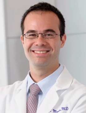 Luc G. T. Morris合作论文数Laboratory of Experimental Cancer Immunogenomics, Memorial Sloan Kettering Cancer Center99
 R Michael Tuttle合作论文数David H. Koch Center for Cancer Care at Memorial Sloan Kettering Cancer Center98
 Frank L. Palmer合作论文数80Marc Cohen合作论文数73
 Nadeem Riaz合作论文数Memorial Sloan Kettering Cancer Center69Nora Katabi合作论文数59
 Eric J. Sherman合作论文数David H. Koch Center for Cancer Care, Memorial Sloan Kettering Cancer Center59
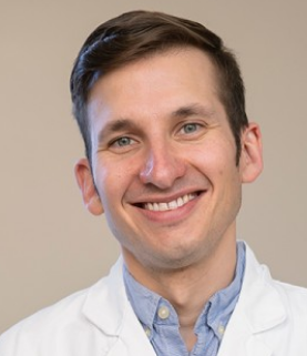 Sean McBride合作论文数Department of Radiation Oncology, Memorial Sloan Kettering Cancer Center;Northwestern University Pritzker School of Law53Migliacci Jocelyn C合作论文数53
 Cristina Valero Mayor合作论文数Mem Sloan Kettering Canc Ctr, Dept Surg, 1275 York Ave, New York, NY 10021 USA51
 Ioanna Nixon合作论文数Department of Head and Neck Surgery, Memorial Sloan Kettering Cancer Center51
 Snjezana Dogan合作论文数50
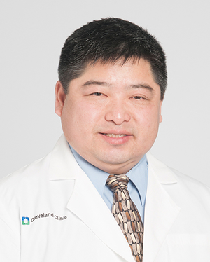 Timothy Chan合作论文数National Center for Regenerative Medicine, School of Medicine, Case Western Reserve University;Case Comprehensive Cancer Center, Case Western Reserve University;Center for Immunotherapy & Precision Immuno-Oncology, Lerner Research Institute, Cleveland Clinic44Bhuvanesh Singh合作论文数Department of Surgery, Memorial Sloan-Kettering Cancer Center, New York, NY42Jennifer R Cracchiolo合作论文数40Dunn Lara合作论文数40Daniella Karassawa Zanoni合作论文数39
 Laura Y Wang合作论文数Liverpool Hosp37
 Alan L. Ho合作论文数David H. Koch Center for Cancer Care, Memorial Sloan Kettering Cancer Center;Division of Solid Tumor Oncology, Department of Medicine, Memorial Sloan Kettering Cancer Center37
 Bin Xu (徐斌)合作论文数36
 Adilbay Dauren合作论文数Head and Neck Oncology, Astana Oncology Center36D W Scholfield合作论文数35Jay O. Boyle合作论文数33
 James A. Fagin合作论文数Division of Subspecialty Medicine, Memorial Sloan Kettering Cancer Center33Montero Pablo H合作论文数32Dennis H. Kraus合作论文数30
 David G Pfister合作论文数Memorial Sloan-Kettering Cancer Center30
 Yao Yu合作论文数29
 Paula Demétrio De Souza França合作论文数Dept Radiol, Mem Sloan Kettering Canc Ctr28
 Vladimir I. Makarov合作论文数23
 Daphna Y. Gelblum合作论文数Memorial Sloan-Kettering Cancer Center23
 Brian R. Untch合作论文数Memorial Sloan Kettering Cancer Center23C Jillian Tsai合作论文数22Danielli Matsuura合作论文数22Michel Loren合作论文数22
 Linda Chen合作论文数Dept Radiat Oncol, Mem Sloan Kettering Canc Ctr21Zakeri Kaveh合作论文数21Alana Eagan合作论文数20Stephen W Dusza合作论文数20Marco Ardigò合作论文数San Gallicano Dermatological Institute IRCCS20
 Nagavara Kishore Pillarsetty合作论文数Department of Radiology, Memorial Sloan Kettering Cancer Center;Weill Cornell Medicine20
 Milind Rajadhyaksha合作论文数Department of Medicine, Dermatology Service, Memorial Sloan Kettering Cancer Center;Department of Dermatology, Weill Cornell Medicine20




