Astrocytomas that harbor recurrent genomic alterations in MYB or MYBL1 are a group of Pediatric-type diffuse low-grade gliomas that were newly recognized in the 2021 WHO Classification of Tumors of the Central Nervous System. These tumors are described in the WHO classification as harboring fusions in MYB or MYBL1. In this report, we examine 14 consecutive cases in which a MYB or MYBL1 alteration was identified, each with diagnostic confirmation by genome-wide DNA methylation profiling (6 Angiocentric gliomas and 8 Diffuse astrocytomas, MYB- or MYBL1-altered), for their specific genomic alterations in these genes. Using RNA sequencing, we find productive in-frame fusions of the MYB or MYBL1 genes in only 5/14 cases. The remaining 9 cases show genomic alterations that result in truncation of the gene, without evidence of an in-frame fusion partner. Gene expression analysis showed overexpression of the MYB(L1) genes, regardless of the presence of a productive fusion. In addition, QKI, a recognized fusion partner common in angiocentric glioma, was generally up-regulated in these 14 cases, compared to a cohort comprising >1000 CNS tumors of various types, regardless of whether a genomic alteration in QKI was present. Overall, the results show that truncations, in the absence of a productive fusion, of the MYB(L1) genes can likely drive the tumors and have implications for the analysis and diagnosis of Angiocentric glioma and Diffuse astrocytoma, MYB- or MYBL1-altered, especially for cases that are tested on panels designed to focus on fusion detection.
Tumor/Normal paired whole exome sequencing (T/N-WES) testing permits the identification of actionable somatic variants for therapeutic decision-making, in addition to clinically significant germline variants. The identification of germline pathogenic/likely pathogenic (P/LP) variants associated with heritable conditions enables the development of a systematic management plan for the patient as well as cascade testing of at-risk relatives. However, limited resources in molecular labs specialized in the detection of somatic variants, including clinical data and personnel trained in clinical genetics, can impede germline variant curation and implementation of a comprehensive care plan.
Indeterminate dendritic cell histiocytosis (IDCH) is a rare and poorly understood entity characterized by accumulation of CD1a(+)/S100(+) histiocytes (as Langerhans cell histiocytosis [LCH]) but with reduced-absent expression of Langerin/CD207. We assembled 43 cases of IDCH (defined by CD1a(+)/CD207(<20%) immunophenotypic profile) examining the clinical, pathologic, and molecular landscape. Median age at presentation was 70 years (interquartile range, 44-80) with cutaneous (31/43; 72%) and nodal (11/43; 26%) involvement predominating. Eighteen (42%) individuals had an associated nonhistiocytic hematopoietic neoplasm ("secondary" IDCH) whereas 7 of 43 (16%) had a concurrent non-IDCH histiocytosis ("mixed" histiocytosis). Most cases exhibited morphology indistinguishable from LCH but with a CD1c(+)/CSF1R(CD115)(-) phenotype, mirroring the signature of normal indeterminate cells and conventional DC type 2. Mutational analysis revealed frequent KRAS (13/32; 41%) and BRAF p.V600E (11/36, 31%) mutations that were nearly mutually exclusive. RNA-sequencing analysis uncovered ETV3::NCOA2 fusion in 6 other patients presenting as a sole genetic alteration without any other concurrent histiocytic or hematopoietic neoplasm. BRAF and MAP2K1 alterations were significantly associated with partial/retained (1%-20%) Langerin expression (P = .005) and mixed histiocytosis (P = .002). Remarkably, myeloid alterations (DNMT3A, TET2, and SRSF2) co-occurred in IDCH tissues of several individuals. Paired sequencing of IDCH and concurrent non-IDCH hematopoietic neoplasm in 4 individuals revealed shared mutations. Age at diagnosis and any nodal involvement at diagnosis predicted inferior overall survival, but BRAF/RAS pathway alterations did not affect outcome. These data have implications for the diagnostic evaluation, classification, and therapeutic management of IDCH.
Primary cutaneous follicle center lymphoma has been distinguished from nodal follicular lymphoma (FL) based on genomic and clinical features. The nature of other extranodal FLs is not well defined. We report 15 cases of follicle center lymphoma involving the lower female genital tract. Cases were evaluated using an immunohistochemical panel for B-cell lymphoma, B-cell clonality, fluorescence in situ hybridization for BCL2 gene rearrangement, and next-generation sequencing. All patients had localized disease with no evidence of bone marrow involvement. Most cases (12/15, 80%) had a follicular pattern, at least focally. Large centrocytes were a prominent feature leading to concern for diffuse large B-cell lymphoma by referring pathologists. Neoplastic cells were positive for CD20 and BCL-6, while BCL-2 was positive in 2/15 (13%) cases. Fluorescence in situ hybridization for BCL2 gene rearrangement was negative in 10/11 (91%) cases. Next-generation sequencing performed in 10 cases revealed TNFRSF14 as the most frequently mutated gene in 6/10 (60%) cases. No case had CREBBP or KMT2D mutations as seen in nodal FL. None of the patients had progressive disease with durable complete remission achieved in 10/12 (83%) cases. The median follow-up period was 7.8 years (range: 0.2 to 20.5 y) with a 5-year overall survival of 100%. We conclude that follicle center lymphoma of the lower female genital tract is a novel variant of primary cutaneous follicle center lymphoma. Despite a frequent component of large cells, it is characterized by localized disease and low risk for dissemination. Awareness and recognition are important to distinguish these lesions from aggressive B-cell lymphomas.
Ozkaya, Neval MD; Lee, Ina MD, PhD; Johnson, Terrica S. BS; Jaffe, Elaine S. MD Author Information
A host of signature genetic alterations have been demonstrated in Spitz neoplasms, most notably fusions of kinase genes (including BRAF, ALK, ROS1, NTRK1, NTRK3, RET, MET, MAP3K8) or variants in HRAS. While there are multiple reports of rearrangements involving NTRK1 and NTRK3 in Spitz tumors, there are very few reports of NTRK2-rearranged Spitz nevi in the literature. This report presents an NTRK2-rearranged atypical Spitz tumor with spindled cell features. The patient was a 6-year-old female with a growing pigmented papule on the back. Histopathological evaluation revealed an asymmetric, biphasic, compound proliferation of melanocytes featuring an epithelioid cell population arranged as variably sized nests and single cells along the basal layer with extension down adnexa, as well as a population of spindled melanocytes with desmoplastic features and loss of Melan-A expression in the dermis. There was partial loss of p16 expression in the epidermal component and diffuse loss in the dermal component. Immunohistochemistry for PRAME, ALK, NTRK1, HRAS Q61R, p53, and BRAF V600E were negative. A SQSTM1::NTRK2 fusion was identified by RNA sequencing. No TERT promoter hotspot variants were detected. This case report expands the known histopathologic spectrum of genetic alterations in Spitz neoplasms.
Tumors of the central nervous system (CNS) often display a wide morphologic spectrum that has, until recently, been the sole basis for tumor classification. The introduction of the integrated histomolecular diagnostic approach in CNS tumors has facilitated a classification system that is increasingly data-driven and with improved alignment to clinical outcome. Here, we report a previously uncharacterized glioma type (n = 31) using unsupervised clustering analysis of DNA methylation array data from approximately 14,000 CNS tumor samples. Histologic examination revealed circumscribed growth and morphologic similarities to pleomorphic xanthoastrocytoma (PXA), astroblastoma, ependymoma, polymorphous neuroepithelial tumor of the young (PLNTY), and IDH-wildtype glioblastoma (GBM). Median age (46.5 years) was significantly older than other circumscribed gliomas and younger than GBM. Dimensionality reduction with uniform manifold approximation and projection (UMAP) and hierarchical clustering confirmed a methylation signature distinct from known tumor types and methylation classes. DNA sequencing revealed recurrent mutations in TP53 (57%), RB1 (26%), NF1 (26%), and NF2 (14%). BRAF V600E mutations were detected in 3/27 sequenced cases (12%). Copy number analysis showed increased whole chromosome aneuploidy with recurrent loss of chromosome 13 (28/31 cases, 90%). CDKN2A/B deletion (2/31, 6%) and MGMT promoter methylation (1/31, 3%) were notably rare events. Most tumors showed features of a high-grade glioma, yet survival data showed significantly better overall survival compared to GBM (p < 0.0001). In summary, we describe a previously uncharacterized glioma of adults identified by a distinct DNA methylation signature and recurrent loss of chromosome 13.
 Mark Raffeld合作论文数Laboratory of Pathology, Center for Cancer Research, National Cancer Institute8
 Liqiang Xi合作论文数5
 Elaine S. Jaffe合作论文数Laboratory of Pathology, Center for Cancer Research, National Cancer Institute, National Institutes of Health5
 Svetlana D. Pack合作论文数Center for Cancer Research, National Cancer Institute5
 Stefania Pittaluga合作论文数Laboratory of Pathology, Center for Cancer Research, National Cancer Institute, National Institutes of Health4
 Manoj Tyagi合作论文数Laboratoire de Biochimie et Génétique Moléculaire;Université de La Réunion;Laboratoire de Biochimie et Génétique Moléculaire, Université de La Réunion4
 Neval Ozkaya合作论文数NCI3Antonios Papanicolau-Sengos合作论文数3
 Kenneth Aldape合作论文数Laboratory of Pathology, Center for Cancer Research, National Cancer Institute, National Institutes of Health3
 Kim Jung合作论文数Feinberg School of Medicine, Northwestern University3
 Maxime Battistella合作论文数2Hye-Jung Chung合作论文数2
 Annapurna Saksena合作论文数Center for Cancer Research (CCR),, National Cancer Institute (NCI),2Jean-François Emile合作论文数Université de Versailles Saint-Quentin-en-Yvelines2Hélias-Rodzewicz Zofia合作论文数2
 Ashish Jain合作论文数Clinical Immunology Unit, Laboratory of Host Defenses,2
 Zied Abdullaev合作论文数Laboratory of Pathology, Center for Cancer Research, National Cancer Institute2Anne Moreau合作论文数Centre Hospitalier Universitaire De Nantes2Jean Donadieu合作论文数2
 Sylvie Fraitag合作论文数Departement de Pathologie, Assistance Publique – Hôpitaux de Paris;Service d'Anatomie et Cytologie Pathologique, Hôpital Necker-Enfants Malades2Julien Haroche合作论文数2
 Martha Quezado合作论文数Laboratory of Pathology, Center for Cancer Research, National Cancer Institute, National Institutes of Health2François Le Pelletier合作论文数1Gregory S. Phillips合作论文数1Laszlo J. Karai合作论文数1Courtney Ketchum合作论文数1Stephanie Mengden Koon合作论文数1Grace-Ann Fasaye合作论文数1Joshua Klonoski合作论文数1
 Pavalan Selvam合作论文数1Sharika Rajan合作论文数From the Department of Neurology (C.G., N.W., R.F.),, Beth Israel Deaconess Medical Center;Department of Pathology (S.R.),, NIH;Department of Neurology (D.K.),, University of Colorado;and Department of Neurology (J.-A.P., H.K.),, NYU Grossman School of Medicine;NYU Grossman School of Medicine, University of Colorado1Jina Chung合作论文数1Julie Dhossche合作论文数1
 Matthew McCord合作论文数Feinberg School of Medicine, Northwestern University1
 Christina K Ferrone合作论文数Department of Pathology and Molecular Medicine, Queen's University1
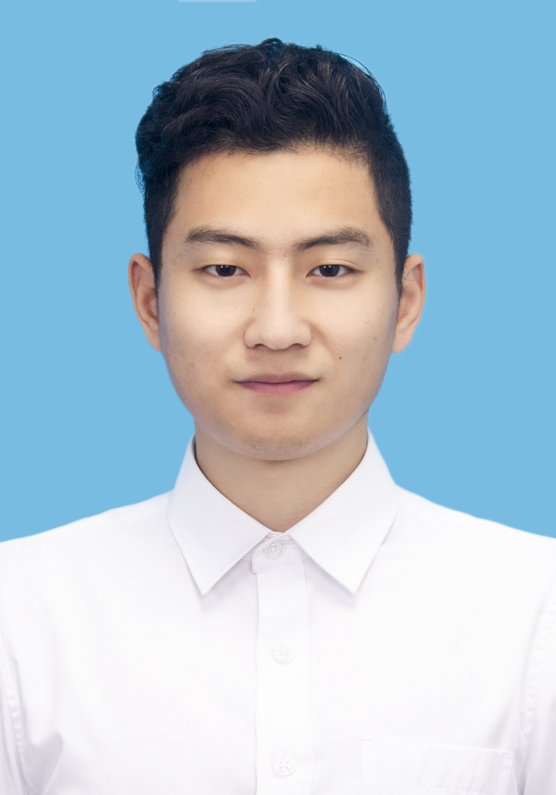 Yi Liu (刘易)合作论文数Department of Occupational Medicine and Environmental Health, School of Public Health, Nanjing Medical University1Christopher H Dampier合作论文数1
 Jerome Razanamahery合作论文数1Michael R Sargen合作论文数National Cancer Institute, National Institutes of Health1
 Craig M Horbinski合作论文数Department of Pathology, Feinberg School of Medicine, Northwestern University1
 Drew W. Pratt合作论文数National Cancer Institute, National Institutes of Health/University of Michigan1
 Philip E. LeBoit合作论文数School of Medicine, University of California, San Francisco1
 Kathleen A Calzone合作论文数NCI, NIH1Lebensohn Alexandra合作论文数1
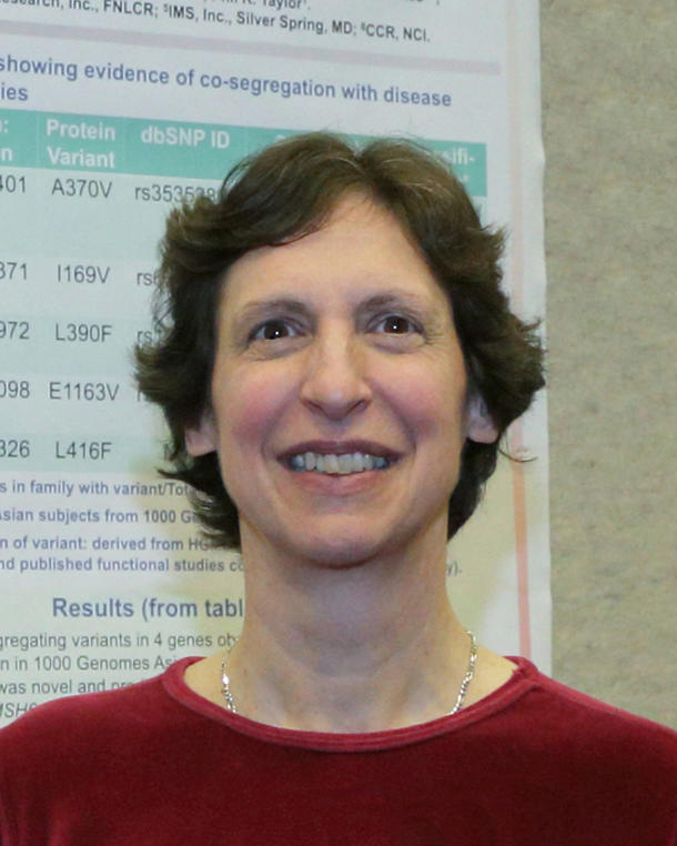 Alisa M. Goldstein合作论文数Division of Cancer Epidemiology and Genetics, National Cancer Institute1
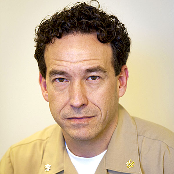 Stephen M. Hewitt合作论文数Laboratory of Pathology, Center for Cancer Research, National Cancer Institute, National Institutes of Health1
 Kesserwan Chimene合作论文数National Human Genome Research Institute, National Institutes of Health1Borys Ewa合作论文数1
 Arie Perry合作论文数Division of Neuropathology, Departments of Pathology and Laboratory Medicine, School of Medicine, University of California, San Francisco1




