BACKGROUND:Early-onset hearing loss is mostly of genetic origin. The complexity of the hearing process is reflected by its extensive genetic heterogeneity, with probably many causative genes remaining to be identified. Here, we aimed at identifying the genetic basis for autosomal dominant non-syndromic hearing loss (ADNSHL) in a large German family. METHODS:A panel of 66 known deafness genes was analyzed for mutations by next-generation sequencing (NGS) in the index patient. We then conducted genome-wide linkage analysis, and whole-exome sequencing was carried out with samples of two patients. Expression of Osbpl2 in the mouse cochlea was determined by immunohistochemistry. Because Osbpl2 has been proposed as a target of miR-96, we investigated homozygous Mir96 mutant mice for its upregulation. RESULTS:Onset of hearing loss in the investigated ADNSHL family is in childhood, initially affecting the high frequencies and progressing to profound deafness in adulthood. However, there is considerable intrafamilial variability. We mapped a novel ADNSHL locus, DFNA67, to chromosome 20q13.2-q13.33, and subsequently identified a co-segregating heterozygous frameshift mutation, c.141_142delTG (p.Arg50Alafs*103), in OSBPL2, encoding a protein known to interact with the DFNA1 protein, DIAPH1. In mice, Osbpl2 was prominently expressed in stereocilia of cochlear outer and inner hair cells. We found no significant Osbpl2 upregulation at the mRNA level in homozygous Mir96 mutant mice. CONCLUSION:The function of OSBPL2 in the hearing process remains to be determined. Our study and the recent description of another frameshift mutation in a Chinese ADNSHL family identify OSBPL2 as a novel gene for progressive deafness.
Determination of variant pathogenicity represents a major challenge in the era of high-throughput sequencing. Erroneous categorization may result if variants affect genes that are in fact dispensable. We demonstrate that this also applies to rare, apparently unambiguous truncating mutations of an established disease gene. By whole-exome sequencing (WES) in a consanguineous family with congenital non-syndromic deafness, we unexpectedly identified a homozygous nonsense variant, p.Arg1066*, in AHI1, a gene associated with Joubert syndrome (JBTS), a severe recessive ciliopathy. None of four homozygotes expressed any signs of JBTS, and one of them had normal hearing, which also ruled out p.Arg1066* as the cause of deafness. Homozygosity mapping and WES in the only other reported JBTS family with a homozygous C-terminal truncation (p.Trp1088Leufs*16) confirmed AHI1 as disease gene, but based on a more N-terminal missense mutation impairing WD40-repeat formation. Morpholinos against N-terminal zebrafish Ahi1, orthologous to where human mutations cluster, produced a ciliopathy, but targeting near human p.Arg1066 and p.Trp1088 did not. Most AHI1 mutations in JBTS patients result in truncated protein lacking WD40-repeats and the SH3 domain; disease was hitherto attributed to loss of these protein interaction modules. Our findings indicate that normal development does not require the C-terminal SH3 domain. This has far-reaching implications, considering that variants like p.Glu984* identified by preconception screening ('Kingsmore panel') do not necessarily indicate JBTS carriership. Genomes of individuals with consanguineous background are enriched for homozygous variants that may unmask dispensable regions of disease genes and unrecognized false positives in diagnostic large-scale sequencing and preconception carrier screening.
Although many genes have been identified for the autosomal recessive cerebellar ataxias (ARCAs), several patients are unlinked to the respective loci, suggesting further genetic heterogeneity. We combined homozygosity mapping and exome sequencing in a consanguineous Egyptian family with congenital ARCA, mental retardation and pyramidal signs. A homozygous 5-bp deletion in SPTBN2, the gene whose in-frame mutations cause autosomal dominant spinocerebellar ataxia type 5, was shown to segregate with ataxia in the family. Our findings are compatible with the concept of truncating SPTBN2 mutations acting recessively, which is supported by disease expression in homozygous, but not heterozygous, knockout mice, ataxia in Beagle dogs with a homozygous frameshift mutation and, very recently, a homozygous SPTBN2 nonsense mutation underlying infantile ataxia and psychomotor delay in a human family. As there was no evidence for mutations in 23 additional consanguineous families, SPTBN2-related ARCA is probably rare.
The mechanisms underlying retinal dystrophy in Usher syndrome type I (USH1) remain unknown because mutant mice lacking any of the USH1 proteins-myosin VIIa, harmonin, cadherin-23, protocadherin-15, sans-do not display retinal degeneration. We found here that, in macaque photoreceptor cells, all USH1 proteins colocalized at membrane interfaces (i) between the inner and outer segments in rods and (ii) between the microvillus-like calyceal processes and the outer segment basolateral region in rods and cones. This pattern, conserved in humans and frogs, was mediated by the formation of an USH1 protein network, which was associated with the calyceal processes from the early embryonic stages of outer segment growth onwards. By contrast, mouse photoreceptors lacked calyceal processes and had no USH1 proteins at the inner-outer segment interface. We suggest that USH1 proteins form an adhesion belt around the basolateral region of the photoreceptor outer segment in humans, and that defects in this structure cause the retinal degeneration in USH1 patients.
BACKGROUND:Usher syndrome (USH) is an autosomal recessive genetically heterogeneous disorder with congenital sensorineural hearing impairment and retinitis pigmentosa (RP). We have identified a consanguineous Lebanese family with two affected members displaying progressive hearing loss, RP and cataracts, therefore clinically diagnosed as USH type 3 (USH3). Our study was aimed at the identification of the causative mutation in this USH3-like family.METHODS:Candidate loci were identified using genomewide SNP-array-based homozygosity mapping followed by targeted enrichment and next-generation sequencing.RESULTS:Using a capture array targeting the three identified homozygosity-by-descent regions on chromosomes 1q43-q44, 20p13-p12.2 and 20p11.23-q12, we identified a homozygous nonsense mutation, p.Arg65X, in ABHD12 segregating with the phenotype.CONCLUSION:Mutations of ABHD12, an enzyme hydrolyzing an endocannabinoid lipid transmitter, cause PHARC (polyneuropathy, hearing loss, ataxia, retinitis pigmentosa, and early-onset cataract). After the identification of the ABHD12 mutation in this family, one patient underwent neurological examination which revealed ataxia, but no polyneuropathy. ABHD12 is not known to be related to the USH protein interactome. The phenotype of our patient represents a variant of PHARC, an entity that should be taken into account as differential diagnosis for USH3. Our study demonstrates the potential of comprehensive genetic analysis for improving the clinical diagnosis.
Joubert syndrome (JBTS) is characterized by a specific brain malformation with various additional pathologies. It results from mutations in any one of at least 10 different genes, including NPHP1, which encodes nephrocystin-1. JBTS has been linked to dysfunction of primary cilia, since the gene products known to be associated with the disorder localize to this evolutionarily ancient organelle. Here we report the identification of a disease locus, JBTS12, with mutations in the KIF7 gene, an ortholog of the Drosophila kinesin Costal2, in a consanguineous JBTS family and subsequently in other JBTS patients. Interestingly, KIF7 is a known regulator of Hedgehog signaling and a putative ciliary motor protein. We found that KIF7 co-precipitated with nephrocystin-1. Further, knockdown of KIF7 expression in cell lines caused defects in cilia formation and induced abnormal centrosomal duplication and fragmentation of the Golgi network. These cellular phenotypes likely resulted from abnormal tubulin acetylation and microtubular dynamics. Thus, we suggest that modified microtubule stability and growth direction caused by loss of KIF7 function may be an underlying disease mechanism contributing to JBTS.
Das Usher Syndrom ist eine genetisch heterogene Erkrankung, die sich mit Hörverlust und Renitis pigmentosa manifestiert. Häufig zeigen sich unerklärliche, oft innerfamiliäre Unterschiede des retinalen Phänotyps. Obwohl 9 ursächliche Usher Gene bekannt sind, können bei vielen der Patienten keine Mutationen in diesen Genen nachgewiesen werden, so dass es wahrscheinlich verschiedene weitere, bisher nicht identifizierte Usher Gene gibt. Wir zeigen hier, dass Mutationen im Gen PDZ domain containing 7 (PDZD7), das für ein Homolog der Usher Proeine USH1C und USH2D kodiert, zum Usher Syndrom beitragen. Mutationen in PDZD7 fanden sich interessanterweise nur bei Patienten mit Mutationen in anderen Usher Genen. In zwei Schwestern, die jeweils eine homozygote USH2A Mutation aufwiesen, fand sich eine frame-shift Mutation in der Schwester, die den schwereren Phänotyp und den früheren Krankheitsbeginn aufwies. Des Weiteren fanden sich heterozygote Mutatonen in Patienten mit trunkierenden Mutationen in den Genen USH2A, USH2C (GPR98) und einem weiteren unidentifizierten Locus. Interessanterweise fanden wir, dass das Protein PDZD7 mit den Proteinen USH2A und USH2C interagiert. Bestätigt wurden unsere Befunde außerdem im Zebrafisch Tiermodell. Der Knockdown von pdzd7 im Zebrafisch führte zu einem Usher-ähnlichen Phänotyp, in Kombination mit ush2a oder gpr98 Knockdown zu verstärkter Apoptose in der Retina und zu einer verringerten Lokalisation des Proteins Gpr98 am Verbindungszilium des Photorezeptors. Die neuen Daten deuten auf eine digenische Vererbung von PDZD7 und USH2C hin, sowie auf eine Rolle von PDZD7 als Modifikator der retinalen Erkrankung bei USH2A Patienten. Beim Usher Syndrom handelt es sich somit nicht immer um eine rein mendelische, sondern auch um eine oligogenetisch vererbte Erkrankung.
Usher syndrome is a genetically heterogeneous recessive disease characterized by hearing loss and retinitis pigmentosa (RP). It frequently presents with unexplained, often intrafamilial, variability of the visual phenotype. Although 9 genes have been linked with Usher syndrome, many patients do not have mutations in any of these genes, suggesting that there are still unidentified genes involved in the syndrome. Here, we have determined that mutations in PDZ domain-containing 7 (PDZD7), which encodes a homolog of proteins mutated in Usher syndrome subtype 1C (USH1C) and USH2D, contribute to Usher syndrome. Mutations in PDZD7 were identified only in patients with mutations in other known Usher genes. In a set of sisters, each with a homozygous mutation in USH2A, a frame-shift mutation in PDZD7 was present in the sister with more severe RP and earlier disease onset. Further, heterozygous PDZD7 mutations were present in patients with truncating mutations in USH2A, G protein-coupled receptor 98 (GPR98; also known as USH2C), and an unidentified locus. We validated the human genotypes using zebrafish, and our findings were consistent with digenic inheritance of PDZD7 and GPR98, and with PDZD7 as a retinal disease modifier in patients with USH2A. Pdzd7 knockdown produced an Usher-like phenotype in zebrafish, exacerbated retinal cell death in combination with ush2a or gpr98, and reduced Gpr98 localization in the region of the photoreceptor connecting cilium. Our data challenge the view of Usher syndrome as a traditional Mendelian disorder and support the reclassification of Usher syndrome as an oligogenic disease.
Purpose: It has been demonstrated that mutations in deafness, autosomal recessive 31 (DFNB31), the gene encoding whirlin, is responsible for nonsyndromic hearing loss (NSHL; DFNB31) and Usher syndrome type II (USH2D). We screened DFNB31 in a large cohort of patients with different clinical subtypes of Usher syndrome (USH) to determine the prevalence of DFNB31 mutations among USH patients.Methods: DFNB31 was screened in 149 USH2, 29 USH1, six atypical USH, and 11 unclassified USH patients from diverse ethnic backgrounds. Mutation detection was performed by direct sequencing of all coding exons.Results: We identified 38 different variants among 195 patients. Most variants were clearly polymorphic, but at least two out of the 15 nonsynonymous variants (p.R350W and p.R882S) are predicted to impair whirlin structure and function, suggesting eventual pathogenicity. No putatively pathogenic mutation was found in the second allele of patients with these mutations.Conclusions: DFNB31 is not a major cause of USH.
Mutations in the large GPR98 gene underlie Usher syndrome type 2C (USH2C), and all patients described to date have been female. It was speculated that GPR98 mutations cause a more severe, and eventually lethal, phenotype in males. We describe for the first time two male patients with USH2 with novel GPR98 mutations. Clinical characterization of a male patient and his affected sister revealed a typical USH2 phenotype in both. GPR98 may have been excluded from systematic investigation in previous studies, and the proportion of patients with USH2C probably underestimated. GPR98 should be considered in patients with USH2 of both sexes.
Aim: To characterise the ocular phenotype of a family segregating the splice site mutation c.2189+1G>T in the tyrosine kinase receptor gene MERTK.Methods: Five affected children of a consanguineous Moroccan family were investigated by ophthalmic examinations, including fundus photography, autofluorescence (FAF) imaging, optical coherence tomography (OCT), psychophysical and electrophysiological methods.Results: Affected children were between 5 and 19 years of age, allowing an estimation of disease progression. Electroretinography demonstrated loss of scotopic and photopic function in the first decade of life. Younger siblings showed drusen-like deposits with focal relatively increased FAF in the macular area. With increasing age, a yellowish lesion with relatively increased FAF and subsequent macular atrophy developed. Visual acuity deteriorated with age and ranged between 20/50 in the best eye of the youngest affected and 20/400 in the worst eye of the oldest affected sibling. Spectral-domain OCT revealed debris-like material in the subneurosensory space.Conclusion: The splice site mutation c.2189+1G>T in MERTK causes rod-cone dystrophy with a distinct macular phenotype. The debris in the subneurosensory space resembles that in the Royal College of Surgeons (RCS) rat being the mertk animal model. Patients might therefore benefit from advances in gene therapy that were previously achieved in the RCS rat.
We have earlier described a syndrome characterized by microcephaly, cutis verticis gyrata, retinitis pigmentosa, cataracts, hearing loss and mental retardation (Mendelian inheritance in man (MIM) no: 605685) in two brothers from a non-consanguineous Lebanese family. In view of the rarity of the disorder and the high rate of inbreeding in the Lebanese population, we assumed an autosomal recessive trait inherited from a common ancestor. A genomewide scan was performed. The single locus on the long arm of chromosome 8 that showed homozygosity by descent comprised the gene responsible for Cohen syndrome (CS), VPS13B. We then sequenced VPS13B in the patients and found a homozygous splice site mutation. Several possible explanations for the overlap between CS and the clinical features observed in our patients are discussed. Our data highlight the potential of high-resolution homozygosity mapping in small populations with a high rate of inbreeding.
Congenital hearing loss affects approximately one child in 1000. About 10% of the deaf population have Usher syndrome (USH). In USH, hearing loss is complicated by retinal degeneration with onset in the first (USH1) or second (USH2) decade. In most populations, diagnostic testing is hampered by a multitude of mutations in nine genes. We have recently shown that in French Canadians from Quebec, USH1 largely results from a single USH1C founder mutation, c.216G>A (‘Acadian allele’). The genetic basis of USH2 in Canadians of French descent, however, has remained elusive. Here, we have investigated nine USH2 families from Quebec and New Brunswick (the former Acadia) by haplotype analyses of the USH2A locus and sequencing of the three known USH2 genes. Seven USH2A mutations were identified in eight patients. One of them, c.4338_4339delCT, accounts for 10 out of 18 disease alleles (55.6%). This mutation has previously been reported in an Acadian USH2 family, and it was found in homozygous state in the three Acadians of our sample. As in the case of c.216G>A (USH1C), a common haplotype is associated with c.4338_4339delCT. With a limited number of molecular tests, it will now be possible in these populations to estimate whether children with congenital hearing impairment of different degrees will develop retinal disease – with important clinical and therapeutic implications. USH2 is the second example that reveals a significant genetic overlap between Quebecois and Acadians: in contrast to current understanding, other genetic disorders present in both populations are likely based on common founder mutations as well.
Usher syndrome (USH) is an autosomal recessive condition characterized by sensorineural hearing loss, vestibular dysfunction, and visual impairment due to retinitis pigmentosa. Truncating mutations in the cadherin‐23 gene (CDH23) result in Usher syndrome type 1D (USH1D), whereas missense mutations affecting strongly conserved motifs of the CDH23 protein cause non‐syndromic deafness (DFNB12). Four missense mutations constitute an exception from this genotype‐phenotype correlation: they have been described in USH1 patients in homozygous state. Using a minigene assay, we have investigated these changes (c.1450G>C, p.A484P; c.3625A>G, p.T1209A; c.4520G>A, p.R1507Q; and c.5237G>A, p.R1746Q) for a possible impact on mRNA splicing which could explain the syndromic phenotype. While in silico analysis suggested impairment of splicing in all four cases, we found aberrant splicing for only one mutation, p.R1746Q. However, splicing was normal in case of p.A484P, p.T1209A and p.R1507Q. These three latter CDH23 missense mutations could interfere with functions of both, the auditory and the visual system. Alternatively, they could represent rare non‐pathogenic polymorphisms. © 2008 Wiley‐Liss, Inc.
Congenital indifference to pain (CIP, MIM #243000) is rare (<30 cases reported). We describe a consanguineous family from Egypt with the index patient displaying lesions typical of CIP. In addition, he had short stature, myopathy, and liver dysfunction—a combination typical of Navajo neurohepatopathy (NNH, MIM #256810). No mutation was found in MPV17 , the gene primarily involved in NNH.1 Pain perception was normal in a sister with congenital liver disease. Detailed genetic and clinical investigation revealed two separate rare disorders in the index patient, glycogen storage disease type 3 (GSD III; MIM #232400) and CIP, based on homozygous mutations in the genes AGL and SCN9A . ### Case report. The index patient (IV-10, age 11 years) and his sister (IV-11, age 1 year 10 months) underwent detailed clinical assessment. Starting at 2 years of age, IV-10 developed frequent painless fractures of legs and arms, bruises, cuts, and abdominal enlargement. On recent examination, he had short stature (<3 SD), corneal ulcerations, self-mutilative injuries of lips, skin, and fingers, and limb deformities due to untreated fractures (figure). There was mild hypotonia in lower limbs, flexors of hips and trunk muscles, and hypesthesia in both arms. Liver enzymes (AST: 260 U/L, ALT: 607 U/L), creatine phosphokinase (CPK: 341 U/L), and serum triglycerides (214 mg%) were markedly elevated. Generalized decrease of bone density was noted (dual energy X-ray absorptiometry). Hepatosplenomegaly, hepatocyte swelling, and periportal fibrosis were consistent with GSD (ultrasonography, …
Usher syndrome is an autosomal recessive condition characterized by sensorineural hearing loss, variable vestibular dysfunction, and visual impairment due to retinitis pigmentosa (RP). The seven proteins that have been identified for Usher syndrome type 1 (USH1) and type 2 (USH2) may interact in a large protein complex. In order to identify novel USH genes, we followed a candidate strategy, assuming that mutations in proteins interacting with this “USH network” may cause Usher syndrome as well. The DFNB31 gene encodes whirlin, a PDZ scaffold protein with expression in both hair cell stereocilia and retinal photoreceptor cells. Whirlin represents an excellent candidate for USH2 because it binds to Usherin (USH2A) and VLGR1b (USH2C). Genotyping of microsatellite markers specific for the DFNB31 gene locus on chromosome 9q32 was performed in a German USH2 family that had been excluded for all known USH loci. Patients showed common haplotypes. Sequence analysis of DFNB31 revealed compound heterozygosity for a nonsense mutation, p.Q103X, in exon 1, and a mutation in the splice donor site of exon 2, c.837+1G>A. DFNB31 mutations appear to be a rare cause of Usher syndrome, since no mutations were identified in an additional 96 USH2 patients. While mutations in the C-terminal half of whirlin have previously been reported in non-syndromic deafness (DFNB31), both alterations identified in our USH2 family affect the long protein isoform. We propose that mutations causing Usher syndrome are probably restricted to exons 1–6 that are specific for the long isoform and probably crucial for retinal function. We describe a novel genetic subtype for Usher syndrome, which we named USH2D and which is caused by mutations in whirlin. Moreover, this is the first case of USH2 that is allelic to non-syndromic deafness.
BACKGROUND:Usher syndrome type 1 (USH1) is the leading cause of deafblindness. In most populations, many private mutations are distributed across the five known USH1 genes. We investigated patients from the French Canadian population of Quebec (approximately 6 million people) that descends from about 8,500 French settlers who colonized the St Lawrence River valley between 1608 and 1759. We hypothesized that founder mutations in USH1 genes exist in this population.RESULTS:We have genetically characterized 15 patients from different regions of Quebec who were clinically diagnosed as USH1. Of these cases, 60% carried mutations of the USH1C gene, a genetic subtype that is rare outside the Acadian population. We have discovered a founder effect of the c.216G>A mutation, which has previously been designated the 'Acadian allele' because it accounts for virtually all Acadian USH1 cases. It represents 40% of disease alleles in Quebec, and a carrier of c.216G>A was identified in the general population. Mutations in other genes, except CDH23, are very rare.CONCLUSION:Based on our findings, approximately 0.5% of congenitally deaf children in Quebec are at risk of developing retinal degeneration due to homozygosity for c.216G>A. Although the Acadians and French Canadians from Quebec are descended from French ancestors, they have always been considered genetically distinct. The genetic conditions common in Quebec are generally not found in Acadians, or they are due to different mutations. Our results, however, show that carriers of the c.216G>A allele haplotype belonged to the early founders of both the Acadian and the Quebec population.
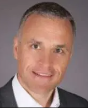 Hanno Bolz合作论文数Institute of Human Genetics, University Hospital of Cologne20
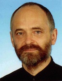 Peter Nürnberg合作论文数Cologne Center for Genomics, University of Cologne10
 Gudrun Nürnberg合作论文数10Irma Lopez合作论文数6
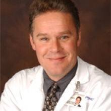 Robert K. Koenekoop合作论文数Department of Pediatric Surgery, Faculty of Medicine and Health Sciences, McGill University5
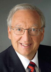 Eberhart Zrenner合作论文数Werner Reichardt Centrum für Integrative Neurowissenschaften (CIN) Institute for Ophthalmic Research Centre for Ophthalmology, University of Tübingen5
 Max C Liebau合作论文数Medical Faculy, University of Cologne;Social-Pediatric Center, Department of Pediatrics, University Hospital of Cologne;University Hospital RWTH Aachen4
 Jennifer B Phillips合作论文数Institute of Molecular Biology, University of Oregon4
 Thomas Benzing合作论文数Clinic II for Internal Medicine, University Hospital of Cologne;Department of Medicine II, University of Cologne;Kidney Research Center, University of Cologne4Claudia Dafinger合作论文数4
 Monte Westerfield合作论文数Department of Biology, College of Arts and Sciences, University of Oregon;Institute of Neuroscience, Office of the Vice President for Research and Innovation, University of Oregon;Zebrafish International Resource Center, University of Oregon4Solaf M. Elsayed合作论文数Yassin Abdelghaffar Charity Center for Liver Disease and Research4
 Bernhard Schermer合作论文数Renal Division;University Hospital Freiburg;Department of Molecular and Cellular Biology;Baylor College of Medicine;Baylor College of Medicine, University Hospital Freiburg4
 Rima Slim合作论文数Departments of Human Genetics and Obstetrics and Gynecology, McGill University Health Centre Research Institute3Thoenes Michaela合作论文数3
 Peter Charbel Issa合作论文数Department of Ophthalmology, University of Bonn3
 Ezzat Elsobky合作论文数3Friederike Koerber合作论文数2
 Jose M Millan合作论文数2Elena Aller合作论文数2
 Maha S Zaki合作论文数Natl Res Ctr, Human Genet & Genome Res Div, Dept Clin Genet, Cairo 12311, Egypt2
 Holger Thiele合作论文数Department of Internal Medicine/Cardiology, Heart Center Leipzig, University of Leipzig2Heller Raoul合作论文数2Eisenberger Tobias合作论文数2Carsten Bergmann合作论文数University of Freiburg2R Pigeon合作论文数2
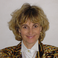 Mireille Claustres合作论文数Laboratoire de Genetique de Maladies Rares, EA 7402 Universite de Montpellier2
 Eugen Boltshauser合作论文数Department of Neuropaediatrics, Children’s Hospital Zürich2Anne-Françoise Roux合作论文数2
 Hendrik Scholl合作论文数Institute of Molecular and Clinical Ophthalmology Basel;Department of Ophthalmology, University of Basel;Department of Clinical Research, University of Basel;Department of Ophthalmology, University Hospital Basel;European Vision Institute2Antje Bernd合作论文数2Ellen Schäfer合作论文数2
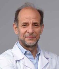 Andre Megarbane合作论文数Department of Human Genetics, Gilbert and Rose-Marie Chagoury School of Medicine, Lebanese American University2
 Elvir Becirovic合作论文数2Christian Becker合作论文数1E Elsobky合作论文数1H. Loewenheim合作论文数1Bernhard Jurklies合作论文数1Iman Sahly合作论文数1Amrit Estivalet合作论文数1Morag A Lewis合作论文数1María José Aparisi合作论文数1G. Nuernberg合作论文数1
 Vincent Michel合作论文数1
 Christian Decker合作论文数1
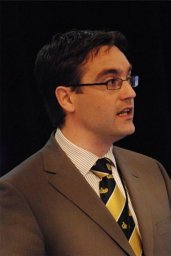 Ronald Pennings合作论文数Donders Inst Brain Cognit & Behav, Radboudumc1Daniel Swan合作论文数1
 Maria Iribarne合作论文数1Gema García-García合作论文数1




