Opal and CODEX multiplexed tissue imaging data shows infiltration of proliferative CD8+ T-cells to the rejected SCCs.
Differentially expressed genes in tumor compartment at days 0, 6, 10, and 16 post-inoculation.
CellChat and CellPhoneDB analyses reveal increased MIF signaling from tumor cells to macrophages in non-rejected clones.
Immunopeptidomics and TCR sequencing analyses showed similar results for the rejected and non-rejected SCCs.
Cell types from scRNA-seq of tumors from mice inoculated with rejected or non-rejected SCCs.
CyTOF analysis comparing tumors derived from Mif WT and Mif KO validating the scRNAseq findings.
Rejection and non-rejection gene sets identified in the scRNA sequencing analysis can stratify patients with high/low ITH and hot/cold tumor annotations.
 Shifra Ben-Dor合作论文数The Department of Life Sciences Core Facilities, Faculty of Biochemistry, Weizmann Institute of Science23
 Liat Fellus Alyagor合作论文数Dept Vet Resources, Weizmann Inst Sci21
 Roni Oren合作论文数Weizmann Inst Sci, Dept Vet Resources, Rehovot, Israel21
 Lipika R. Pal合作论文数21
 Bartok Osnat合作论文数21
 Golani Ofra合作论文数21Ronen Levy合作论文数21
 Yardena Samuels合作论文数The Sidney Kimmel Comprehensive Cancer Center and The Howard Hughes Medical Institute, The Johns Hopkins University Medical Institutions21Julia Martínez Gómez合作论文数20
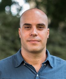 Dahan Rony合作论文数Faculty of Biology, Technion-Israel Institute of Technology20
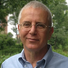 Eytan Ruppin合作论文数Center for Bioinformatics and Computational Biology , University of Maryland;Center for Cancer Research, National Cancer Institute, The National Institutes of Health19
 Ofer Yifrach合作论文数10Lewin Limor合作论文数4
 Uri Abdu合作论文数Department of Life Sciences, Ben-Gurion University of the Negev4
 Tamar Sapir合作论文数Department of Molecular Genetics;Weizmann Institute of Science;Department of Molecular Genetics, Weizmann Institute of Science4Nitzan Zandany合作论文数4Talia Levy合作论文数3Elhanan Magidovich合作论文数3Orly Reiner合作论文数3Joy A Pai合作论文数2
 Tzilhav shem-ad合作论文数2
 Shir Marciano合作论文数2Esraa Nsasra合作论文数2Valerie Nirenberg合作论文数2Yingying Cao合作论文数2Yajie Yin合作论文数2Peter P Du合作论文数2Lihi Radomir合作论文数Weizmann Inst Sci, Dept Immunol, IL-76100 Rehovot, Israel2Inbar Cohen-Gihon合作论文数2
 Ansuman Satpathy合作论文数Parker Institute for Cancer Immunotherapy, Stanford University;Wing Venture Capital;Arpelos Biosciences;Santa Ana Bio, Inc.;Cartography Biosciences;Immunai2
 Gregor Eichele合作论文数Max Planck Institute for Multidisciplinary Sciences, Max-Planck-Gesellschaft;Göttingen Graduate Center for Neurosciences, Biophysics, and Molecular Biosciences, Georg-August-Universität Göttingen2Cheng Kuoyuan合作论文数School of Pharmaceutical Sciences, Peking University2Yehezkel Rinat合作论文数2
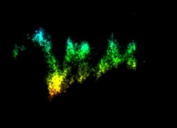 Goliand Inna合作论文数Department of Life Sciences and the National Institute for Biotechnology in the Negev (NIBN),, Ben-Gurion University of the Negev2
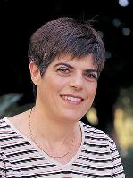 Naama Barkai合作论文数Department of Molecular Genetics, Weizmann Institute of Science2Shirly Becker-Herman合作论文数2
 Michal Shapira合作论文数Department of Chemistry, Technion - Israel Institute of Technology2Ofir Israeli合作论文数2
 Idit Shachar合作论文数2Matthias P. Kramer合作论文数Cell Cycle Regulation Group, Department of Medicine I, Division: Institute of Cancer Research, Medical University of Vienna, Borschkegasse 8 a, A-1090 Vienna, Austria2
 Alejandro A. Schäffer (Alejandro Schaffer)合作论文数National Center for Biotechnology Information, National Institutes of Health;Center for Cancer Research, National Cancer Institute, National Institutes of Health2Sven Bergmann合作论文数2
 Frederic M. Coquelle合作论文数Department of Molecular Genetics, Weizmann Institute of Science;Equipe SDM, Université de Rennes 12Federica Sella合作论文数1Michèle Welti合作论文数1Eszter Bakos合作论文数1Amir Pri-Or合作论文数1Winnie Yao合作论文数1Maya David合作论文数1




