Conventional polarimetry, including schemes leveraging entangled light, characterizes optical samples through linear transformations of polarization states. We introduce a two-photon probing approach in which both photons of an entangled pair interact with the same depolarizing medium simultaneously. In this regime, the transformation of the two-photon polarization correlations becomes quadratic in the Mueller matrix, enabling access to second-order polarization information beyond conventional polarimetry. We develop a theoretical framework linking the Mueller matrix to the evolution of the two-photon polarization correlation tensor and show that depolarization induces quadratic degradation of entanglement and state purity. Experiments using polarization-entangled photon pairs transmitted through controlled scattering media confirm the predicted response and reveal enhanced sensitivity to polarization scrambling compared with single-photon probing. These results establish two-photon probing as a higher-order quantum polarimetric modality for characterizing polarization channels.
We report the implementation of a flow-imaging platform suitable for characterization of cerebral vascular function of small animals. Consisting of an ultra-high-speed streaming camera that ensures throughput up to 200 000 frames per second and high performance PC workstation with an in-house developed processing software, this platform is currently being deployed as part of the infrastructure at the Biocenter Oulu, allowing researchers to conduct detailed examination of cerebral circulation alterations which can be caused by various factors. By utilizing top level hardware and the most recent developments in imaging with dynamic light scattering methods, such as Diffuse Correlation Spectroscopy (DCS), the spatial and temporal resolution of transcranial blood flow imaging is significantly enhanced, allowing to perform close to real-time data visualization and analysis with high efficiency. The proposed platform is designed to serve a diverse range of target groups, each of whom can benefit from its innovative technology, such as pharmaceutical companies, HealthTech startups, research institutions, medical device manufacturers and wearable technology companies. By catering to these key sectors, the platform aims to drive advancements in healthcare, research, and technology.
We investigate twisted light propagation in turbid media, demonstrating phase memory retention despite microscopic scattering. Simulations and experiments reveal high refractive index sensitivity (10(-6)) and a novel phase preservation framework, benefiting biomedical imaging, secure optics, and precision applications. (c) 2025 The Authors
Conical refraction (CR) is a phenomenon observed when light is transformed into a hollow slanted cone propagating along an optical axis of a biaxial crystal and emerges as a cylinder of light behind the exit facet of the crystal [1]. The beam shape can display different patterns depending on the propagation length of the focused light (Fig. 1a). To study the CR effect and explore the propagation of the vortex-structured laser beam-shaped light carrying orbital angular momentum (OAM) through a scattering medium, we have developed a Mach-Zehnder interferometer (Fig. 1 b).
This study explores OAM beams for analyte sensing in biological tissues. Demonstrating robust phase memory, our approach enables non-invasive glucose detection in scattering media, offering a highly sensitive optical technique for biomedical diagnostics. (c) 2025 The Authors
We investigate propagation and phase stability of Orbital Angular Momentum beams in turbid tissue-like scattering medium, demonstrating its robustness for high-precision diagnosis of tissue samples, with potential for refractive index sensing in complex biological environments. (c) 2025 The Authors
Recent advancements in wavefront shaping techniques have facilitated the study of complex structured light's propagation with orbital angular momentum (OAM) within various media. The introduction of a spiral phase modulation to the Laguerre-Gaussian (LG) beam during its paraxial propagation is facilitated by the negative gradient of the medium's refractive index's temporal change, resulting in an accelerated retardation in OAM twist. This approach attains remarkable sensitivity to even the slightest variations in the medium's refractive index (10^(-6)). The phase memory of OAM is revealed as the ability of twisted light preserving initial helical phase even propagating through the turbid tissue-like multiple scattering medium. The results confirm fascinating opportunities of the exploiting OAM light in biomedical applications, e.g. such as non-invasive trans-cutaneous glucose diagnosis and optical communication through biological tissues and other optically dense media.
We perform comprehensive experimental studies of the behavior of polarization entangled photon pairs when probing realistic samples using tissue-mimicking phantoms and show the applicability of polarization-entangled photons for detecting diagnosis-relevant properties despite scattering losses.
Significance: Phase retardation of circularly polarized light (CPL), backscattered by biological tissue, is used extensively for quantitative evaluation of cervical intraepithelial neoplasia, presence of senile Alzheimer's plaques, and characterization of biotissues with optical anisotropy. The Stokes polarimetry and Mueller matrix approaches demonstrate high potential in definitive non-invasive cancer diagnosis and tissue characterization. The ultimate understanding of CPL interaction with tissues is essential for advancing medical diagnostics, optical imaging, therapeutic applications, and the development of optical instruments and devices. Aim: We investigate propagation of CPL within turbid tissue-like scattering medium utilizing a combination of Jones and Stokes-Mueller formalisms in a Monte Carlo (MC) modeling approach. We explore the fundamentals of CPL memory effect and depolarization formation. Approach: The generalized MC computational approach developed for polarization tracking within turbid tissue-like scattering medium is based on the iterative solution of the Bethe-Salpeter equation. The approach handles helicity response of CPL scattered in turbid medium and provides explicit expressions for assessment of its polarization state. Results: Evolution of CPL backscattered by tissue-like medium at different conditions of observation in terms of source-detector configuration is assessed quantitatively. The depolarization of light is presented in terms of the coherence matrix and Stokes-Mueller formalism. The obtained results reveal the origins of the helicity flip of CPL depending on the source-detector configuration and the properties of the medium and are in a good agreement with the experiment. Conclusions: By integrating Jones and Stokes-Mueller formalisms, the combined MC approach allows for a more complete representation of polarization effects in complex optical systems. The developed model is suitable to imitate propagation of the light beams of different shape and profile, including Gaussian, Bessel, Hermite-Gaussian, and Laguerre-Gaussian beams, within tissue-like medium. Diverse configuration of the experimental conditions, coherent properties of light, and peculiarities of polarization can be also taken into account. (c) The Authors.
We explore the application of structured vortex laser beams, or shaped light with orbital angular momentum (OAM), in the diagnosis of cell and cell cultures and the quantitative characterization of biological tissues. To examine the conservation of spin and orbital angular momenta during propagation, we constructed a Mach-Zehnder-like interferometer, equipped with a spatial light modulator (SLM), to generate Laguerre-Gaussian (LG) beams with varying momenta. As the LG beam traverses tissue samples, its interference with a reference plane wave is captured by a camera. Our findings reveal that the OAM of the LG beam is maintained through both normal and cancerous tissue samples, exhibiting a distinct phase shift – or twist of light – which is significantly more sensitive (up to ~1000 times) to changes in the tissue's refractive indices compared to conventional methods. We conclude that leveraging OAM in biomedical diagnosis presents exciting prospects for both groundbreaking biological research and enhanced clinical applications.
SignificanceThe study of the effect of aging on the optical properties of biological tissues, in particular polarization, is important in the development of new diagnostic approaches.AimThis work aims to provide a comprehensive analysis of the factors and mechanisms that contribute to the alteration of skin polarization properties caused by aging, using polarization-sensitive hyperspectral imaging measurements and Monte Carlo simulation.ApproachOur investigation involved both experimental studies of in vivo human skin of volunteers of different ages and computational modeling that accounted for changes in the absorption and scattering properties of the skin model. Specifically, we analyzed alterations in the degree of linear polarization (DOLP) to better understand the impact of aging on skin polarization properties.ResultsA statistically significant increase in the DOLP was found for the elderly group. At the same time, there was no correlation between changes in polarization and the calculated blood volume fraction parameter for different ages. According to the simulation results, it was also found that a change in the scattering properties of biological tissues has a more significant effect on the change in polarizing light compared to the change in absorption.ConclusionsThe results of the work prove that the sensitivity of polarization imaging to age- or pathological-related skin changes may be primarily due to changes in scattering, which in turn is associated with changes in the collagen fibers of the dermis. The proposed technique shows promise for in vivo non-invasive real-time assessment of age-associated skin changes and can also be extended to monitor changes associated with the development of age-related pathologies.
We explore the propagation of structured vortex laser beams-shaped light carrying orbital angular momentum (OAM)-through complex multiple scattering medium. These structured vortex beams consist of a spin component, determined by the polarization of electromagnetic fields, and an orbital component, arising from their spatial structure. Although both spin and orbital angular momenta are conserved when shaped light propagates through a homogeneous, low-scattering medium, we investigate the conservation of these angular momenta during the propagation of Laguerre-Gaussian (LG) beams with varying topological charges through a turbid multiple scattering environment. Our findings demonstrate that the OAM of the LG beam is preserved, exhibiting a distinct phase shift indicative of the 'twist of light' through the turbid medium. This preservation of OAM within such environments is confirmed by in-house developed Monte Carlo simulations, showing strong agreement with experimental studies. Our results suggest exciting prospects for leveraging OAM in sensing applications, opening avenues for groundbreaking fundamental research and practical applications in optical communications and remote sensing.
In turbid tissue-like scattering medium the conventional polarised light, scattered multiple number of times, is depolarised, and the depolarisation rate depends strongly on the size and shape of scattering particles, as well as on the number of scattering events. In fact, the structure of light can be more complicated when the polarisation of light across the laser beam can be radially or azimuthally polarised and carry orbital angular momentum (OAM). We use both conventional polarisation and shaped light with OAM for characterisation of biological tissues and their structural malformations associated with dangerous diseases, including cancer, dementia, diabetes and other.
We explore the potential of using shaped light carrying orbital angular momentum (OAM) in diagnosis of cells and biological tissues. So far, the spin angular momentum (SAM) of light has been employed extensively in various diagnostic applications [1], [2]. Since recently much attention has been drawn to the laser beams carrying OAM. It has been demonstrated that this, so-called, complex structured light is able penetrate deeper into the turbid tissue-like scattering medium providing higher visibility contrast [3], [4]. Nevertheless, the potential of OAM for practical biomedical diagnosis and tissue characterization is far from being fully explored.
In recent years, special attention of scientific society has been paid to investigation of Laguerre-Gaussian (LG) beams carrying orbital angular momentum (OAM) (known also as ‘vortex’ light). It was shown that this ‘vortex’ light with OAM penetrates deeper into tissue-like scattering medium comparing to the Gaussian light [1]. Therefore, ‘vortex’ light has a high potential for a purpose of biomedical diagnosis tissue biopsy [2].
 Igor Meglinski合作论文数Aston University17
 Anton Sdobnov合作论文数Saratov, Chernyshevskii State Univ12
 Alexander Bykov合作论文数11
 Edik Rafailov合作论文数School of Engineering and Applied Science, Aston University5
 Oleksii Sieryi合作论文数University of Oulu3
 Yu. A. Eremin合作论文数3
 Diana Galiakhmetova合作论文数College of Engineering and Physical Sciences, Aston University2
 Frank Setzpfandt合作论文数Friedrich-Schiller-Univ. Jena;c;Friedrich-Schiller-Univ. Jena2
 Doronin Alexander合作论文数Jack Dodd Centre for Quantum Technology, University of Otago2Vira R. Besaga合作论文数1Mika Kaakinen合作论文数1I. N. Zavestovskaya合作论文数1N. L. Kartashkina合作论文数1Elena V. Zharkikh合作论文数1
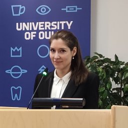 Mariia Borovkova合作论文数Optoelectronics and Measurement Techniques Unit, University of Oulu1
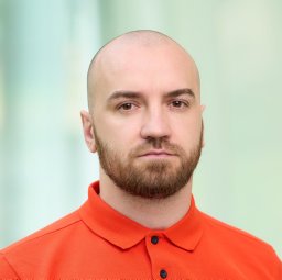 Viktor Dremin合作论文数1
 Pahnke Jens合作论文数1
 Zbignevs Marcinkevics合作论文数1
 Jinyong Ma (马进勇)合作论文数College of Physics and Optoelectronic Engineering, Shenzhen University1Aleksei Georgievich Sveshnikov合作论文数1Irina N Zavestovskaia合作论文数1Lauri Eklund合作论文数1Thomas Wriedt合作论文数1




