In processes where ionic concentrations vary significantly, the standard cable equation fails to accurately predict the transmembrane potential. Such processes call for a mathematical description able to account for the spatiotemporal variations in ion concentrations as well as the subsequent effects of these variations on the membrane potential. We here derive a general electrodiffusive formalism for consistently modeling the dynamics of ion concentration and the transmembrane potential in a one-dimensional geometry, including both the intra- and extracellular domains. Unlike standard cable theory, the electrodiffusive formalism accounts for diffusive currents and concentration-dependent variation of the longitudinal resistivities.
The coupling between the water channel aquaporin-4 (AQP4) and K+ transport has attracted much interest. In this study, we assessed the effect of Aqp4 deletion on activity-induced [K+]o changes in acute slices from hippocampus and corpus callosum of adult mice. We show that Aqp4 deletion has a layer-specific effect on [K+]o that precisely mirrors the known effect on extracellular volume dynamics. In CA1, the peak [K+]o in stratum radiatum during 20 Hz stimulation of Schaffer collateral/commissural fibers was significantly higher in Aqp4 −/− mice than in wild types, whereas no differences were observed throughout the [K+]o recovery phase. In stratum pyramidale and corpus callosum, neither peak [K+]o nor post-stimulus [K+]o recovery was affected by Aqp4 deletion. Our data suggest that AQP4 modulates [K+]o during synaptic stimulation through its effect on extracellular space volume.
Event Abstract Back to Event Electrodiffusion in neural tissue at long timescales Geir Halnes1*, Klas Pettersen1, Stig W. Omholt2, Gaute T. Einevoll1 and Ivar Østby1 1 Department of Mathematical Sciences and Technology, Norwegian University of Life Sciences, Ã…s, Norway, Norway 2 Centre for Integrative Genetics (CIGENE), Department of Animal Science, Norwegian University of Life Sciences, Ã…s, Norw, Norway Electrical signaling in neurons is typically modeled at the timescales of integration of synaptic inputs, i.e., < 100 ms. In standard models based on the cable-equation, the key dynamical variable is the membrane potential. With the possible exception of the signal molecule Ca2+, intra- and extracellular ion concentrations are typically assumed to be constant. As synaptic activity and action-potential firing induce relatively small concentration changes of main charge carriers, this simplification is often warranted. Commonly used measurement techniques such as fMRI based on hemodynamics and vascular dynamics probe the system at timescales of seconds or more. At these longer timescales, other neural processes become relevant: Ion pumps and membrane co-transporters actively regulate ion concentrations in the neural tissue, and also diffusion becomes an important transport mechanism (e.g. for funneling out excess potassium from regions with high neural activity). In order to model key long-timescale neural processes, we need models that couple electrical dynamics and ionic diffusion, and that explicitly incorporate the ion concentrations in all parts of the neural tissue (neurons, astrocytes, extracellular space, vasculature). As a step in this direction, we here present an electrodiffusive scheme for modeling ion dynamics in a one-dimensional geometry for an astrocyte exchanging ions with the extracellular space through transmembrane currents. Our scheme essentially models the extra- and intracellular concentrations (C_k(x)) of all ion species (k), and the membrane potential that follows from the resulting charge densities (Ï(x)). Compared to previous, related approaches [e.g. 1,2], our framework ensures (i) global particle/charge conservation, (ii) consistency between charge density and concentration of ion concentrations (charge carriers), and (iii) that any constraint on charges/currents (such as, e.g., ρ(x)_outside = - ρ(x)_inside) is properly translated to corresponding constraints on concentrations/particle fluxes (and vice versa). We identify the conditions under which our framework can be reduced to standard cable theory without severely violating points (i-iii). References: [1] Qian, N. & Sejnowski, TJ. (1989). Biological Cybernetics 62, 1-15. [2] Chen, KC & Nicholson, C. (2000). Biophysical journal 78(6), 2776-97. DOI:10.1016/S0006-3495(00)76822-6 Keywords: computational neuroscience, Neural tissue engineering, neuron models, ionic diffusion, measurement techniques Conference: 5th INCF Congress of Neuroinformatics, Munich, Germany, 10 Sep - 12 Sep, 2012. Presentation Type: Poster Topic: Neuroinformatics Citation: Halnes G, Pettersen K, Omholt S, Einevoll G and Østby I (2014). Electrodiffusion in neural tissue at long timescales. Front. Neuroinform. Conference Abstract: 5th INCF Congress of Neuroinformatics. doi: 10.3389/conf.fninf.2014.08.00070 Copyright: The abstracts in this collection have not been subject to any Frontiers peer review or checks, and are not endorsed by Frontiers. They are made available through the Frontiers publishing platform as a service to conference organizers and presenters. The copyright in the individual abstracts is owned by the author of each abstract or his/her employer unless otherwise stated. Each abstract, as well as the collection of abstracts, are published under a Creative Commons CC-BY 4.0 (attribution) licence (https://creativecommons.org/licenses/by/4.0/) and may thus be reproduced, translated, adapted and be the subject of derivative works provided the authors and Frontiers are attributed. For Frontiers’ terms and conditions please see https://www.frontiersin.org/legal/terms-and-conditions. Received: 21 Mar 2013; Published Online: 27 Feb 2014. * Correspondence: Dr. Geir Halnes, Department of Mathematical Sciences and Technology, Norwegian University of Life Sciences, Ã…s, Norway, Ås, Norway, geih@nmbu.no Login Required This action requires you to be registered with Frontiers and logged in. To register or login click here. Abstract Info Abstract The Authors in Frontiers Geir Halnes Klas Pettersen Stig W. Omholt Gaute T. Einevoll Ivar Østby Google Geir Halnes Klas Pettersen Stig W. Omholt Gaute T. Einevoll Ivar Østby Google Scholar Geir Halnes Klas Pettersen Stig W. Omholt Gaute T. Einevoll Ivar Østby PubMed Geir Halnes Klas Pettersen Stig W. Omholt Gaute T. Einevoll Ivar Østby Related Article in Frontiers Google Scholar PubMed Abstract Close Back to top Javascript is disabled. Please enable Javascript in your browser settings in order to see all the content on this page.
The cable equation is a proper framework for modeling electrical neural signalling that takes place at a timescale at which the ionic concentrations vary little. However, in neural tissue there are also key dynamic processes that occur at longer timescales. For example, endured periods of intense neural signaling may cause the local extracellular K+-concentration to increase by several millimolars. The clearance of this excess K+ depends partly on diffusion in the extracellular space, partly on local uptake by astrocytes, and partly on intracellular transport (spatial buffering) within astrocytes. These processes, that take place at the time scale of seconds, demand a mathematical description able to account for the spatiotemporal variations in ion concentrations as well as the subsequent effects of these variations on the membrane potential. Here, we present a general electrodiffusive formalism for modeling of ion concentration dynamics in a one-dimensional geometry, including both the intra- and extracellular domains. Based on the Nernst-Planck equations, this formalism ensures that the membrane potential and ion concentrations are in consistency, it ensures global particle/charge conservation and it accounts for diffusion and concentration dependent variations in resistivity. We apply the formalism to a model of astrocytes exchanging ions with the extracellular space. The simulations show that K+-removal from high-concentration regions is driven by a local depolarization of the astrocyte membrane, which concertedly (i) increases the local astrocytic uptake of K+, (ii) suppresses extracellular transport of K+, (iii) increases axial transport of K+ within astrocytes, and (iv) facilitates astrocytic relase of K+ in regions where the extracellular concentration is low. Together, these mechanisms seem to provide a robust regulatory scheme for shielding the extracellular space from excess K+.
Little is known about the physiological roles of aquaporin‐4 (AQP4) in the central nervous system. AQP4 water channels are concentrated in endfeet membranes of astrocytes but also localize to the fine astrocytic processes that abut central synapses. Based on its pattern of expression, we predicted that AQP4 could be involved in controlling water fluxes and changes in extracellular space (ECS) volume that are associated with activation of excitatory pathways. Here, we show that deletion of Aqp4 accentuated the shrinkage of the ECS that occurred in the mouse hippocampal CA1 region during activation of Schaffer collateral/commissural fibers. This effect was found in the stratum radiatum (where perisynaptic astrocytic processes abound) but not in the pyramidal cell layer (where astrocytic processes constitute but a minor volume fraction). For both genotypes the ECS shrinkage was most pronounced in the pyramidal cell layer. Our data attribute a physiological role to AQP4 and indicate that this water channel regulates extracellular volume dynamics in the mammalian brain. © 2012 Wiley Periodicals, Inc.
Exposed to a sufficiently high extracellular potassium concentration ([K + ]o), the neuron can fire spontaneous discharges or even become inactivated due to membrane depolarisation (`depolarisation block'). Since these phenomena likely are related to the maintenance and propagation of seizure discharges, it is of considerable importance to understand the conditions under which excess [K + ]o causes them. To address the putative effect of glial buffering on neuronal activity under elevated [K + ]o conditions, we combined a recently developed dynamical model of glial membrane ion and water transport with a Hodgkin---Huxley type neuron model. In this interconnected glia-neuron model we investigated the effects of natural heterogeneity or pathological changes in glial membrane transporter density by considering a large set of models with different, yet empirically plausible, sets of model parameters. We observed both the high [K + ]o-induced duration of spontaneous neuronal firing and the prevalence of depolarisation block to increase when reducing the magnitudes of the glial transport mechanisms. Further, in some parameter regions an oscillatory bursting spiking pattern due to the dynamical coupling of neurons and glia was observed. Bifurcation analyses of the neuron model and of a simplified version of the neuron-glia model revealed further insights about the underlying mechanism behind these phenomena. The above insights emphasise the importance of combining neuron models with detailed astroglial models when addressing phenomena suspected to be influenced by the astroglia-neuron interaction. To facilitate the use of our neuron-glia model, a CellML version of it is made publicly available.
Event Abstract Back to Event Modeling of astrocytic mechanisms explaining neural activity-induced shrinkage of extracellular space and clearance of excess extracellular potassium Ivar Ostby1*, Leiv Oyehaug1, Gaute T. Einevoll1, Ole P. Ottersen2 and Stig W. Omholt2 1 Norwegian University of Life Sciences, Centre for Integrative Genetics (CIGENE), Norway 2 University of Oslo, Norway Traditionally, astrocytes have been considered passive bystanders of neural activity. It is now recognized that astrocytes are critically involved in modulation of synapses by removal, metabolism, and release of neurotransmitters and of extracellular K+, H+ and glutamate. Using various models, we investigate (i) the neural-activity induced shrinkage of the extracellular space (ECS) between neurons and surrounding astrocytes, (ii) the effect of adding a spatial dimension on astrocytic K+ uptake and the role of K+ spatial buffering. Neuronal stimulation causes ~30% shrinkage of the ECS in grey and white matter under experimental conditions. Despite its possible implications for a proper understanding of basic aspects of potassium clearance and astrocyte function, the phenomenon remains unexplained. We first present results from investigations with a dynamic model with a simplified point-like structure which nevertheless can account for current experimental data related to the shrinkage phenomenon [1]. Specifically, we find that neuronal release of potassium and uptake of sodium during stimulation, astrocyte uptake of potassium, sodium and chloride in passive channels, action of the Na/K/ATPase pump and osmotically driven transport of water through the astrocyte membrane together seem sufficient for generating ECS shrinkage as such. However, in order to explain results regarding changes of ECS volume and astrocyte ion concentrations observed in connection with neuronal stimulation, actions of the Na+/K+/2Cl- and the Na+/HCO3– cotransporters appear to be critical determinants for achieving observed quantitative levels of ECS shrinkage. Next, a spatially extended model of an astrocyte was used to assess geometrical effects of glial K+ uptake and ECS shrinkage during enhanced neuronal activity. When comparing results from the spatially extended model with the analogous point-like model, it is seen that the relative spatial extension of the zone of [K+]o buildup during excitation is a crucial determinant for the characteristics of K+ uptake in terms of maximal [K+]o values and ECS shrinkage during excitation. We show results from comparisons between the K+ uptake characteristics of the simplified and the spatially extended model. References 1. I.Ostby, L.Oyehaug, G. Einevoll, E. Nagelhus, E. Plahte, T. Zeuthen, C. Lloyd, O. Ottersen, and S. Omholt. 2009. Astrocytic mechanisms explaining neural-activity-induced shrinkage of extraneuronal space. PLOS Computational Biology 5:1-12. Conference: Neuroinformatics 2009, Pilsen, Czechia, 6 Sep - 8 Sep, 2009. Presentation Type: Poster Presentation Topic: Computational neuroscience Citation: Ostby I, Oyehaug L, Einevoll GT, Ottersen OP and Omholt SW (2019). Modeling of astrocytic mechanisms explaining neural activity-induced shrinkage of extracellular space and clearance of excess extracellular potassium. Front. Neuroinform. Conference Abstract: Neuroinformatics 2009. doi: 10.3389/conf.neuro.11.2009.08.108 Copyright: The abstracts in this collection have not been subject to any Frontiers peer review or checks, and are not endorsed by Frontiers. They are made available through the Frontiers publishing platform as a service to conference organizers and presenters. The copyright in the individual abstracts is owned by the author of each abstract or his/her employer unless otherwise stated. Each abstract, as well as the collection of abstracts, are published under a Creative Commons CC-BY 4.0 (attribution) licence (https://creativecommons.org/licenses/by/4.0/) and may thus be reproduced, translated, adapted and be the subject of derivative works provided the authors and Frontiers are attributed. For Frontiers’ terms and conditions please see https://www.frontiersin.org/legal/terms-and-conditions. Received: 25 May 2009; Published Online: 09 May 2019. * Correspondence: Ivar Ostby, Norwegian University of Life Sciences, Centre for Integrative Genetics (CIGENE), Akershus, Norway, ivar.ostby@umb.no Login Required This action requires you to be registered with Frontiers and logged in. To register or login click here. Abstract Info Abstract The Authors in Frontiers Ivar Ostby Leiv Oyehaug Gaute T Einevoll Ole P Ottersen Stig W Omholt Google Ivar Ostby Leiv Oyehaug Gaute T Einevoll Ole P Ottersen Stig W Omholt Google Scholar Ivar Ostby Leiv Oyehaug Gaute T Einevoll Ole P Ottersen Stig W Omholt PubMed Ivar Ostby Leiv Oyehaug Gaute T Einevoll Ole P Ottersen Stig W Omholt Related Article in Frontiers Google Scholar PubMed Abstract Close Back to top Javascript is disabled. Please enable Javascript in your browser settings in order to see all the content on this page.
Neuronal stimulation causes approximately 30% shrinkage of the extracellular space (ECS) between neurons and surrounding astrocytes in grey and white matter under experimental conditions. Despite its possible implications for a proper understanding of basic aspects of potassium clearance and astrocyte function, the phenomenon remains unexplained. Here we present a dynamic model that accounts for current experimental data related to the shrinkage phenomenon in wild-type as well as in gene knockout individuals. We find that neuronal release of potassium and uptake of sodium during stimulation, astrocyte uptake of potassium, sodium, and chloride in passive channels, action of the Na/K/ATPase pump, and osmotically driven transport of water through the astrocyte membrane together seem sufficient for generating ECS shrinkage as such. However, when taking into account ECS and astrocyte ion concentrations observed in connection with neuronal stimulation, the actions of the Na(+)/K(+)/Cl(-) (NKCC1) and the Na(+)/HCO(3) (-) (NBC) cotransporters appear to be critical determinants for achieving observed quantitative levels of ECS shrinkage. Considering the current state of knowledge, the model framework appears sufficiently detailed and constrained to guide future key experiments and pave the way for more comprehensive astroglia-neuron interaction models for normal as well as pathophysiological situations.
A stochastic model of cancer initiation is considered. The model is used to evaluate whether a bystander effect may be important in the pre-malignant and malignant stages of carcinogenesis, and furthermore, on the basis of epidemiological data, to estimate the mutation rates of genes involved in the development of oral leukoplakias. The bystander effect is defined here as the capability of oncogenic mutations to increase the mutation probability of neighbouring (bystander) cells, thus leading potentially to a cascade of neighbouring mutated and neoplastic cells as a pre-stage in the development to leukoplakias and cancer. We find that incidence data for oral cancer are indeed in accordance with a significant bystander effect, operating either alone or in combination with genomic instability in the early stages of carcinogenesis, i.e. the development of neoplasia. Simulations performed gave a picture of how mutations and neoplasia may spread in a tissue, to form characteristic leukoplakias with a core of neoplastic cells. The model also showed that the probability of finding at least one neoplastic cell in the tissue after a given number of years is more sensitive to changes in genomic instability within the cell itself than to changes in a bystander effect. Based on epidemiological data we also calculate the maximum number of oncogenic genes that may be involved in the bystander effect and development of genomic instability. Even if capable of explaining the initial development of oncogenic mutations towards neoplastic cells, the bystander model could not reproduce the observed incidence rates of leukoplakia without assuming a carcinogen mutation probability per cell per year of neoplastic cells practically equal to one. This means that the bystander effect, to be of substantial importance in the final development of neoplastic cells towards leukoplakias, requires a very significant increase in mutation probabilities for bystanders to neoplastic cells. Alternatively, additional mechanisms such as abnormal cell differentiation and uncontrolled proliferation and apoptotis in the neoplastic stage may be of major importance during the development to cancerization.
A class of mathematical models involving a convection-reaction partial differential equation (PDE) is introduced with reference to recovering human granulopoiesis after high dose chemotherapy with stem cell support. The stability properties of the model are addressed by means of numerical investigations and analysis. A simplified model with proliferation rate and mobilization rate independent of maturity shows that the model is stable as the maturation rate grows without bounds, but may go through stable and non-stable regimens as the maturation rate varies. It is also shown that the system is stable when parameters are chosen to approximate a real physiological situation. System characteristics do not change profoundly by introduction of a maturity-dependent proliferation and mobilization rate, as is necessary to make the model operate more in accordance with hematological observations. However, by changing the system mitotic responsiveness with respect to changes in cytokine level, the system is still stable but may show persistent oscillations much resembling clinical observations of cyclic neutropenia. Furthermore, in these cases, changes in the model feedback signal caused by, for instance, an impaired effective cytokine elimination by cell receptors may enforce these oscillations markedly.
High dose chemotherapy supported with hematopoietic progenitor cells gives a characteristic neutropenic period (blood neutrophils <0.5⋅109 c/l) ranging from 10 to 16 days. The question of a correlation between the CFU-GM content of the transplanted CD34+ cells and time to neutrophil recovery by patients having been given high-dose chemotherapy (HD-CT) with stem cell support was addressed by means of a mathematical model of granulopoiesis. The model utilizes a convection-reaction partial differential equation (PDE) with feedback from a cytokine compartment on proliferation, maturation, and mobilization of granulocytes from bone marrow to blood. The observed number of CFU-GM cells in the transplanted CD34+ cell autograft was used as input to the model. Using this approach, the observed gross relationship between CFU-GM content in the reinfused blood product and engraftment time could be reproduced. At the same time, the effects of assumed physiological mechanisms, especially some of the effects of G-CSF on proliferation rate, maturation rate, mobilization, and cell death, could be investigated and discussed relative to observed engraftment. The model makes it possible to explain how cytokines interfere with progenitor cell mobilization from bone marrow to blood, and it points out the implications of a regulating mechanism for the granulocyte maturation rate.
Steady state human granulopoiesis was modeled by a convection-reaction differential equation of the Rubinow type for the bone marrow granulocyte precursors and an ordinary differential equation for the blood granulocytes. Measured values reported from several laboratories were used as sources for the model proliferation, maturation, and mobilization rates.Due to the large variability in the measured input data, four alternative models were constructed initially, each one with a specific combination of proliferation rate and maturation rate. They were all able to produce output values for the bone marrow neutrophil count and turnover rate close to accepted data, but neither of them could reproduce good values for the differential fractions of the neutrophil precursor stages. The model output was especially sensitive to changes in transit time in the mitotic relative to the postmitotic precursor compartments.When the net proliferation rate was modeled to optimize the bone marrow differential fractions according to published data, the total bone marrow neutrophil count would not fit with published data. However, a composite model optimizing differential fractions, bone marrow neutrophil count, and turnover rate yielded plausible output values and a reduced proliferation rate in the myelocyte stage. This result opens for a possibly substantial apoptosis rate at the myelocyte stage in accordance with results from earlier investigators. However, the result was based on a special choice of precursor transit times, taken from the literature. More precise data concerning granulocyte precursor cycle times, transit times, and differential fractions would radically improve the model's ability to clarify the role of apoptosis during granulocyte production and storage. (C) 2003 Elsevier Inc. All rights reserved.
 Stig William Omholt合作论文数Department of Circulation and Medical Imaging, Norwegian University of Science and Technology7
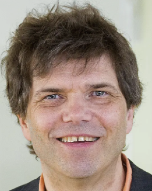 Gaute Einevoll合作论文数University of Oslo;Department of Physics, Norwegian University of Life Sciences;International Neuroinformatics Coordinating Facility6
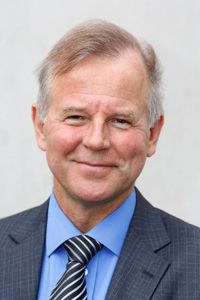 Ole Petter Ottersen合作论文数Division of Anatomy, Institute of Basic Medical Sciences, Faculty of Medicine, University of Oslo4
 Leiv Oyehaug合作论文数4
 Erlend A. Nagelhus合作论文数Institute of Basic Medical Sciences, University of Oslo;Fax: (47) 22851299;e-mail: o.p.ottersen@basalmed.uio.no;Institute of Basic Medical Sciences, University of Oslo3
 Geir Halnes合作论文数Royal Institute of Technology, AlbaNova University3
 Klas H. Pettersen合作论文数Department of Mathematical Sciences and Technology, Norwegian University of Life Sciences3
 Nadia Nabil Haj-Yasein合作论文数2Catherine M. Lloyd合作论文数2Vidar Jensen合作论文数2Oivind Hvalby合作论文数2Per Grøttum合作论文数1Ls Rusten合作论文数1H B Benestad合作论文数1
 Juha Voipio合作论文数1
 Thomas Zeuthen合作论文数Department of Neuroscience, University of Copenhagen1
 Ragnar Winther合作论文数Department of Mathematics, The Faculty of Mathematics and Natural Sciences, University of Oslo1
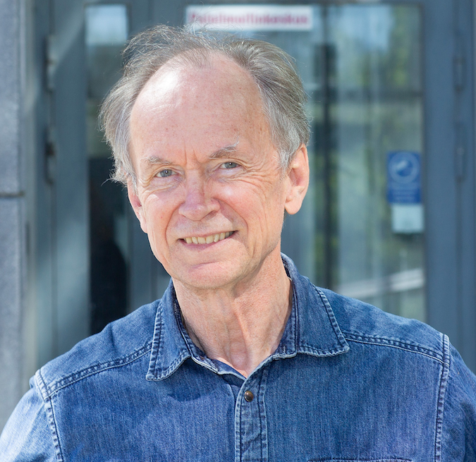 Kai Kaila合作论文数Laboratory of Neurobiology, Department of Biosciences & Neuroscience Center, Faculty of Biological and Environmental Sciences, University of Helsinki1Gunnar Kvalheim合作论文数1Erik Plahte合作论文数1Harald B. Steen合作论文数1P GROTTUM合作论文数1




