BACKGROUND:HFE (homeostatic iron regulator) p.S65C (rs1800730) is associated with European ancestry, although there is no comprehensive geographic study of p.S65C in Europe. METHODS:We tabulated published p.S65C genotypes in European population/control cohorts, depicted allele frequency ranges in a map, and computed correlations of p.S65C allele frequencies with latitudes and longitudes of cohort recruitment sites. RESULTS:We identified 44 cohorts from 24 of the 44 countries of Europe. These 24 countries comprise ~62.9% of Europe's population. There were 233,528 subjects, including 38 HFE p.S65C homozygotes (~1 in 6145 subjects) and 4347 p.S65C heterozygotes (~1 in 54 subjects). Genotypes in 42 cohorts (95.5%) were consistent with Hardy-Weinberg equilibrium. The median allele frequency was 0.0129 (range 0-0.0350). p.S65C was not reported in Bulgaria, although the allele frequency in non-Bulgarian cohorts did not differ significantly (0/200 vs. 4423/462,432, respectively; p = 0.2728). Allele frequency ranges (means) were 0-0.0100 (0.0087), 0.0101-0.0200 (0.0146), and ≥0.0201 (0.0253) in 7, 14, and 3 respective aggregate country cohorts (p < 0.0001, all mean comparisons). The seven countries with low-range frequencies extend from north to south (Estonia to Italy). The three countries with high-range frequencies are east of ~14.1° E (western-most border of Poland). Correlations of allele frequencies with latitudes and longitudes in 44 cohorts were not significant (p = 0.1745 and 0.6031, respectively). CONCLUSIONS:We conclude that the median HFE p.S65C allele frequency in Europe is 0.0129, allele frequencies differ between some countries, and there is no linear north-to-south or west-to-east allele frequency gradient.
BACKGROUND:HFE p.C282Y (rs1800562) homozygotes, including those with normal iron phenotypes, have increased risks of infection and death from infections, although serum IgA and IgM in cohorts of adults with p.C282Y homozygosity are not reported. METHODS:We compiled serum IgA and IgM levels at diagnosis of hemochromatosis in probands with p.C282Y homozygosity, investigated associations of IgA and IgM with clinical characteristics, blood count measures, and iron phenotypes, and compared mean IgA and IgM of probands with combined/weighted means of published adult European cohorts not selected for hemochromatosis. RESULTS:There were 73 probands (36 men, 37 women; mean age 51 ± 13 y). Fifty probands (68.5%) had human leukocyte antigen (HLA)-A*03. Mean IgA ± standard deviation [95% confidence interval] was 2.11 ± 1.06 g/L [1.87, 2.35]. Mean IgM was 1.11 ± 0.75 g/L [0.94, 1.28]. IgM was inversely associated with age (Pearson's r73 = -0.2733; p = 0.019). A multiple regression on IgA revealed no significant association with other characteristics. A regression on IgM revealed one positive association (daily alcohol intake; p = 0.036) and one negative association (age; p = 0.016). Mean IgA of male and female probands and corresponding mean IgA of Europeans in two cohorts (918 men, 458 women) did not differ significantly. Mean IgM of probands was lower than the mean IgM of Europeans in four cohorts (men 1.03 ± 0.84 g/L vs. 1.35 ± 0.55 g/L (n = 1084), respectively (p < 0.001); women 1.18 ± 0.67 g/L vs. 1.57 ± 0.68 g/L (n = 622), respectively (p < 0.001)). CONCLUSIONS:There is no significant association of serum IgA in HFE p.C282Y homozygotes with the clinical and laboratory characteristics we studied. Serum IgM levels are positively associated with daily alcohol intake, are inversely associated with age, and are lower than those of Europeans not selected for hemochromatosis.
BACKGROUND:The elevated average mean corpuscular volume (MCV) of adults with hemochromatosis and HFE p.C282Y (rs1800562) homozygosity is incompletely understood. METHODS:We retrospectively measured percentages of small (100.0-109.9 fL) and large (110.0-200.0 fL) macrocytes in automated hematology analyzer erythrocyte volume histograms of hemochromatosis probands with p.C282Y homozygosity and iron overload without cirrhosis. We evaluated associations of small and large macrocyte percentages with age, sex, body mass index (BMI), daily alcohol consumption, diabetes, hemochromatosis arthropathy, transferrin saturation (TS), and serum ferritin (SF) at diagnosis. RESULTS:There were 69 probands (46 men, 23 women) of mean age 49 ± 15 y. Median BMI was 27.5 kg/m2 (16.9, 46.3). Ten probands (14.5%) had diabetes and seven (10.1%) had arthropathy. Mean TS, SF, and MCV were 81 ± 14%, 791 ± 480 µg/L, and 95.5 ± 4.1 fL, respectively. Mean small and large macrocyte percentages were 17.9 ± 14.3 % and 31.3 ± 8.2 %, respectively. Correlations of small and large macrocyte percentages with age were significant (r69 = 0.3937; p = 0.0008 and r69 = 0.2634; p = 0.0288, respectively). The mean small macrocyte percentage of 11 probands who reported daily alcohol consumption was greater than that of the 58 other probands (20.6 ± 2.9 % and 17.4 ± 4.4 %, respectively; p = 0.0070). The correlation of large macrocyte percentages with TS was significant (r69 = 0.2661; p = 0.0271). Linear regressions confirmed these positive associations: small macrocyte percentages with age (p = 0.0012) and daily alcohol consumption (p = 0.0355); and large macrocyte percentages with age (p = 0.0306) and TS (p = 0.0247). CONCLUSIONS:In HFE p.C282Y homozygotes, small macrocyte percentages are associated with age and daily alcohol consumption and large macrocyte percentages are associated with age and TS.
Background:The aim of this study was to define the relationships between mean corpuscular volume (MCV) and 12 clinical and laboratory variables in HFE p.C282Y (rs1800562)/p.H63D (rs1799945) compound heterozygotes. Methods:We retrospectively studied self-reported non-Hispanic white adult compound heterozygotes with transferrin saturation (TS) > 50% and serum ferritin (SF) > 300 µg/L (men) or TS > 45% and SF > 200 µg/L (women) who participated in primary care-based screening. In post-screening evaluations, we excluded participants with anemia, pregnancy, or medication use that increases MCV. We defined heavy alcohol intake as > 28 g/day in men and > 14 g/day in women. We determined associations of MCV with the following clinical and laboratory variables: age, sex, body mass index (BMI), diabetes, daily intakes of heme, non-heme, and supplemental iron, daily intakes of alcohol, swollen or tender second/third metacarpophalangeal (MCP) joints, reports of therapeutic phlebotomy, TS, and SF. Results:There were 74 participants (37 men, 37 women) of mean age 59 ± 12 (SD) years. Mean screening TS and SF were 65±13% and 529 ± 169 µg/L (men) and 59 ± 14% and 376 ± 195 µg/L (women). Post-screening values did not differ significantly. Mean MCV was 95.7 ± 4.0 fL. There was a negative correlation of MCV with BMI (P = 0.0488) and positive correlations of MCV with age (P = 0.0098), daily heme iron intake (P = 0.0333), and daily alcohol intake (P = 0.0113). Mean MCVs of 19 participants with and 55 without heavy alcohol intake were 97.8 ± 3.8 fL and 95.0 ± 3.9 fL, respectively; P = 0.0074). Linear regression on MCV confirmed positive associations with age (P = 0.0064) and daily alcohol intake (P = 0.0151). MCV was not significantly associated with sex, diabetes, daily intakes of non-heme and supplemental iron, swollen or tender second/third MCP joints, reports of therapeutic phlebotomy, TS, or SF. Conclusion:MCV in HFE p.C282Y/p.H63D compound heterozygotes with high iron phenotypes is positively associated with age and daily alcohol intake, after adjustment for other variables.
BACKGROUND:We estimated European ancestry in U.S. Hispanics using HFE p.C282Y (c.845G > A; rs1800562), a highly informative autosomal marker of European ancestry. METHODS:We tabulated published p.C282Y allele frequencies in U.S. Hispanic control/population region/city cohorts. We assumed that Hispanics inherited p.C282Y from European ancestors only. We defined European ancestry (M) of each cohort as the quotient of its published p.C282Y frequency by the published aggregate p.C282Y frequency in Iberian Spaniards. We compared the present average region/city values of M and the aggregate M with previous European ancestries of U.S. Hispanics estimated using multiple ancestry-informative markers (AIMs). RESULTS:There were 14,472 Hispanics (nine region/city cohorts; aggregate p.C282Y frequency 0.0176 [509/28,944] [95% confidence interval: 0.0159, 0.0189]). There were 12,297 Spaniards (34 population/control cohorts; aggregate p.C282Y frequency 0.0291 (716/24,594) [0.0271, 0.0313]). Nine region/city estimates of M differed 2.6-fold: Alabama 1.0000, California/Irvine 0.5395, California/Oakland 0.6186; California/San Diego 0.9381; Connecticut 0.6873; District of Columbia 0.3883; Hawaii 0.6976; New York 0.4330; and Oregon 1.0000. The average of these nine region/city estimates of M was 0.7003. The aggregate estimate of M in the present 14,472 Hispanics was 0.6048 [0.5369, 0.6728]. The range of estimated European ancestries in 29,365 Hispanics in eight previous studies that used multiple AIMs was 0.502-0.683. CONCLUSIONS:European ancestry in U.S. Hispanics estimated using p.C282Y frequencies varies across regions/cities. The average nine region/city and aggregate European ancestry estimates in the present U.S. Hispanics are similar to those in eight previous studies that used multiple AIMs.
Background: We sought to analyze the geographic distribution of HFE p.C282Y (homeostatic iron regulator c.845G>A; rs1800562) allele frequencies in Iberia. Methods: We analyzed published population/control cohorts of 50 or more subjects in mainland Spain and mainland Portugal and determined whether or not the p.C282Y genotypes in each cohort deviated from Hardy-Weinberg equilibrium (HWE) proportions. We defined combined p.C282Y allele frequencies from Spain and Portugal as representative of Iberia. We computed linear regressions (Pearson's correlations) of allele frequencies vs. latitudes and longitudes of cohort recruitment sites, defined significant regressions as allele frequency gradients, and mapped regional allele frequencies. Results: There were 34 Iberian cohorts: 25 Spanish (12,297 subjects; 11 autonomous communities) and 9 Portuguese (1024 subjects; five administrative regions). p.C282Y genotypes in one of 34 cohorts (2.9%) deviated significantly from HWE proportions. Aggregate allele frequency in Iberia was 0.0292 (778/26,642) [95% confidence interval: 0.0272, 0.0313]. The correlation of allele frequencies with latitude in Iberia was significant (r34 = 0.4184; p = 0.0138). The correlation of allele frequencies with longitude was not significant (r34 = 0.0014; p = 0.9936). The range of 16 regional allele frequencies in Iberia was 0.0068 (Murcia) to 0.5000 (Galicia). Frequencies were highest in regions adjacent to the north and northwest coasts (Cantabria, Galicia, Norte) and lowest in the south (Algarve, Murcia). Conclusions: There is a significant decreasing linear north-to-south gradient of HFE p.C282Y allele frequencies in Iberia. p.C282Y allele frequencies are highest in regions adjacent to the north and northwest coasts.
There are few reports of non-malignant hemorrhagic pericardial effusion and cardiac tamponade in patients with B-chronic lymphocytic leukemia (B-CLL) who were not concurrently treated with an anticoagulant or an anti-platelet agent. We report the case of a 57-year-old woman with B-CLL treated for 54 months with single-agent ibrutinib who developed non-malignant hemorrhagic pericardial effusion and cardiac tamponade. She recovered after pericardiocentesis and the discontinuation of ibrutinib treatment. We conclude that the pathogenesis of her hemorrhagic pericardial effusion involved a combination of abnormal coagulation and platelet dysfunction typical of B-CLL, off-target kinase effects of ibrutinib on platelets, and serosal inflammation.
Background:We sought to determine associations of serum ferritin (SF) with live birth numbers and other iron-related variables in women with HFE p.C282Y (rs1800562) homozygosity. Methods:We studied non-pregnant, non-Hispanic white women in post-screening evaluations to determine associations of SF with age, pregnancy and live birth numbers, dichotomous menopause and therapeutic phlebotomy reports, daily food and supplemental iron intakes, and transferrin saturation (TS). Results:There were 136 women with mean age 51 ± 13 (SD) years and median SF 238 µg/L (range: 8, 2960). There were 376 pregnancies (median: 3/woman (1, 8)) and 296 live births (median: 2/woman (0, 6)). A total of 70 women (51.5%) reported menopause and 31 women (22.8%) reported phlebotomy. Median heme + non-heme food iron intake was 13.4 mg/d (3.1, 57.3). Mean TS was 62 ± 25%. Pearson's coefficient of ln SF versus age was 0.1955 (p = 0.0226). Median SF of women with and without menopause was 386 µg/L (8, 2960) and 165 µg/L (8, 1894), respectively (p < 0.0002). SF associations with phlebotomy and iron intakes were not significant. Spearman's coefficient of SF versus TS was 0.5079 (p < 0.0001). Four mean ln SF values of 66 women without menopause subgrouped by live birth numbers were similar (one-way ANOVA p = 0.4460). Multiple regressions on SF using pregnancy numbers revealed positive associations with menopause (p = 0.0157) and TS (p < 0.0001) and using live birth numbers revealed positive associations with live births (p = 0.0389), menopause (p = 0.0305), and TS (p < 0.0001). Conclusions:SF levels in 136 women with p.C282Y homozygosity are positively associated with age, numbers of live births, menopause reports and TS.
Background IgG subclass levels in hemochromatosis are incompletely characterized. Methods We characterized IgG subclass levels of referred hemochromatosis probands with HFE p.C282Y/p.C282Y (rs1800562) and human leukocyte antigen (HLA)-A and -B typing/haplotyping and compared them with IgG subclass levels of eight published cohorts of adults unselected for hemochromatosis. Results There were 157 probands (82 men, 75 women; mean age 49 +/- 13 y). Median serum ferritin, mean body mass index (BMI), median IgG4, and median phlebotomy units to achieve iron depletion were significantly higher in men. Diabetes, cirrhosis, and HLA-A*03,-B*44, -A*03,B*07, and -A*01,B*08 prevalences and median absolute lymphocyte counts in men and women did not differ significantly. Mean IgG subclass levels [95% confidence interval] were: IgG1 5.31 g/L [3.04, 9.89]; IgG2 3.56 g/L [1.29, 5.75]; IgG3 0.61 g/L [0.17, 1.40]; and IgG4 0.26 g/L [<0.01, 1.25]. Relative IgG subclasses were 54.5%, 36.6%, 6.3%, and 2.7%, respectively. Median IgG4 was higher in men than women (0.34 g/L [0.01, 1.33] vs. 0.19 g/L [<0.01, 0.75], respectively; p = 0.0006). A correlation matrix with Bonferroni correction revealed the following positive correlations: IgG1 vs. IgG3 (p<0.01); IgG2 vs. IgG3 (p<0.05); and IgG2 vs. IgG4 (p<0.05). There was also a positive correlation of IgG4 vs. male sex (p<0.01). Mean IgG1 was lower and mean IgG2 was higher in probands than seven of eight published adult cohorts unselected for hemochromatosis diagnoses. Conclusions Mean IgG subclass levels of hemochromatosis probands were 5.31, 3.56, 0.61, and 0.26 g/L, respectively. Median IgG4 was higher in men than women. There were positive associations of IgG subclass levels. Mean IgG1 may be lower and mean IgG2 may be higher in hemochromatosis probands than adults unselected for hemochromatosis.
ImportanceThe prevalence of iron deficiency varies widely according to how it is defined. ObjectiveTo compare the prevalence of iron deficiency among women using 3 different definitions. Design, Setting, and ParticipantsThe cross-sectional Hemochromatosis and Iron Overload Screening Study (HEIRS; 2000-2006) evaluated the prevalence, determinants, and outcomes of hemochromatosis and other iron-related disorders. Multiethnic, primary care-based screening (2001-2003) was performed at 5 field centers (4 in the US and 1 in Canada). Volunteer women aged 25 years and older were recruited at primary care venues associated with the field centers. Data were analyzed from June to December 2023. Main Outcomes and MeasuresMeasures included transferrin saturation, serum ferritin level, and self-reported age, pregnancy, and race and ethnicity. Three iron deficiency definitions were studied: (1) combined transferrin saturation less than 10% and serum ferritin less than 15 ng/mL (HEIRS), (2) serum ferritin less than 15 ng/mL (World Health Organization [WHO]), and (3) serum ferritin less than 25 ng/mL (a threshold for iron-deficient erythropoiesis [IDE]). ResultsAmong 62 685 women (mean [SD] age, 49.58 [14.27] years), 1957 women (3.12%) had iron deficiency according to the HEIRS definition, 4659 women (7.43%) had iron deficiency according to the WHO definition, and 9611 women (15.33%) had iron deficiency according to the IDE definition. Among 40 381 women aged 25 to 54 years, 1801 women (4.46%) had iron deficiency according to HEIRS, 4267 women (10.57%) had iron deficiency according to WHO, and 8573 women (21.23%) had iron deficiency according to IDE. Prevalence rates of iron deficiency among 2039 women aged 25 to 44 years who reported pregnancy were 5.44% (111 women) according to HEIRS, 18.05% (368 women) according to WHO, and 36.10% (736 women) according to IDE. Iron deficiency prevalence by the 3 respective definitions increased significantly in each racial and ethnic group and was significantly higher among Black and Hispanic participants than Asian and White participants. The relative iron deficiency prevalence among the 62 685 women increased 2.4-fold (95% CI, 2.3-2.5; P < .001) using the WHO definition and increased 4.9-fold (95% CI, 4.7-5.2; P < .001) using the IDE definition. Conclusions and RelevanceThree definitions of iron deficiency were associated with significantly different prevalence of iron deficiency in women, regardless of self-reported age, pregnancy, or race and ethnicity. Using higher serum ferritin thresholds to define iron deficiency could lead to diagnosis and treatment of more women with iron deficiency and greater reduction of related morbidity.
AbstractIntroductionThe aim of this study was to define the relationships between factors other than transferrin saturation (TS) to mean corpuscular volume (MCV) and macrocytosis (MCV > 100 fL) in HFE p.C282Y (rs1800562) homozygotes.MethodsWe studied white post‐screening participants with p.C282Y homozygosity who did not have anemia, report cirrhosis or pregnancy, or use medications that increase MCV. We analyzed relations of MCV and macrocytosis with age, sex, diabetes reports, daily alcohol consumption, swollen or tender 2nd/3rd metacarpophalangeal (MCP) joints, TS, and serum ferritin (SF).ResultsThere were 257 participants (110 men and 147 women). Median alcohol consumption, median TS, median SF, and macrocytosis prevalence were significantly greater in men. Relative risk of macrocytosis in men was 2.81. In men and women, there were significant positive Pearson's correlations of MCV versus age and Spearman's correlations of MCV versus alcohol consumption and TS. Mean MCV and macrocytosis prevalence were significantly greater in participants with than without swollen or tender 2nd/3rd MCP joints. Linear regression on MCV revealed positive associations: age (p < 0.0001), alcohol consumption (p = 0.0007), and TS (p < 0.0001). Logistic regression on macrocytosis revealed an odds ratio for age of 1.04 (95% confidence interval: 1.00, 1.07).ConclusionsMCV in HFE p.C282Y homozygotes is positively related to age, daily alcohol consumption, TS, and swollen or tender 2nd/3rd MCP joints.
Background: We sought to evaluate height in white adults with hemochromatosis.Methods: We analyzed the height of (1) post-screening examination participants with HFE p.C282Y/p.C282Y (rs1800562) and wt/wt (absence of p.C282Y and p.H63D (rs1799945)) and (2) referred hemochromatosis probands with p.C282Y/p.C282Y.Results: There were 762 participants (270 p.C282Y/p.C282Y, 492 wt/wt; 343 men, 419 women) and 180 probands (104 men, 76 women). Median height of male participants with p.C282Y/p.C282Y or wt/wt was 177.8 cm. Median height of female participants was greater in those with p.C282Y/p.C282Y than wt/wt (165.1 cm vs 162.6 cm, respectively; p = 0.0298). Median height of p.C282Y/p.C282Y participants and probands was the same (men 177.8 cm; women 165.1 cm). Regressions on height of male and female participants revealed no associations with HFE genotype and inverse and positive associations with age and weight, respectively. Height of female participants was positively and inversely associated with transferrin saturation and serum ferritin, respectively. Regressions on height of male and female probands revealed positive associations with weight.Conclusions: The height of men with HFE p.C282Y/p.C282Y and wt/wt does not differ significantly. The height of female participants was greater in those with p.C282Y/p.C282Y than wt/wt. We found no independent association of HFE genotype with the height of men or women.
BACKGROUND:IgG subclass levels in hemochromatosis are incompletely characterized. METHODS:We characterized IgG subclass levels of referred hemochromatosis probands with HFE p.C282Y/p.C282Y (rs1800562) and human leukocyte antigen (HLA)-A and -B typing/haplotyping and compared them with IgG subclass levels of eight published cohorts of adults unselected for hemochromatosis. RESULTS:There were 157 probands (82 men, 75 women; mean age 49±13 y). Median serum ferritin, mean body mass index (BMI), median IgG4, and median phlebotomy units to achieve iron depletion were significantly higher in men. Diabetes, cirrhosis, and HLA-A*03,-B*44, -A*03,B*07, and -A*01,B*08 prevalences and median absolute lymphocyte counts in men and women did not differ significantly. Mean IgG subclass levels [95% confidence interval] were: IgG1 5.31 g/L [3.04, 9.89]; IgG2 3.56 g/L [1.29, 5.75]; IgG3 0.61 g/L [0.17, 1.40]; and IgG4 0.26 g/L [<0.01, 1.25]. Relative IgG subclasses were 54.5%, 36.6%, 6.3%, and 2.7%, respectively. Median IgG4 was higher in men than women (0.34 g/L [0.01, 1.33] vs. 0.19 g/L [<0.01, 0.75], respectively; p = 0.0006). A correlation matrix with Bonferroni correction revealed the following positive correlations: IgG1 vs. IgG3 (p<0.01); IgG2 vs. IgG3 (p<0.05); and IgG2 vs. IgG4 (p<0.05). There was also a positive correlation of IgG4 vs. male sex (p<0.01). Mean IgG1 was lower and mean IgG2 was higher in probands than seven of eight published adult cohorts unselected for hemochromatosis diagnoses. CONCLUSIONS:Mean IgG subclass levels of hemochromatosis probands were 5.31, 3.56, 0.61, and 0.26 g/L, respectively. Median IgG4 was higher in men than women. There were positive associations of IgG subclass levels. Mean IgG1 may be lower and mean IgG2 may be higher in hemochromatosis probands than adults unselected for hemochromatosis.
Horse bites are common non-fatal injuries in the United States. Infections of horse bite wounds in humans are usually due to bacteria that correspond to the oropharyngeal bacterial flora of horses. We report the novel case of a 25-year-old woman who sustained a horse bite wound that was infected with Prevotella bivia, a Gram-negative, non-pigmented anaerobe. We discuss the epidemiology, bacteriology, and clinical management of horse bites.
BackgroundCharacteristics of cohorts of individuals aged <= 17 years with homeostatic iron regulator (HFE) p.C282Y (rs1800562) homozygosity, a common hemochromatosis genotype, have not been reported.MethodologyWe retrospectively tabulated characteristics of white individuals aged <= 17 years with p.C282Y homozygosity. Individuals were not recruited for this study. We defined transferrin saturation (TS) >45%, serum ferritin (SF) >300 mu g/L (M) and >200 mu g/L (F) as elevated and liver iron grade 3 or 4, hepatic iron index >1.9 mu mol Fe/g dry weight liver/y, and phlebotomy-mobilized iron >1.0 g (M) and >0.3 g (F) as increased.ResultsThere were nine males and six females with a mean age of 12 +/- 4 years (range = 5-17 years). The mean age of 10 probands (13 +/- 3 years) was greater than that of five individuals discovered in family studies (9 +/- 4 years) (p = 0.0403). Presenting manifestations of probands included fatigue/lethargy (5), elevated TS (2), and polycystic ovary syndrome, amenorrhea, and diabetes (2). In 15 individuals, the mean TS was 65 +/- 23%. TS was elevated in 11 (73.3%) individuals aged 5-17 years. In 14 individuals, the mean SF was 262 +/- 289 mu g/L. SF was elevated and liver and phlebotomy-mobilized iron were increased in two male and three female probands aged 13-16 years (5/14 individuals, 35.7%). No individual had advanced hepatic fibrosis, arthropathy, hypogonadism, cardiomyopathy, or hyperpigmentation.ConclusionsWe conclude that five individuals aged 13-16 years (5/14 individuals, 35.7%) had increased liver and phlebotomy-mobilized iron.
Abstract Our aim was to document the effects of genotype HFE p.C282Y/p.C282Y and hemochromatosis-associated laboratory and clinical manifestations on platelet counts (PC). We compiled genotype (p.C282Y/p.C282Y or HFE wt/wt (absence of p.C282Y and p.H63D (rs1799945)), age, sex, body mass index, presence/absence of chronic fatigue, swelling/tenderness of second/third metacarpophalangeal joints, and hyperpigmentation, transferrin saturation (TS), serum ferritin (SF), hemoglobin levels, absolute neutrophil, lymphocyte, and monocyte counts, C-reactive protein levels, and PC of non-Hispanic white participants in a hemochromatosis and iron overload post-screening clinical examination. There were 171 men and 254 women (141 p.C282Y/p.C282Y, 284 wt/wt) of median age 53 y. Median TS and SF were higher in p.C282Y/p.C282Y than wt/wt participants grouped by sex (p < .0001, all comparisons). Median PC by genotype was lower in men than women (p < .0001, both comparisons). Regression on PC using 14 independent variables identified these significant positive associations: absolute neutrophil, lymphocyte, and monocyte counts and C-reactive protein levels and these significant inverse associations: age, TS, and hemoglobin levels. We conclude that PC is significantly associated with absolute neutrophil, lymphocyte, and monocyte counts, and C-reactive protein (positive) and age, TS, and hemoglobin (inverse), after adjustment for other variables. HFE genotypes we studied were not significantly associated with PC. Plain Language Summary What is the context? Hemochromatosis is typically associated with inheritance of two copies of p.C282Y, a common mutation of the HFE gene on chromosome 6p that regulates iron absorption. Platelet counts, age, and serum levels of liver enzymes have been used to estimate risks of cirrhosis in adults with hemochromatosis. Lower platelet counts in Europeans are significantly associated with a mutation in CARMIL1, a gene on chromosome 6p close to HFE. Clinical and laboratory associations of normal platelet counts in adults with HFE p.C282Y/p/C282Y and wt/wt uncomplicated by cirrhosis are unreported. What is new? We studied normal platelet counts in 425 white adults who participated in a primary care-based hemochromatosis screening program. These participants did not have cirrhosis or other conditions that often influence platelet counts. Our analyses of 14 variables identified these significant positive associations with platelet counts, after adjustment for other variables: absolute neutrophil, lymphocyte, and monocyte counts and C-reactive protein levels; and these significant inverse associations: age, TS, and hemoglobin levels. What is the impact? Laboratory and clinical factors significantly associated with platelet counts in adults with HFE p.C282/p.C282Y or wt/wt are similar to those in persons unselected for HFE genotypes or hemochromatosis. It is unlikely that genes that influence platelet counts are closely linked to HFE on chromosome 6p. Adults with hemochromatosis and HFE p.C282/p.C282Y who have abnormal platelet counts should be evaluated for cirrhosis or non-iron platelet disorders.
INTRODUCTION:We sought to quantify percentages of microcytes and macrocytes in archived automated hematology analyzer (AHA) red blood cell (RBC) volume histogram images.METHODS:In preliminary studies, we demonstrated that an on-line application of Gauss' area formula (SketchAndCalc™) measured percentage areas of 20 segments under a computer-generated normal distribution curve (-3.0 standard deviations [SD] to +3.0 SD) with accuracy and precision (Pearson's correlation of measured areas with corresponding theoretical areas r [20] = 0.9962 [p < 0.0001]). Thus, we used SketchAndCalc™ to quantify percentages of microcytes (50-80 fL) and macrocytes (110-200 fL) in archived AHA histogram images in women with previously untreated iron-deficiency anemia (IDA) and previously untreated hemochromatosis.RESULTS:Median microcyte percentages in 13 women with IDA and 13 women with hemochromatosis were 63.6% (range 13.5-76.8) and 6.7% (range 3.4-24.8), respectively (p < 0.0001). Mean macrocyte percentages in women with IDA and hemochromatosis were 8.8% ± 6.1 SD and 33.8% ± 11.7 SD, respectively (p < 0.0001). Spearman's correlations of microcyte percentages with macrocyte percentages, mean corpuscular volume, and mean corpuscular hemoglobin in 26 women were rs [26] = -0.9485, rs [26] = -0.9641, and rs [26] = -0.9036, respectively (each p < 0.0001).CONCLUSIONS:This method of quantifying microcyte and macrocyte percentages could enable other studies of RBC volume subpopulations in archived AHA histogram images.
This dataset consists of observations at diagnosis of 180 referred Alabama hemochromatosis probands with HFE p.C282Y/p.C282Y, including sex, age, transferrin saturation, serum ferritin, height, and weight.
The aim of this study was to identify characteristics of non-alcoholic fatty liver disease (NAFLD) in adults with HFE p.C282Y/p.C282Y. We retrospectively studied non-Hispanic white hemochromatosis probands with iron overload (serum ferritin (SF) > 300 µg/L (M), > 200 µg/L (F)) and p.C282Y/p.C282Y at non-screening diagnosis who did not report alcohol consumption > 14 g/d, have cirrhosis or other non-NAFLD liver disorders, use steatogenic medication, or have diagnoses of heritable disorders that increase NAFLD risk. We identified NAFLD-associated characteristics using univariate and multivariable analyses. There were 66 probands (31 men, 35 women), mean age 49 ± 14 (SD) y, of whom 16 (24.2
BACKGROUND:Little is known about the prevalence of HFE (homeostatic iron regulator) hemochromatosis in African Americans (AA).METHODS:We defined AA as self-identified AA, blacks, or non-Hispanic blacks. We defined hemochromatosis-associated HFE genotypes as p.C282Y/p.C282Y and p.C282Y/p.H63D. We compiled prevalences of these genotypes in AA using published population and cohort data and numbers of men and women ≥18 y in 2018 U.S. Census estimates. We defined iron overload (IO) and IO-related disease by genotype as previously reported in population and cohort studies of hemochromatosis in whites of European ancestry. We used these definitions to estimate prevalences and numbers of AA with IO and IO-related disease associated with hemochromatosis-associated HFE genotypes.RESULTS:There were ∼16,287,599 men and ∼17,644,898 women. HFE genotypes and their respective prevalences were: p.C282Y/p.C282Y, 0.00017 (6/34,905) [95% confidence interval 0.000034, 0.00031] and p.C282Y/p.H63D, 0.0012 (41/33,596) [0.000084, 0.0016]. IO prevalences were: men 0.000076 [0.000072, 0.000081] and women 0.0000061 [0.0000050, 0.0000073]. IO-related disease prevalences were: men 0.000063 [0.000059, 0.000067] and women 0.0000021 [0.0000014, 0.0000027]. There were ∼1021 [961, 1091] men and ∼36 [25, 48] women with IO-related disease.CONCLUSIONS:We conclude that ∼1/25,061 AA >18 y have a hemochromatosis-associated HFE genotype and IO and that ∼1/32,103 AA >18 y have a hemochromatosis-associated HFE genotype and IO-related disease.
 Ronald T Acton合作论文数90
 Paul C. Adams合作论文数85
 Prad Phatak合作论文数83Corwin Q. Edwards合作论文数82
 Bruce R. Bacon合作论文数Saint Louis University School of Medicine;B.R. Bacon Consulting, LLC63Gordon D. Mclaren合作论文数Department of Medicine, University of California59Robert S. Britton合作论文数55
 Christine E. McLaren合作论文数School of Medicine, University of California, Irvine;Genetic Epidemiology Research Institute, University of California, Irvine;Chao Family Comprehensive Cancer Center, University of California, Irvine54Luigi F. Bertoli合作论文数Southern Iron Disorders Center, Birmingham, AL 35209, USA36R T Acton合作论文数35David M. Reboussin合作论文数35
 Victor Gordeuk合作论文数Department of Medicine, Howard University33
 Mark Speechley合作论文数Department of Epidemiology and Biostatistics, Western University32Fitzroy W. Dawkins合作论文数31Pauline Lee合作论文数26Catherine Leiendecker-Foster合作论文数25
 John Eckfeldt合作论文数Department of Laboratory Medicine and Pathology, Medical School, University of Minnesota24Lf Bertoli合作论文数22Emily L. Harris合作论文数Division of Cancer Control and Population Sciences, National Cancer Institute, National Institutes of Health21
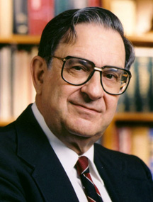 Ernest Beutler合作论文数Department of Molecular and Experimental Medicine, The Scripps Research Institute19
 Lawrie Powell合作论文数Queensland Institute of Medical Research17Phyliss Sholinsky合作论文数17Grant A. Ramm合作论文数15Jh Eckfeldt合作论文数14Gregory J Anderson合作论文数QIMR Berghofer Medical Research Institute;Iron Metabolism Laboratory, Queensland Institute of Medical Research, PO Royal Brisbane Hospital14Charles A. Rivers合作论文数14
 V. Nathan Subramaniam合作论文数The Queensland Institute of Medical Research14
 Lyle Gurrin合作论文数Faculty of Medicine, Dentistry and Health Sciences, University of Melbourne;Melbourne School of Population and Global Health, University of Melbourne13Snively Beverly M合作论文数12Emily L Harris合作论文数11Jackson Clayborn Barton合作论文数11Barry E. Rothenberg合作论文数10
 Mary Emond合作论文数School of Public Health, University of Washington10Richard D. Press合作论文数10Wen-Pin Chen合作论文数8Carol West合作论文数8
 Katrina J Allen合作论文数Murdoch Children's Research Institute;School of Inflammation and Repair, University of Manchester;Royal Children’s Hospital7Thomas W. Butler合作论文数7
 Oswaldo Castro合作论文数7Jacob A. Reiss合作论文数7Bm Snively合作论文数6Britton Robert合作论文数6
 Deborah A. Nickerson合作论文数University of Washington6Thomas M. Vogt合作论文数6Laura Lovato合作论文数6Terri Gelbart合作论文数6Howard W. Wiener合作论文数6Sreenivas V. Rao合作论文数6




