Extracellular vesicles (EVs) released by neurons (nEVs) provide an opportunity to measure biomarkers from the brain circulating in the periphery. No study yet has directly compared molecular cargo in brain tissue to nEVs found in circulation in humans. In 5 matched sets of brain tissue, serum, total EVs, and nEVs, obtained from the Bartoli Brain Tumor Laboratory at Columbia University, we compared the levels microRNAs and environmental chemicals because microRNAs are one of the most studied nEV cargoes and offer great potential as biomarkers and environmental chemical load in nEVs is understudied and could reveal chemical burden in the brain. We also compared metabolomic profiles in a different set of matched serum, total EVs, and nEVs since metabolites in nEVs are also understudied but could offer potential biomarkers. Highly expressed brain tissue miRNAs showed stronger correlations with nEVs than serum or total EVs. We detected several environmental chemical pollutant classes in nEVs. The chemical pollutant concentrations in nEVs were more strongly correlated with brain tissue levels (r = 0.72, P = 7.2e-16) than those observed between brain tissue and serum (r = 0.7, P = 5.8e-15) or total EVs (r = 0.58, P = 1.5e-09). Compared to serum and total EVs, we observed an enrichment of metabolites with known signaling roles, such as bile acids, oleic acid, phosphatidylserine, and isoprenoids in nEVs. We provide evidence that nEV cargo is closely correlated with brain tissue content, further supporting their utility as a brain liquid biopsy in humans.
Background. Diffuse midline glioma (DMG) is the most aggressive primary brain tumor in children. All previous studies examining the role of systemic agents have failed to demonstrate a survival benefit; the only standard of care is radiation therapy (RT). Successful implementation of radiosensitization strategies in DMG remains an es-sential and promising avenue of investigation. We explore the use of Napabucasin, an NAD(P)H quinone dehydro-genase 1 (NQO1)-bioactivatable reactive oxygen species (ROS)-inducer, as a potential therapeutic radiosensitizer in DMG. Methods. In this study, we conduct in vitro and in vivo assays using patient-derived DMG cultures to elucidate the mechanism of action of Napabucasin and its radiosensitizing properties. As penetration of systemic therapy through the blood-brain barrier (BBB) is a significant limitation to the success of DMG therapies, we explore fo-cused ultrasound (FUS) and convection-enhanced delivery (CED) to overcome the BBB and maximize therapeutic efficacy. Results. Napabucasin is a potent ROS-inducer and radiosensitizer in DMG, and treatment-mediated ROS produc-tion and cytotoxicity are dependent on NQO1. In subcutaneous xenograft models, combination therapy with RT improves local control. After optimizing targeted drug delivery using CED in an orthotopic mouse model, we estab-lish the novel feasibility and survival benefit of CED of Napabucasin concurrent with RT. Conclusions. As nearly all DMG patients will receive RT as part of their treatment course, our validation of the ef-ficacy of radiosensitizing therapy using CED to prolong survival in DMG opens the door for exciting novel studies of alternative radiosensitization strategies in this devastating disease while overcoming limitations of the BBB.
Background. IDH-wild type (-wt) status is a prerequisite for the diagnosis of glioblastoma (GBM); however, IDH-wt gliomas with low-grade or anaplastic morphology have historically been excluded from GBM trials and may represent a distinct prognostic entity. While alkylating agent chemotherapy improves overall survival (OS) and progression-free survival (PFS) for IDH-wt GBM and also IDH-mutant gliomas, irrespective of grade, the benefit for IDH-wt diffuse histologic lower-grade gliomas is unclear. Methods. We performed a meta-analysis of randomized clinical trials for World Health Organization (WHO) grades 2-3 gliomas (2009 to present) to determine the effect of alkylating chemotherapy on IDH-wt and -mutant gliomas using a random-effects model with inverse-variance pooling. Results. We identified 6 trials with 1204 patients (430 IDH-wt, 774 IDH-mutant) that evaluated alkylating chemoradiotherapy versus radiotherapy alone, allowing us to perform an analysis focused on the value of adding alkylating chemotherapy to radiotherapy. For patients with IDH-wt tumors, alkylating chemotherapy added to radiotherapy was associated with improved PFS (HR:0.77 [95% CI: 0.62-0.97], P = .03) but not OS (HR:0.87 [95% CI: 0.64-1.18], P = .17). For patients with IDH-mutant tumors, alkylating chemotherapy added to radiotherapy improved both OS (HR:0.52 [95% CI: 0.42-0.64], P < .001) and PFS (HR = 0.47 [95% CI: 0.39-0.57], P < .001) compared to radiotherapy alone. The magnitude of benefit was similar for IDH-mutant gliomas with or without 1p19q-codeletion. Conclusions. Alkylating chemotherapy reduces mortality by 48% and progression by 53% for patients with IDH-mutant gliomas. Optimal management of IDH-wt diffuse histologic lower-grade gliomas remains to be determined, as there is little evidence supporting an OS benefit from alkylating chemotherapy. [GRAPHICS]
BACKGROUND/OBJECTIVE:Intracranial epidermoid tumors (ETs) are rare, benign lesions that present significant challenges in neurosurgical management due to their propensity to encase vital neurovascular structures. We aimed to evaluate the impact of clinical, demographic, and tumor-specific factors on surgical decisions (gross total resection [GTR] vs. subtotal resection [STR]) and outcomes and identify patient clusters with distinct profiles and outcomes post-resection. METHODS:We retrospectively analyzed 72 patients with ET treated from 1998 to 2022, employing multivariable logistic regression for GTR versus STR predictors and Kaplan-Meier curves for progression-free survival (PFS). K-prototype clustering classified patients based on clinical data. RESULTS:The mean age of our cohort was 39.8 ± 20.1 years. About 13.9% of patients had a recurrence, with a median PFS of 108 months (interquartile range: 57 -206). Seizures significantly predicted GTR (P < 0.05), whereas adherence to critical structures reduced GTR likelihood (P < 0.05). Initial surgeries more often achieved GTR, correlating with longer PFS (P < 0.0001) and reduced recurrence (P < 0.01) versus re-operations. Cluster analysis identified three distinct groups, with the initial GTR cluster showing superior PFS and the lowest recurrence (P < 0.0001 and P < 0.01, respectively). Statistically significant predictors of PFS included age and preoperative seizure presence, with older age favoring longer PFS (P < 0.01) and seizures associated with reduced PFS (P < 0.01). In addition, patients with previous surgeries showed a trend toward shorter PFS (P < 0.05). CONCLUSIONS:This study emphasizes the importance of tailored surgical strategies in managing intracranial ETs, advocating for GTR to optimize long-term outcomes where possible. Future prospective studies are essential to further refine treatment approaches, enhancing survival for ET patients.
Supplementary Tables 1-6, Figures 1-4 from Randomized Study of Paclitaxel and Tamoxifen Deposition into Human Brain Tumors: Implications for the Treatment of Metastatic Brain Tumors
The molecular classification of gliomas is currently based on the presence of IDH1 mutation, which is associated with better prognosis and longer survival. The hallmark of the IDH1 mutation is the production of D-2-Hydroxyglutarate with subsequent effects that are not fully understood. Ferroptosis, an iron-dependent mechanism of non-apoptotic cell death, is mostly triggered through blocking system Xc- or suppressing the antioxidant enzyme glutathione peroxidase 4 (GPX-4). In this study, we generated a mouse model of glioma harboring the IDH1(R132H) mutation in heterozygous condition, in combination with p53-deletion induced by the expression of PDGFA. We show that IDH1(R132H)-expressing mice survive longer compared to their wild-type counterparts, while histological analysis reveals characteristics of low-grade diffuse gliomas. We generated cell lines from primary tumors and analyzed their metabolic profile and response to mitochondrial stress and Ferroptosis. We treated them in vitro with RSL3, a GPX-4 inhibitor, alone or in combination with Cysteine and Methionine restriction. We show that IDH1(R132H)-expressing cells are far more sensitive to Ferroptosis in vitro. Finally, we show that convection-enhanced delivery of RSL3 in combination with dietary Cysteine and Methionine restriction in vivo, significantly prolongs survival of the IDH1(R132H)-expressing mice. Our findings suggest that Ferroptosis provides promising therapeutic potential worth exploring further and our mouse model could be used to test the efficacy of different treatments for IDH1(R132H) gliomas.
Introduction: Solitary fibrous tumor/hemangiopericytoma (SFT/HPC) of the central nervous system (CNS) is a rare meningeal tumor. Given the absence of prospective or randomized data, there are no standard indications for radiotherapy. Recently, the NRG Oncology and EORTC cooperative groups successfully accrued and completed the first prospective trials evaluating risk-adapted adjuvant radiotherapy strategies for meningiomas. Using a similar framework, we sought to develop prognostic risk categories that may predict the survival benefit associated with radiotherapy, using two large national datasets. Methods: We queried the National Cancer Database (NCDB) and the Surveillance, Epidemiology, and End Results (SEER) databases for all newly diagnosed cases of SFT/HPC within the CNS. Risk categories were created, as follows: low risk—grade 1, with any extent of resection (EOR) and grade 2, with gross–total resection; intermediate risk—grade 2, with biopsy/subtotal resection; high risk—grade 3 with any EOR. The Kaplan–Meier method and Cox proportional hazards regressions were used to determine the association of risk categories with overall and cause-specific survival. We then determined the association of radiotherapy with overall survival in the NCDB, stratified by risk group. Results: We identified 866 and 683 patients from the NCDB and SEER databases who were evaluated, respectively. In the NCDB, the 75% survival times for low- (n = 312), intermediate- (n = 239), and high-risk (n = 315) patients were not reached, 86 months (HR 1.60 (95% CI 1.01–2.55)), and 55 months (HR 2.56 (95% CI 1.68–3.89)), respectively. Our risk categories were validated for overall and cause-specific survival in the SEER dataset. Radiotherapy was associated with improved survival in the high- (HR 0.46 (0.29–0.74)) and intermediate-risk groups (HR 0.52 (0.27–0.99)) but not in the low-risk group (HR 1.26 (0.60–2.65)). The association of radiotherapy with overall survival remained significant in the multivariable analysis for the high-risk group (HR 0.55 (0.34–0.89)) but not for the intermediate-risk group (HR 0.74 (0.38–1.47)). Similar results were observed in a time-dependent landmark sensitivity analysis. Conclusion: Risk stratification based on grade and EOR is prognostic of overall and cause-specific survival for SFT/HPCs of the CNS and performs better than any individual clinical factor. These risk categories appear to predict the survival benefit from radiotherapy, which is limited to the high-risk group and, potentially, the intermediate-risk group. These data may serve as the basis for a prospective study evaluating the management of meningeal SFT/HPCs.
Figure S1. Crizotinib resistant cells reveal a reprogrammed tumor metabolism. Figure S2. Crizotinib modulates fatty acid metabolism and mitochondrial parameters. Figure S3. Tracing analysis reveals distinct modulation of metabolism by MET inhibition. Figure S4. Acute and chronic MET inhibition drives oxygen consumption rate and ECAR. Figure S5. Specific silencing of MET enhances oxidative metabolism in patient-derived xenograft cells. Figure S6. Inhibition of complex I and complex V of the electron transport chain and MET synergistically reduce viability of glioblastoma cells. Figure S7. Gamitrinib and etomoxir synergize with crizotinib to reduce cellular viability in glioblastoma model systems. Figure S8. Combined Met inhibition and Gamitrinib treatment elicits cleavage of caspases and modulate the expression of Bcl2 family members. Figure S9. Crizotinib and gamitrinib elicit cell death with features of apoptosis. Figure S10. Gamitrinib and crizotinib is superior over kinase inhibitor combination treatments to elicit apoptosis. Figure S11. The combination treatment of etomoxir and crizotinib elicits a reduction in tumor proliferation without induction of organ toxicity. Supplementary Table 1. Primer sequences for real time PCR and chromatin immunoprecipitation qPCR
Context Acromegaly presents a unique pattern of lower adiposity and insulin resistance in active disease but reduction in insulin resistance despite a rise in adiposity after surgery. Depot-specific adipose tissue masses and ectopic lipid are important predictors of insulin resistance in other populations, but whether they are in acromegaly is unknown. Long-term persistence of body composition changes after surgery is unknown. Objective To determine how depot-specific body composition and ectopic lipid relate to insulin resistance in active acromegaly and whether their changes with surgery are sustained long-term. Methods Cross-sectional study in patients with active acromegaly and longitudinal study in newly diagnosed patients studied before and in long-term follow-up, 3 (1-8) years (median, range), after surgery. Seventy-one patients with active acromegaly studied cross-sectionally and 28 with newly diagnosed acromegaly studied longitudinally. Main outcome measures were visceral (VAT), subcutaneous (SAT), and intermuscular adipose tissue masses by whole-body magnetic resonance imaging; intrahepatic lipid (IHL) by proton magnetic resonance spectroscopy; insulin resistance measures derived from fasting; and oral glucose tolerance test insulin and glucose levels. Results SAT and insulin-like growth factor 1 level, but not VAT or IHL, were independent predictors of insulin resistance in active acromegaly. VAT, SAT, and IHL gains were sustained long-term after surgery. VAT mass rise with surgery correlated inversely with rise in QUICKI while SAT rise correlated with fall in the Homeostatic Model Assessment score. Conclusion SAT and disease activity are important predictors of insulin resistance in active acromegaly. Adiposity gains are sustained long-term after surgical treatment and impact on the accompanying improvement in insulin resistance.
Suppl. fig. 1: Effect of CP-d/n-ATF5-S1 on ATF5 expression and stability Suppl. fig. 2: Effect of CP-d/n-ATF5-S1 on ASNS mRNA expression Suppl. fig. 3: Effect of CP-d/n-ATF5-S1 on cell viability in HL-60 cells Suppl. fig. 4: Pro-apoptotic activity of CP-d/n-ATF5-S1 in SF188 and GBM12 cells Suppl. fig. 5: Effect of CP-d/n-ATF5-S1 in HCT116 colorectal cancer Suppl. fig. 6: Effect of pan-caspase inhibition on CP-d/n-ATF5-S1 treatment Suppl. fig. 7: Effect of CP-d/n-ATF5-S1 on Bcl-2 family and Usp9X/Bag3 protein expression in A375 and PC3 Suppl. fig. 8: Effect of pan-caspase inhibition on CP-d/n-ATF5-S1-mediated down-regulation of Usp9X Suppl. fig. 9: Effect of Usp9X knock-down on apoptosis in LN229 Suppl. fig. 10: Isobologram for CP-d/n-ATF5-S1 and ABT263 Suppl. fig. 11: Effect of CP-d/n-ATF5-S1 and ABT263 on GBM12 cells Suppl. fig. 12: Knock-down of Mcl-1 sensitizes for ABT263-mediated apoptosis Suppl. fig. 13: Effects of CP-d/n-ATF5-S1 on U87MG xenograft model Suppl. fig. 14: Effects of CP-d/n-ATF5-S1 on PANC-1 and MDA-MB-231 xenograft model Suppl. fig. 15: Effect of CP-d/n-ATF5-S1 on organ toxicity
Suppl. fig. 1: Patient derived-xenograft cells are susceptible to the combination treatment of Gamitrinib and clinically validated BH3-mimetics; Suppl. fig. 2: Transgenically-derived murine glioblastoma cells are susceptible to the combination treatment of Gamitrinib and clinically validated BH3-mimetics; Suppl. fig. 3: Carcinoma cells of different origin are sensitive to the combination treatment of Gamitrinib and clinically validated BH3-mimetics Suppl. fig. 4: The combination treatment of either broad or selective BH3-mimetics with mitochondrial matrix chaperone inhibitors elicits enhanced apoptosis Suppl. fig. 5: The combination treatment of selective BH3-mimetics, ABT199, WEHI-539, A-1210477 with Gamitrinib-TPP elicits enhanced apoptosis Suppl. fig. 6: Selective BH3-mimetics elicit enhanced loss of mitochondrial membrane potential in the presence of Gamitrinib-TPP in a cell-type dependent manner Suppl. fig. 7: The pan-BH3-mimetic GX15-070 and Gamitrinib induce enhanced apoptotic cell death in glioblastoma cells Suppl. fig. 8: Gamitrinib elicits an endoplasmic reticulum stress signature at the level of transcription. Suppl. fig. 9: Knockdown of Usp9X and Mcl-1 is sufficient to sensitize melanoma and glioblastoma cells to the cytotoxic effects of ABT263 Suppl. fig. 10: Suppression of Noxa counteracts Gamitrinib-mediated suppression of Usp9X and Mcl-1 Suppl. fig. 11: Organ toxicity analysis of mice treated with ABT263 and G-TPP Suppl. fig. 12: The combination treatment of Gamitrinib-TPP and ABT263 leads to a reduction in tumor growth in an orthotopic model of triple-receptor negative breast cancer Table S1: Consolidated representation of qualitative combined anti-proliferative effects of G-TPP (G) and ABT263 (A) or GX15-070 (GX) on different cancer cells. Table S2: ABT263 or GX15-070 treatment yields a synergistic antiproliferative effect on U251, GBM6 and GBM39 glioma cells in the presence of G-TPP. Table S3: Consolidated representation of qualitative combined anti-proliferative effects of G-TPP and ABT263 on WC62, MeWo and COLO 829 malignant melanoma cells. Table S4: Consolidated representation of qualitative combined anti-proliferative effects of G-TPP and the selective Bcl-xL inhibitor WEHI-539, the selective Bcl-2 inhibitor ABT199 and the selective Mcl-1 inhibitor A1210477 on U87MG, LN229 and T98G established glioblastoma cells as well as GBM6 primary cultured glioblastoma cells
XLSX - 1231K, Supplementary Table 1: List of genes deleted in 75 to 100% of the end-stage *PTEN tumors Supplementary Table 2: List of genes showing copy number gains in *PTEN end-stage and *PTEN/p53 mouse tumors. Supplementary Table 3: Genes that were deleted in 75-100% of *PTEN end-stage mouse tumors and at least 10% of human tumors within one of the four GBM subtypes mapped to their chromosomal location in the human genome. Supplementary Table 4: List of gene deletions that were specific for one of the four GBM subtypes. Supplementary Table 5: Correlation of proneural-specific genetic alterations and deletions identified by cross-species comparison. Supplementary Table 6: Spearman correlation for Verhaak gene sets show highest correlation for human proneural GBM subtype for 21 dpi *PTEN tumors, end-stage *PTEN tumors and *p53 end-stage tumors. Supplementary Table 7: MR identified from NB versus *PTEN 21 dpi and NB versus *PTEN end-stage mouse MARINas, and human NB versus proneural GBM (TCGA) MARINa.
Melanoma brain metastasis (MBM) frequently occurs in patients with advanced melanoma; yet, our understanding of the underlying salient biology is rudimentary. Here, we performed single-cell/nucleus RNA-seq in 22 treatment-naive MBMs and 10 extracranial melanoma metastases (ECMs) and matched spatial single-cell transcriptomics and T cell receptor (TCR)-seq. Cancer cells from MBM were more chromosomally unstable, adopted a neuronal-like cell state, and enriched for spatially variably expressed metabolic pathways. Key observations were validated in independent patient cohorts, patient-derived MBM/ECM xenograft models, RNA/ATAC-seq, proteomics, and multiplexed imaging. Integrated spatial analyses revealed distinct geography of putative cancer immune evasion and evidence for more abundant intra-tumoral B to plasma cell differentiation in lymphoid aggregates in MBM. MBM harbored larger fractions of monocyte-derived macro-phages and dysfunctional TOX(+)CD8(+) T cells with distinct expression of immune checkpoints. This work provides comprehensive insights into MBM biology and serves as a foundational resource for further discovery and therapeutic exploration.
Only a subset of recurrent glioblastoma (rGBM) responds to anti-PD-1 immunotherapy. Previously, we reported enrichment of BRAF / PTPN11 mutations in 30% of rGBM that responded to PD-1 blockade. Given that BRAF and PTPN11 promote MAPK/ERK signaling, we investigated whether activation of this pathway is associated with response to PD-1 inhibitors in rGBM, including patients that do not harbor BRAF / PTPN11 mutations. Here we show that immunohistochemistry for ERK1/2 phosphorylation (p-ERK), a marker of MAPK/ERK pathway activation, is predictive of overall survival following adjuvant PD-1 blockade in two independent rGBM patient cohorts. Single-cell RNA-sequencing and multiplex immunofluorescence analyses revealed that p-ERK was mainly localized in tumor cells and that high-p-ERK GBMs contained tumor-infiltrating myeloid cells and microglia with elevated expression of MHC class II and associated genes. These findings indicate that ERK1/2 activation in rGBM is predictive of response to PD-1 blockade and is associated with a distinct myeloid cell phenotype.
 Peter Canoll合作论文数Medical Center, Columbia University80
 Michael B Sisti合作论文数College of Physicians and Surgeons, Columbia University28
 Adam M Sonabend Worthalter合作论文数Department of Neurological Surgery, Feinberg School of Medicine, Northwestern University26
 Guy M. McKhann II合作论文数Department of Neurosurgery, Columbia University;Columbia University Irving Medical Center23
 Pamela U. Freda合作论文数20
 Peter A. Sims合作论文数Vagelos College of Physicians & Surgeons, Columbia University Irving Medical Center16
 Markus D. Siegelin合作论文数Department of Pathology and Cell Biology, Columbia University Medical Center16
 Andrew T. Parsa合作论文数Department of Neurological Surgery, Northwestern University14
 Tony J. Wang合作论文数Herbert Irving Comprehensive Cancer Center, Columbia University;Columbia University Irving Medical Center;Center for Radiosurgery, Columbia University13Marcela Assanah合作论文数11
 Aayushi Mahajan合作论文数New York Presbyterian, Columbia Univ11Jonathan Yun合作论文数11Liang Lei合作论文数10Simon Cheng合作论文数Columbia University, Columbia University10
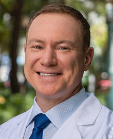 Ricardo Jorge Komotar合作论文数Department of Neurological Surgery, Miller School of Medicine, University of Miami10Alfred T. Ogden合作论文数10Fabio M. Iwamoto合作论文数10
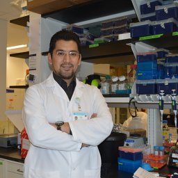 Nelson Humala合作论文数Columbia Univ, Med Ctr, New York, NY USA9Shu Chang合作论文数9
 Georg Karpel-Massler合作论文数Department of Neurosurgery, Ulm University Medical Center, Ulm, Germany9Steven R. Isaacson合作论文数9Benjamin C Kennedy合作论文数Department of Neurological Surgery, The Neurological Institute, Columbia University, 710 West 168th Street, 4th floor, New York, NY 10032, USA9
 Neil A. Feldstein合作论文数Department of Neurological Surgery, Columbia University9
 Zacharia Brad E合作论文数Dept Neurol Surg, Columbia Univ8
 Hani R Malone合作论文数8
 Pavan Upadhyayula合作论文数QB3 Institute, University of California8
 Andrew B. Lassman合作论文数Division of Neuro-Oncology, Department of Neurology, Columbia University Irving Medical Center;Herbert Irving Comprehensive Cancer Center, Columbia University Irving Medical Center8Allen Waziri合作论文数8
 Marc Otten合作论文数Department of Neurosurgery, Columbia University8
 Richard C. E. Anderson合作论文数8
 Jack Grinband合作论文数Program in Imaging and Cognitive Sciences;Columbia University;Program in Imaging and Cognitive Sciences, Columbia University8
 Alexander Khandji合作论文数8Shailendra Joshi合作论文数8Samuel S Bruce合作论文数7Kim A Lopez合作论文数7Randy S D'Amico合作论文数7
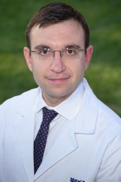 Banu Matei A合作论文数Departments of Neurological Surgery.7
 Michael Argenziano合作论文数7Kalmon D. Post合作论文数7Sharon L. Wardlaw合作论文数7
 Robert L Fine合作论文数7Trang Nguyen合作论文数6Athanassios Dovas合作论文数6
 Richard Leung合作论文数6
 Zhezhen Jin合作论文数Department of Biostatistics, Mailman School of Public Health, Columbia University6D'Ambrosio Anthony L合作论文数6Youngerman Brett E合作论文数Department of Neurosurgery, Columbia University6
 Hannah Goldstein合作论文数6Reyes-Vidal Carlos M合作论文数6




