Abstract Introduction: Tumor-associated neutrophils (TANs) are abundant in glioblastoma (GBM), yet their functions are phase-dependent. We investigated whether TANs are differentially transcribed from peripheral blood neutrophils (PBNs), if they acquire antigen-presenting functions in situ, and if neutrophil depletion timing influences survival and intratumoral immunity Methods: Transcriptomics: Integrated analysis of publicly deposited recent human GBM data sets (bulk RNA-seq and scRNA-seq; Seurat standard pipeline, stringent QC, integration/UMAP, Wilcoxon DE). Prespecified modules: co-stimulation (CD83/CD86/CD40/ICOSLG), MHC-II (HLA-DRB3/A, DPA1/DPB1), and antigen-processing/chaperones (CD74, CALR, PSME2, HLA-DMA/DMB). In vivo survival: Orthotopic CT2A and GL261; anti-Ly6G (1A8) or isotype on two schedules, (day −1 pre-implantation through ≥day 14) and delayed (start day +8). Kaplan-Meier/log-rank; Cox models (HR, 95% CI). Ex vivo flow (day 8): Bead-normalized spectral cytometry; CD45+ leukocytes; CD11b+ myeloids (Ly6G+ neutrophils; Ly6C^hi monocytes), F4/80+ TAMs with MHC-II, CD3+/CD8+ T cells. Two-sided Mann-Whitney; FDR where indicated. Ly6G epitope masking identified; depletion confirmed by CD11b+SSC^hi back-gating and weekly blood counts. Results: Transcriptomics: TANs vs PBNs showed coherent upregulation of APC/co-stimulatory programs (CD83/CD86/CD40/ICOSLG; HLA-DR/DP; CD74/CALR/PSME2/HLA-DMA/DMB), in line with dendritic-like, non-cytotoxic TAN states. Survival (biphasic): Previous anti-Ly6G impaired outcomes, CT2A (n=9/arm) median 17 vs 23 days; HR 1.95 (1.10-3.46), p=0.018. Subsequent depletion abrogated this penalty, CT2A HR 1.23 (0.68-2.22), p=0.49; Flow (day 8): Early-depleted tumors contained more Ly6C^hi monocytes (of CD45+: 29% vs 18%, p=0.006), fewer CD8+ T cells (3.2% vs 6.1%, p=0.011), and lower CD8:Ly6C^hi ratio (0.11 vs 0.36, p=0.004). Directional but non-significant aggregates: CD11b+ myeloids (68% vs 55%, p=0.07), CD3+T cells (9% vs 14%, p=0.09), reduced MHC-II on F4/80+ TAMs (p=0.08), reduced CD11c+MHC-II^hi APCs (q≈0.12). Conclusion: Human TANs take up APC/co-stimulatory programs, and timing is critical: pre-implantation neutrophil depletion imposes a myeloid-skewed, antigen-poor, T-cell-depleted setting, augments survival, while late targeting diminishes this. These observations favor phase-specific TAN modulation. maintenance or re-education of early TANs and suppression of late suppressive programs selectively, to regulate biomarker-based GBM immunotherapies. Citation Format: Matthew Alexander Abikenari, John Choi, Justin Liu, Adam Sjoholm, George Nageeb, James Poe, Brandon Hwa-Lin Bergsneider, Andrew Tran, David Bakalov, Ravi Medikonda, Lily Kim, Rohit Verma, Caren Wu, Kwang Bog Cho, Matei Banu, Michael Lim. Biphasic, time-dependent neutrophil biology in glioblastoma revealed by in vivo survival and flow cytometry with single-cell transcriptomic corroboration [abstract]. In: Proceedings of the American Association for Cancer Research Annual Meeting 2026; Part 1 (Regular Abstracts); 2026 Apr 17-22; San Diego, CA. Philadelphia (PA): AACR; Cancer Res 2026;86(7 Suppl):Abstract nr 170.
Tumor-associated macrophages (TAM) exert essential functions during the immune response to cancer. However, investigations of TAM within a native human tumor microenvironment (TME) have been impeded by a lack of appropriate model systems. Here, patient-derived organoids (PDO) from air-liquid interface (ALI)-grown tumor fragments, containing a human TME that encompassed stroma and immune subsets, robustly preserved TAM that were maintained by endogenous CSF-1 and appropriately responded to polarization signals. Antibody blockade of the CD47 regulatory checkpoint in organoids stimulated phagocytosis and remodeled TAM cytokine secretion profiles that were confirmed in anti-CD47 phase I trial patients. Amongst PDO histologies screened, anti-CD47 tumor killing was notable in clear cell renal cell carcinoma (ccRCC) which was associated with increased TAM infiltration. PDO contained diverse previously described TAM subsets; however, anti-CD47 reprogrammed organoid TAM toward an immunosuppressive SPP1+ phenotype, highlighting a negative feedback mechanism. Our findings uncover a resistance circuit engaged by macrophage checkpoint blockade and position ALI PDO as a robust translational platform for dissecting human macrophage biology and informing precision immunotherapy.
The brain offers a unique environment for cancer, with limited access to nutrients and highly regulated immune surveillance. Here we explore the role of ferroptosis, a form of metabolically regulated cell death driven by iron-mediated lipid peroxidation, in shaping immune-cell composition and function in glial tumours. We review the complex metabolic crosstalk between cell populations regulating ferroptosis in the glioma milieu. Ferroptosis induces polarization of resident microglia and controls the cytotoxic roles of CD8+ T cells and the immunosuppressive effects of regulatory T cells. We discuss recently uncovered mechanisms of ferroptosis-driven immune evasion and the impact on tumour evolution. Additionally, we analyse mechanisms of synergy in combinations incorporating ferroptosis-inducing agents and immunotherapies, including immune checkpoint blockade and adoptive cell therapies, which aim to induce effective immune responses and durable control in gliomas.
Abstract Glioblastoma (GBM) is the most common malignant form of adult brain tumor, with a median survival of around 1.5-2 years. Despite multimodal treatments (tumor resection, radiotherapy, and chemotherapy) achieving an effective cure remains a significant challenge due to its highly aggressive nature. Immune checkpoint inhibitors have emerged as a promising strategy to combat GBM; however, limitations have hindered clinical success, largely due to the “cold” and immunosuppressive tumor microenvironment (TME). Tumor Treating Fields (TTFields) therapy is a non-invasive, FDA approved treatment for GBM that employs specific frequency ranges (100-500 kHz) delivered through transducer arrays placed on the head. Our initial in vitro studies, using the inovitro system, confirmed that TTFields (72 hours, 200kHz) enhanced the immune response in GBM by inducing immunogenic cell death, which led to the recruitment of immune cells through the release of damage-associated molecular patterns (DAMPs). Additionally, we demonstrated that TTFields increased T-cell activity (IFNγ and perforin) and enhanced the motility and phagocytic activity in antigen presenting cells (Raw 264.7) and dendritic cells (JawsII). We then studied the therapeutic potential of directly applying the TTFields (10-14 days, 200kHz), using the inovivo system, to an orthotopic syngeneic GBM mouse model (CT2A-luciferase) with the checkpoint inhibitor anti-PD-1. Consistent with previous findings, the single arm treatments of TTFields and anti-PD-1 reduced tumor volume and slightly modulated the immune environment. Concomitant treatment of TTFields with systemic anti-PD-1 therapy reduced tumor volume and significantly increased immunomodulation, as observed via an increase in T cells and myeloid cells. Our findings suggest that concomitant treatment of TTFields with checkpoint inhibitors has the potential to help transform GBM into a “hot” tumor, making it an attractive target for immunotherapy. Citation Format: Si Yeon Lee, Kwang Bog Cho, Caren Wu, Justin Liu, Joe Ha, Adam Sjoholm, Michael Lim, Gordon Li, Ryan Nitta, . Analyzing Tumor Treating Fields (TTFields) therapy concomitantly with checkpoint inhibitors in a GBM mouse model [abstract]. In: Proceedings of the American Association for Cancer Research Annual Meeting 2026; Part 1 (Regular Abstracts); 2026 Apr 17-22; San Diego, CA. Philadelphia (PA): AACR; Cancer Res 2026;86(7 Suppl):Abstract nr 1565.
Abstract Background: Machine learning-generated segmentations of the trigeminal nerve and nearby blood vessels have the potential to quantify the magnitude of neurovascular compression (NVC) in patients with trigeminal neuralgia (TN). This study applies the nnU-Net machine learning method to create segmentations of the trigeminal nerve region from patient MRIs and correlate resulting quantitative NVC metrics with postoperative TN outcomes. Methods: MRIs from patients undergoing microvascular decompression (MVD) for TN from 2019 to 2022 at a single tertiary care facility were split into training, testing, and inference datasets. The trigeminal nerve and surrounding vasculature were manually labeled (i.e., segmented) in the training and testing datasets to create ground truth (GT) segmentations. nnU-Net was trained on GT segmentations in the training dataset, and predicted segmentations were evaluated using the testing dataset via the F1 score, IoU score, and paired comparison of resulting metrics. To contextualize nnU-Net performance, a manual SE-ResNet152 model was trained and deployed using the same datasets. Predicted nnU-Net segmentations in the inference dataset were then correlated with the rate of post-MVD pain recurrence. Results: Of 366 total GT segmentations, 302 (82.5%) trained the nnU-Net model and 64 (17.5%) validated the predicted segmentations. The nnU-Net model's F1 and IoU scores on the testing dataset were 0.797+/-0.011 and 0.714+/-0.011, respectively, which were higher than those for SE-ResNet152. The sensitivity and specificity of nnU-Net's ability to detect NVC were 91.3% and 66.7%, respectively. Surface area of NVC calculated from nnU-Net and GT segmentations were statistically similar. Deployed on the inference dataset (n=100), higher surface area of NVC was observed in patients without pain recurrence following MVD than patients with pain recurrence (p=0.008). Finally, higher surface area of NVC (hazards ratio [HR] 0.914 per mm2, 95% confidence interval [CI] 0.848-0.985, p=0.019) and presence of NVC (HR 0.369 relative to absent NVC, 95% CI 0.156-0.876, p=0.024) were both associated with a significantly decreased risk of pain recurrence in Cox proportional hazards models. Conclusion: nnU-Net can generate high-fidelity segmentations of the trigeminal nerve region, and the resulting NVC surface area metric is significantly associated with post-MVD pain recurrence. nnU-Net can be a standardized tool to evaluate NVC severity for patients seeking TN treatment. ### Competing Interest Statement The authors have declared no competing interest. ### Funding Statement This study did not receive any funding. ### Author Declarations I confirm all relevant ethical guidelines have been followed, and any necessary IRB and/or ethics committee approvals have been obtained. Yes The details of the IRB/oversight body that provided approval or exemption for the research described are given below: Approval granted by the Johns Hopkins Medicine Institutional Review Boards. I confirm that all necessary patient/participant consent has been obtained and the appropriate institutional forms have been archived, and that any patient/participant/sample identifiers included were not known to anyone (e.g., hospital staff, patients or participants themselves) outside the research group so cannot be used to identify individuals. Yes I understand that all clinical trials and any other prospective interventional studies must be registered with an ICMJE-approved registry, such as ClinicalTrials.gov. I confirm that any such study reported in the manuscript has been registered and the trial registration ID is provided (note: if posting a prospective study registered retrospectively, please provide a statement in the trial ID field explaining why the study was not registered in advance). Yes I have followed all appropriate research reporting guidelines, such as any relevant EQUATOR Network research reporting checklist(s) and other pertinent material, if applicable. Yes All data produced in the present study are available upon reasonable request to the authors.
OBJECTIVE:Intramedullary spinal cord tumors (IMSCTs) are typically treated with maximal safe resection, during which neurosurgeons often monitor for neurological injury using muscle motor evoked potential (mMEP) and direct wave (D-wave) neuromonitoring. The predictive value of changes in D-waves for identifying motor outcomes is underexplored. This study evaluated the utility of D-waves for predicting postoperative motor deficits. METHODS:Patients who underwent resection of a primary IMSCT with mMEP neuromonitoring from 2003 to 2023 at a tertiary care hospital were identified. Patients who underwent D-wave monitoring in addition to mMEP monitoring were compared to those who underwent mMEP monitoring alone using the Mann-Whitney U-test, chi-square test, and Fisher's exact test. Sensitivity, specificity, positive predictive value (PPV), and negative predictive value (NPV) of D-wave and mMEP monitoring for identifying new neurological deficits immediately postoperatively and at 1 month, 6 months, and last follow-up were calculated. RESULTS:After matching, 125 patients were included (median age 42.0 years; 57.6% male; median follow-up 34.0 months), of whom 88 had both mMEP and D-wave data. The most common pathologies were ependymoma (64.0%) and astrocytoma (17.6%). Patients who did and did not undergo D-wave neuromonitoring had similar preoperative neurological function, primary pathology, tumor grade, and tumor location. D-wave use was associated with increased gross-total resection (88.6% vs 64.9%, p = 0.002) and reduced mortality (5.7% vs 24.3%, p = 0.007), length of stay (5.0 vs 6.0 days, p = 0.033), and 30-day readmission (2.3% vs 13.5%, p = 0.013) and reoperation (1.1% vs 10.8%, p = 0.012). At the 6-month follow-up, D-wave monitoring alone was superior to mMEP and combination monitoring for detecting new motor deficits. D-wave monitoring had peak sensitivity (77.8%) and NPV (96.5%) at 6 months and peak specificity (95.8%) and PPV (76.9%) in the immediate postoperative period. CONCLUSIONS:D-wave monitoring was associated with reduced mortality and was more accurate than mMEP monitoring alone or combination monitoring for detecting new postoperative neurological deficits. Further prospective studies are needed to validate these results.
This cross-sectional study characterizes the care pathways of US patients with trigeminal neuralgia from the onset of their pain until their first neurosurgical consultation in terms of symptom duration.
Immune checkpoint inhibitors (ICIs) have transformed outcomes for melanoma brain metastases (MBM), yet early response assessment remains challenging because currently available tools such as magnetic resonance imaging (MRI) lack molecular specificity and are confounded by pseudoprogression. Antibody-based positron emission tomography (immunoPET) is a highly sensitive and specific molecular imaging technique that enables noninvasive, whole-body visualization of immune dynamics in vivo. The inducible T cell costimulatory receptor (ICOS) is a promising biomarker of therapy-induced T cell activation. Here, a previously validated tracer, [89Zr]DFO-ICOS mAb, is used to track activated T cells in a murine MBM model treated with combined anti-PD-1/anti-CTLA-4 therapy, and immunoPET signal is shown to correspond with therapeutic response and intratumoral T cell activation. To establish clinical relevance of ICOS-immunoPET, single-cell RNA sequencing data from 6 human MBM specimens are reanalyzed, revealing significantly higher ICOS expression accompanied by coordinated activation signatures in ICI-treated tumors. These findings support ICOS-immunoPET as a promising strategy for earlier, noninvasive, functionally informative assessment of immunotherapy response, with potential to improve patient stratification in brain metastases.
TPS2096 Background: Glioblastoma remains a lethal disease with limited treatment options. With current standard-of-care, consisting of maximal safe resection followed by radiotherapy with concurrent TMZ, then maintenance TMZ plus TTFields, median overall survival (OS) is 20.9 months, as reported in the pivotal EF-14 trial completed more than a decade ago. While immune checkpoint inhibitors (ICIs) have improved outcomes in multiple malignancies, they have not demonstrated meaningful clinical benefit in glioblastoma, in part because the tumor microenvironment (TME) is typically profoundly immunosuppressive. Strategies that reprogram the TME toward immune activation may therefore enable ICIs to elicit more effective anti-tumor immunity. Beyond antimitotic effects, TTFields have shown preclinical evidence of inducing immunogenic cell death and activating type 1 interferon signaling via DNA sensor inflammasomes, with downstream increases in dendritic cell activation and cytotoxic T cell infiltration. In a single-arm phase 2 study (NCT03405792), TTFields plus TMZ and pembrolizumab was associated with improved progression-free survival (PFS) and OS compared to historical controls in newly diagnosed glioblastoma. Here, we describe the design of a randomized phase 3 trial evaluating this regimen. Methods: EF-41/KEYNOTE D58 is a randomized, double-blind, placebo-controlled, phase 3 study (NCT06556563). Eligible patients have newly diagnosed glioblastoma (WHO 2021 Classification), have completed concurrent chemoradiotherapy, and can initiate treatment 4–7 weeks thereafter. Additional criteria include ECOG performance status 0–1 and availability of tumor tissue for central MGMT methylation analysis. Key exclusion criteria include prior anti-PD(L)1 or anti-PDL2 therapy and ongoing dexamethasone >2 mg/day. Patients are randomized 2:1 to TTFields (200 kHz for ≥18 hours/day) plus maintenance TMZ (150–200 mg/m 2 /day PO, days 1-5 of each 28 day cycle for 6-12 cycles) with either pembrolizumab 200 mg IV Q3W (up to 35 cycles) or matching placebo. TTFields is continued until second progression. At first progression, TTFields therapy is maintained and patients may receive standard salvage therapy, including re-resection and/or radiotherapy as well as systemic therapy. The target enrollment is 741 patients, providing 85% power to detect an OS improvement at a two-sided alpha of 0.05 using a 2-sided log-rank test. The primary endpoint is OS. Secondary endpoints include PFS per RANO 2.0 and RANO, PFS6 and PFS12 rates (RANO 2.0), PFS2 (RANO 2.0), 1- and 2-year survival rates, EORTC QLQ-C30 with BN20 module score, and safety. MRI assessments are performed every 9 weeks and evaluated per RANO 2.0. Adverse events are monitored throughout the study. Enrollment is ongoing. Clinical trial information: NCT03405792 .
Poor treatment response in brain metastases is largely attributed to anti-tumor T-cell suppression through the modulation of tumor-associated myeloid cells (TAMCs), resulting in immune evasion. Triggering receptor expressed on myeloid cells-1 (TREM1) is a membrane receptor highly expressed on TAMCs that is associated with poor clinical outcomes and of interest as a potential imaging biomarker of myeloid cell function, prognosis, and treatment response. Here we evaluate TREM1-targeted positron emission tomography (PET) tracer, [64Cu]TREM1-mAb, for TAMC detection in a murine model of intracranial metastatic melanoma. Forty-eight hours after tracer administration, PET imaging revealed significantly higher [64Cu]TREM1-mAb signal in implanted tumors compared to contralateral brain parenchyma or sham brains. Ex vivo gamma counting and autoradiography confirmed significantly elevated, tumor-localized signal, while markedly lower uptake with [64Cu]-isotype control-mAb confirmed tracer specificity. Similar patterns were seen in the lymphoid organs, including bone marrow and spleen. Flow cytometry confirmed TREM1 expression in myeloid cells alone in brain and spleen. We conclude that [64Cu]TREM1-mAb is a promising PET tracer for the detection of increased TREM1+ myeloid cells in the tumor microenvironment and peripheral tissues.
Malignant melanotic nerve sheath tumors (MMNSTs) are rare tumors with uncertain clinical behavior. Standard treatment includes surgical resection; however, the potential benefit of adjuvant radiotherapy remains unclear. We present four patients with MMNSTs who were treated with stereotactic radiosurgery following either partial or gross total resection. The series included two females and two males, ranging in age from 18 to 72 years, with tumors located in the spine or intracranially. At follow-up intervals of 1.6, 1.8, 3.5, and 6.0 years after radiosurgery, all tumors remained controlled, with radiographic stability and no evidence of metastatic disease.
Figure S3: Ccr6-/- Tregs are transcriptionally and metabolically distinct from WT Tregs.
Abstract Introduction: Tumor-associated neutrophils (TANs) in glioblastoma (GBM) are heterogeneous and poorly captured by the classical N1/N2 dichotomy. We characterized TAN states relative to peripheral blood neutrophils (PBNs) and wondered if a discrete, targetable program drives their pro-tumoral phenotype. Methods: Public scRNA-seq datasets were reanalyzed (GSM8380727, GSM8380728: TANs 15,000 cells; PBNs 10,000 cells; 36,601 genes). Standard Seurat workflow was applied (stringent QC; SCTransform; integration; PCA/UMAP; shared-nearest-neighbor clustering). Differential expression used Wilcoxon rank-sum with Bonferroni FDR (adj. p<0.05, |log2FC|>0.25). Module scoring examined prespecified programs: antigen presentation/co-stimulation, interferon/cytotoxicity, lipid/stress adaptation. Functional enrichment and protein-protein networks were assessed via GSEA and STRING. Results: Integration revealed six tumor-associated Neutrophil states beyond the classical N1/N2 paradigm. Among them, an SPP1+(osteopontin-high) (avg log2FC ∼10.7; adj. p≈0) population was particularly transcriptionally associated with lipid processing genes (APOE, APOC1, APOC2) and chemokine master regulators (CCL3, CCL4) of immune cell recruitment and metabolic reprogramming. SPP1+ TANs featured repression of cytotoxicity pathways and induction of oxidative and lipid metabolism modules, pointing toward a shift from antimicrobial to tissue-remodeling and tumor-supporting activities. Network analysis revealed two large hubs: SPP1-APOE/APOC (lipid remodeling) and CCL3/CCL4 (immune signaling) that are bridged by metabolic stress genes (CTSB, EIF1B) into a cohesive immunometabolic circuit. Conclusion: Single-cell analysis characterizes GBM TAN heterogeneity and implicates an SPP1-centered, APC-like neutrophil axis bridging lipid regulation (APOE/APOCs) and chemokine signaling (CCL3/CCL4). This osteopontin-driven axis establishes a mechanistic basis for TAN-mediated tumor support and nominates SPP1 and its lipid-chemokine network as actionable targets to reprogram TANs for anti-tumor activity.This is among the first studies to define an osteopontin-driven immunometabolic axis in pro-tumoral neutrophils, establishing a mechanistic framework for future neutrophil-targeted immunotherapies in glioblastoma. Citation Format: Matthew Alexander Abikenari, John Hyunkuk Choi, Ravi Medikonda, Lily Kim, Rohit Verma, Justin Liu, Adam Sjoholm, George Nageeb, Brandon Hwa-Lin Bergsneider, Caren Yu-Ju Wu, Kwang Bog Cho, Andrew Tran, David Bakalov, Matei Banu, Michael Lim. SPP1-driven immunometabolic reprogramming of tumor-associated neutrophils in glioblastoma [abstract]. In: Proceedings of the American Association for Cancer Research Annual Meeting 2026; Part 1 (Regular Abstracts); 2026 Apr 17-22; San Diego, CA. Philadelphia (PA): AACR; Cancer Res 2026;86(7 Suppl):Abstract nr 3879.
Glioblastoma (GBM) persists as one of the greatest challenges in the treatment of human cancer, despite extensive efforts to leverage the therapeutic potential of immunotherapy. While checkpoint blockade and other forms of immunotherapy have revolutionized the treatment of various cancers, their therapeutic efficacy in GBM has been hindered by the profound immunosuppressive environment, spatial heterogeneity, and dynamic immune metabolic challenges associated with the tumor microenvironment. In this review, we will synthesize recent advances and insights to develop a next-generation framework for GBM immunotherapy based on systems biology approaches to understanding the complex interplay between GBM and the immune system, as opposed to single-axis approaches to immune activation and modulation. We will discuss how the functional competence of the interferon system, myeloid antigen presentation status, T-cell clone status, spatial organization of the immune microenvironment, and resource competition between GBM and the immune system dictate therapeutic responsiveness. Furthermore, the current paper elucidates how recent advances in spatial transcriptomics, single-cell analysis, and high-parameter imaging enable us to understand how immune phenotype status varies across GBM regions and treatment status, and how this information can be used to develop predictive and pharmacodynamic biomarkers of therapeutic efficacy and failure. We will then discuss how these advances form the basis for rational combination approaches to GBM immunotherapy, which involve the integration of checkpoint blockade with metabolic reprogramming, myeloid modulation, and interferon system reactivation, and how artificial intelligence-based analytics and adaptive clinical trial design can guide the development of biomarker-based therapeutic selection approaches.
 Christina Jackson合作论文数Dept Neurosurg, Johns Hopkins Univ143
 Bettegowda Chetan合作论文数The Johns Hopkins University111
 Henry Brem合作论文数Department of Neurosurgery, School of Medicine, The Johns Hopkins University90
 Lawrence Kleinberg合作论文数Department of Radiation Oncology, Johns Hopkins Hospital84
 John S. Choi合作论文数jet propulsion laboratory70Kristin J. Redmond合作论文数65
 Gordon Li合作论文数Department of Neurosurgery, School of medicine, Stanford University;Department of Neurology, School of medicine, Stanford University;Department of Otolaryngology - Head & Neck Surgery, School of medicine, Stanford University62
 Drew Mark Pardoll合作论文数Department of Oncology, School of Medicine, Johns Hopkins University;Bloomberg~Kimmel Institute for Cancer Immunotherapy, School of Medicine, Johns Hopkins University;Sidney Kimmel Comprehensive Cancer Center, School of Medicine, Johns Hopkins University60J D Weingart合作论文数51Ayush Pant合作论文数47
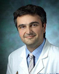 Mathios Dimitrios合作论文数45
 Risheng Xu合作论文数45
 Gary L. Gallia合作论文数School of Medicine, Johns Hopkins University44
 Kim Lily H合作论文数San Jose Gastroenterology, Stanford University Medical Center42Ravi Medikonda合作论文数38
 Timothy Cloughesy合作论文数Department of Molecular and Medical Pharmacology, David Geffen School of Medicine, University of California, Los Angeles;Brain Research Institute, University of California, Los Angeles38
 David Reardon合作论文数Center for Neuro-Oncology, Dana-Farber Cancer Institute;Harvard Medical School37
 Charles Drake合作论文数Herbert Irving Comprehensive Cancer Center, Columbia University;The Janssen Pharmaceutical Companies of Johnson & Johnson37Alessandro Olivi合作论文数35
 Judy Huang合作论文数Department of Neurosurgery, School of Medicine, Johns Hopkins University34
 Xiaobu Ye合作论文数Departments of Neurology and Neurosurgery, School of Medicine, Biomedical Engineering33Garzon-Muvdi Tomas合作论文数Department of †Neurosurgery;‡Oncology Center-Chemical Therapeutics, The Johns Hopkins University School of Medicine;§Technion Faculty of Medicine, Technion-Israel Institute of Technology;Technion-Israel Institute of Technology, The Johns Hopkins University School of Medicine33
 Betty Tyler合作论文数Departments of Neurology and Neurosurgery, School of Medicine, Biomedical Engineering32
 Daniele Rigamonti合作论文数Johns Hopkins University;KHDR Consulting32
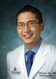 Yuanxuan Xia合作论文数Dept Neurosurg, Johns Hopkins Univ30
 Rohit Verma合作论文数Stanford University School of Medicine, Stanford University30Ruzevick Jacob合作论文数29
 Debebe Theodros合作论文数Department of Neurosurgery, School of Medicine, Stanford University28
 Anita Kalluri合作论文数Division of Gastroenterology and Hepatology, Perelman School of Medicine25
 Michael Weller合作论文数Department of Neurology, University Hospital Zurich25
 Alfredo Quinones-Hinojosa合作论文数Department of Neurosurgery, Mayo Clinic25Eileen Kim合作论文数24Jillian Phallen合作论文数24
 Jennifer Kim合作论文数Department of Neurology, Yale School of Medicine, Yale University24Zineb Belcaid合作论文数Department of Neurosurgery, Brain Tumor Center, Erasmus University Medical Center24
 Antonio Omuro合作论文数Department of Neurology and Neurological Sciences, Stanford University School of Medicine23
 Kaisorn Chaichana合作论文数Johns Hopkins University School of Medicine23
 Patrick Y. Wen合作论文数Department of Neurology, Harvard Medical School;Center For Neuro-Oncology, Dana-Farber Cancer Institute23Laura Saleh合作论文数22Sumil K Nair合作论文数Department of Neurosurgery, Johns Hopkins University School of Medicine22Sahebjam Solmaz合作论文数Shriners Hospital for Children, McGill University20
 Kisha Patel合作论文数Dept Neurosurg, Johns Hopkins Univ19
 Steven D. Chang合作论文数Department of Neurosurgery, School of Medicine, Stanford University19
 John Howard Sampson合作论文数School of Medicine, Duke University18
 Jon Weingart合作论文数Department of The Sidney Kimmel Comprehensive Cancer Center, School of Medicine, Johns Hopkins University18Taube Janis M合作论文数18Ryan Nitta合作论文数17
 Todd R. McNutt合作论文数Radiation Oncology and Molecular Radiation Sciences, School of Medicine, Johns Hopkins University17
 Phuoc T. Tran合作论文数Department of Radiation Oncology, School of Medicine, University of Maryland, Baltimore17




