The importance of virus maturation has been appreciated for nearly 70 years as it provides models for large-scale protein reorganization resulting in functional activation as well as being a target for antiviral therapies. However, a detailed description of the pathway from the initial assembly product (procapsid) to the mature, infectious particle (virion) has been elusive. This is due to the "in cell" nature of the natural process, the 2-state behavior of maturation (no detectable intermediates) in some viruses in vitro and heterogeneous populations of particle intermediates that are only partially matured in other systems. The non-enveloped, T=4, ssRNA-containing, Nudaurelia capensis omega virus (NωV), is a highly accessible model system that exemplifies the maturation process of a eukaryotic virus. During maturation the particle shrinks in outer diameter from 482 Angstroms (pH 7.5) to 428 Angstroms (pH 5.0). It is possible to mimic the maturation process in vitro by lowering the pH of a population of procapsids produced in heterologous systems. Indeed, by controlling the pH in vitro it is possible to produce homogenous populations of intermediate NωV virus-like particles (VLPs) that occur too fleetingly to be observed in vivo. Here we report structural models, based on cryo-electron microscopy (cryo-EM), of five intermediates in the NωV maturation process. The structures of the intermediate particles reveal unique, quaternary position-dependent trajectories and refolding of subunit N and C-terminal regions, including the formation of the autocatalytic cleavage site at N570. The detailed structures reported here, coupled with previously determined structures of the procapsids and mature particles, allows the maturation pathway to be described in detail for the first time for a eukaryotic virus. ### Competing Interest Statement The authors have declared no competing interest.
BACKGROUND:Genetic recombination is a tremendous source of intrahost diversity in viruses and is critical for their ability to rapidly adapt to new environments or fitness challenges. While viruses are routinely characterized using high-throughput sequencing techniques, characterizing the genetic products of recombination in next-generation sequencing data remains a challenge. Viral recombination events can be highly diverse and variable in nature, including simple duplications and deletions, or more complex events such as copy/snap-back recombination, intervirus or intersegment recombination, and insertions of host nucleic acids. Due to the variable mechanisms driving virus recombination and the different selection pressures acting on the progeny, recombination junctions rarely adhere to simple canonical sites or sequences. Furthermore, numerous different events may be present simultaneously in a viral population, yielding a complex mutational landscape. FINDINGS:We have previously developed an algorithm called ViReMa (Virus Recombination Mapper) that bootstraps the bowtie short-read aligner to capture and annotate a wide range of recombinant species found within virus populations. Here, we have updated ViReMa to provide an "error density" function designed to accurately detect recombination events in the longer reads now routinely generated by the Illumina platforms and provide output reports for multiple types of recombinant species using standardized formats. We demonstrate the utility and flexibility of ViReMa in different settings to report deletion events in simulated data from Flock House virus, copy-back RNA species in Sendai viruses, short duplication events in HIV, and virus-to-host recombination in an archaeal DNA virus.
EMPIAR, the Electron Microscopy Public Image Archive centered at EMBL-EBI, is a public resource for raw electron microscopy images related to EMDB, contains micrographs, particle sets and tilt-series.
EMPIAR, the Electron Microscopy Public Image Archive centered at EMBL-EBI, is a public resource for raw electron microscopy images related to EMDB, contains micrographs, particle sets and tilt-series.
There are many methods for characterizing the heterogeneity of an ensemble of particles from single-particle cryo EM images. This talk concerns a method [1-3] based on describing the electron scattering intensity of the particle as a Fourier series and describing the coefficients of the Fourier series as random variables that are independent and identically distributed from instance to instance of the particle. The heterogeneity is characterized by estimating the mean and variance of the coefficients from the image data by a maximum likelihood estimator. The mean results give a reconstruction and the variance results give a spatially-resolved characterization of the heterogeneity of the ensemble of particles. When symmetry is present, the method can allow each instance of the particle to lack symmetry while imposing the symmetry on the statistics of the particle. This avoids anomalous peaks in the variance map located on and near symmetry axes of the particle [4]. Imposing symmetry on the statistics also allows the computation of ensemble averages of the product of the electron scattering intensity at two different locations which can be used to detect allosteric interactions between the different locations. We demonstrate the method on the bacteriophage HK97 where we show that binding of the maturation protease on the inner surface of the capsid has wide-ranging effects on the heterogeneity of the outer surface of the capsid.
Many virus capsids undergo exquisitely choreographed maturation processes in their host cells to produce infectious virions, and these remain poorly understood. As a tool for studying virus maturation, we transiently expressed the capsid protein of the insect virus Nudaurelia capensis omega virus (NωV) in Nicotiana benthamiana and were able to purify both immature procapsids and mature capsids from infiltrated leaves by varying the expression time. Cryo-EM analysis of the plant-produced procapsids and mature capsids to 6.6 Å and 2.7 Å resolution, respectively, reveals that in addition to large scale rigid body motions, internal regions of the subunits are extensively remodelled during maturation, creating the active site required for autocatalytic cleavage and infectivity. The mature particles are biologically active in terms of their ability to lyse membranes and have a structure that is essentially identical to authentic virus. The ability to faithfully recapitulate and visualize a complex maturation process in plants, including the autocatalytic cleavage of the capsid protein, has revealed a ~30 Å translation-rotation of the subunits during maturation as well as conformational rearrangements in the N and C-terminal helical regions of each subunit.
The structural study of icosahedral viruses has a long and impactful history in both crystallographic methodology and molecular biology. The evolution of the Protein Data Bank has paralleled and supported these studies providing readily accessible formats dealing with novel features associated with viral particle symmetries and subunit interactions. This overview describes the growth in size and complexity of icosahedral viruses from the first early studies of small RNA plant viruses and human picornaviruses up to the larger and more complex bacterial phage, insect, and human disease viruses such as Zika, hepatitis B, Adeno and Polyoma virus. The analysis of icosahedral viral capsid protein domain folds has shown striking similarities, with the beta jelly roll motif observed across multiple evolutionarily divergent species. The icosahedral symmetry of viruses drove the development of noncrystallographic symmetry averaging as a powerful phasing method, and the constraints of maintaining this symmetry resulted in the concept of quasi-equivalence in viral structures. Symmetry also played an important early role in demonstrating the power of cryo-electron microscopy as an alternative to crystallography in generating atomic resolution structures of these viruses. The Protein Data Bank has been a critical resource for assembling and disseminating these structures to a wide community, and the virus particle explorer (VIPER) was developed to enable users to easily generate and view complete viral capsid structures from their asymmetric building blocks. Finally, we share a personal perspective on the early use of computer graphics to communicate the intricacies, interactions, and beauty of these virus structures.
Abstract VIrus Particle ExploreR data base (VIPERdb) (http://viperdb.scripps.edu) is a curated repository of virus capsid structures and a database of structure-derived data along with various virus specific information. VIPERdb has been continuously improved for over 20 years and contains a number of virus structure analysis tools. The release of VIPERdb v3.0 contains new structure-based data analytics tools like Multiple Structure-based and Sequence Alignment (MSSA) to identify hot-spot residues within a selected group of structures and an anomaly detection application to analyze and curate the structure-derived data within individual virus families. At the time of this writing, there are 931 virus structures from 62 different virus families in the database. Significantly, the new release also contains a standalone database called ‘Virus World database’ (VWdb) that comprises all the characterized viruses (∼181 000) known to date, gathered from ICTVdb and NCBI, and their capsid protein sequences, organized according to their virus taxonomy with links to known structures in VIPERdb and PDB. Moreover, the new release of VIPERdb includes a service-oriented data engine to handle all the data access requests and provides an interface for futuristic data analytics using machine leaning applications.
Michael Rossmann passed away on May 14, 2019 at nearly 89 years of age following a courageous 5-year fight with cancer. During that time he maintained the arduous schedule of laboratory leadership, preparing grant proposals (he had four R01 NIH grants at the time of his death), traveling and publishing that characterized his previous 55 years in structural biology. Although there were many medical challenges associated with his illness few people were aware of the monumental effort that was required to maintain the “Rossmann persona” that we saw at conferences and gatherings during that time. He would not have appreciated that descriptor because it implies a pretense. His “persona” came from doing the best science possible in his unique style and demanding the same from others. Many graduate students, post docs, and principal investigators recall the penetrating questions (putting it nicely) that Michael would raise during presentations at conferences, graduate student committee meetings and seminars. His passion was maintained to the end. Three weeks before he died he was working on manuscripts and grant proposals and there were already six publications in print for 2019. The first word that comes to mind when colleagues talk about Michael is energy. Throughout his life he exuded mental and physical energy and a passion for getting things done. The second word is impatience. Things rarely happened fast enough for Michael because he could not imagine others lacking the same gifts, focus, and discipline that he had. This could lead to edginess in interactions with lab members, collaborators, and administrators and Michael frequently experienced frustration with those around him, myself included. The upside for those on the receiving end was motivation, accomplishments, and exceptionally good science; the downside was sleepless nights when you were not living up to expectation. Meetings of lab alumni and collaborators often result in sharing “special moments” of dealing with these aspects of Michael G. Rossmann. The other side of Michael contrasts dramatically with the description earlier. He could be warm, considerate, and compassionate with colleagues, providing insightful, nonscientific, advice, as I experienced during difficult times in my personal and professional life. There is widespread respect from those that worked with Michael or knew him well and gratitude for how he shaped our professional lives. Forty years after leaving his lab his opinion of my choice and execution of projects was still very important to me. What follows is a commentary, mostly in chronological order, on some of Michael's contributions to structural biology most of which are easily found by going to PubMed and following the 446 (of over 600 total) publications listed there. I finish with some personal remembrances of times with Michael. Michael's contributions to structural biology span 60 years and started when he joined the Medical Research Council (MRC) laboratory of Max Perutz in 1958. He made pivotal contributions to the structure determination of hemoglobin and, with David Blow, developed the practical approaches that lead to the widespread use of molecular replacement (MR). The 1962 rotation function paper was prescient in describing the use of noncrystallographic symmetry (NCS) applied to virus structure determination, well before high resolution crystallographic data were collected from virus crystals. Rossmann moved from the MRC to the Department of Biological Sciences at Purdue University in 1964. Henry Koefler, the head of Biosciences at Purdue, was determined to build a molecular biology department and recruiting Rossmann was a high priority. The laboratory was running in a short time and produced the structure of dogfish Lactate Dehydrogenase (LDH) in 1970, one of the largest, early protein structures determined. Ironically the subunits formed a functional enzyme with 222 symmetry, but the symmetry site was on a crystallographic 222 site, so NCS could not be used in the structure determination. Three years later Rossmann's group determined the structure of lobster Glyceraldehyde-3-Phosphate Dehydrogenase (GPD), employing NCS 222 symmetry locate heavy atoms and to improve the multiple isomorphous replacement (MIR) phases. Rossmann immediately recognized the conserved nucleotide-binding site in GPD when compared to LDH (I recall arriving at the lab the morning after the team had spent the night tracing the GPD chain and seeing “GPD is LDH” on the blackboard) (Figure 1). The paper describing the Rossmann Received: 24 June 2019 Accepted: 24 June 2019
In this letter, we report on the ability of functional fusion proteins presenting a lytic gamma peptide, to promote interactions with HeLa cells and delivery of large hybrid nanostructures.
Luteovirids rank among the most destructive viruses of economically important crops. Until now their structures have only been inferred by inadequate homology models due to their phloem-limited infection and inadequate yields. Employing virus-like particles, Byrne et al. (2019) now report near-atomic resolution structures of two family members providing important functional insights.
Michael George Rossmann, who made monumental contributions to science, passed away peacefully in West Lafayette, Indiana on 14 May 2019 at the age of 88, following a courageous five-year battle with cancer. Michael was born in Frankfurt, Germany on 30 July 1930. As a young boy, he emigrated to England with his mother just as World War II ignited. Michael was a highly innovative and energetic person, well known for his intensity, persistence and focus in pursuing his research goals. Michael was a towering figure in crystallography as a highly distinguished faculty member at Purdue University for 55 years. Michael made many seminal contributions to crystallography in a career that spanned the entirety of structural biology, beginning in the 1950s at Cambridge where the first protein structures were determined in the laboratories of Max Perutz (hemoglobin, 1960) and John Kendrew (myoglobin, 1958). Michael's work was central in establishing and defining the field of structural biology, which amazingly has described the structures of a vast array of complex biological molecules and assemblies in atomic detail. Knowledge of three-dimensional biological structure has important biomedical significance including understanding the basis of health and disease at the molecular level, and facilitating the discovery of many drugs.
was delighted to accept the ACA invitation to give an opening plenary talk at this meeting, but before having the opportunity, very sadly passed away, in West Lafayette, Indiana on May 14 th 2019 , following a courageous five-year battle with cancer.We alumni of his research group will present memories of Michael's legacy
The information content of cryo EM data sets exceeds that of the electron scattering potential (cryo EM) density initially derived for structure determination. Previously we demonstrated the power of data variance analysis for characterizing regions of cryo EM density that displayed functionally important variance anomalies associated with maturation cleavage events in Nudaurelia Omega Capensis Virus and the presence or absence of a maturation protease in bacteriophage HK97 procapsids. Here we extend the analysis in two ways. First, instead of imposing icosahedral symmetry on every particle in the data set during the variance analysis, we only assume that the data set as a whole has icosahedral symmetry. This change removes artifacts of high variance along icosahedral symmetry axes, but retains all of the features previously reported in the HK97 data set. Second we present a covariance analysis that reveals correlations in structural dynamics (variance) between the interior of the HK97 procapsid with the protease and regions of the exterior (not seen in the absence of the protease). The latter analysis corresponds well with hydrogen deuterium exchange studies previously published that reveal the same correlation.
Genome ejection proteins are required to facilitate transport of bacteriophage P22 double-stranded DNA safely through membranes of Salmonella. The structures and locations of all proteins in the context of the mature virion are known, with the exception of three ejection proteins. Furthermore, the changes that occur to the proteins residing in the mature virion upon DNA release are not fully understood. We used cryogenic electron microscopy to obtain what is, to our knowledge, the first asymmetric reconstruction of mature bacteriophage P22 after double-stranded DNA has been extruded from the capsid—a state representative of one step during viral infection. Results of icosahedral and asymmetric reconstructions at estimated resolutions of 7.8 and 12.5 Å resolutions, respectively, are presented. The reconstruction shows tube-like protein density extending from the center of the tail assembly. The portal protein does not revert to the more contracted, procapsid state, but instead maintains an extended and splayed barrel structure. These structural details contribute to our understanding of the molecular mechanism of P22 phage infection and also set the foundation for future exploitation serving engineering purposes.
The VIrus Particle ExploreR database (VIPERdb) ( http://viperdb.scripps.edu ) is a database and web portal for primarily icosahedral virus capsid structures that integrates structure-derived information with visualization and analysis tools accessed through a set of web interfaces. Our aim in developing VIPERdb is to provide comprehensive structure-derived information on viruses comprising simple to detailed attributes such as size (diameter), architecture ( T number), genome type, taxonomy, intersubunit association energies, and surface-accessible residues. In addition, a number of web-based tools are provided to enable users to interact with the structures and compare and contrast structure-derived properties between different viruses. Recently, we have constructed a series of data visualizations using modern JavaScript charting libraries such as Google Charts that allow users to explore trends and gain insights based on the various data available in the database. Furthermore, we now include helical viruses and nonicosahedral capsids by implementing modified procedures for data curation and analysis. This article provides an up-to-date overview of VIPERdb, describing various data and tools that are currently available and how to use them to facilitate structure-based bioinformatics analysis of virus capsids.
ABSTRACT Our understanding of archaeal virus diversity and structure is just beginning to emerge. Here we describe a new archaeal virus, tentatively named Metallosphaera turreted icosahedral virus (MTIV), that was isolated from an acidic hot spring in Yellowstone National Park, USA. Two strains of the virus were identified and were found to replicate in an archaeal host species closely related to Metallosphaera yellowstonensis. Each strain encodes a 9.8- to 9.9-kb linear double-stranded DNA (dsDNA) genome with large inverted terminal repeats. Each genome encodes 21 open reading frames (ORFs). The ORFs display high homology between the strains, but they are quite distinct from other known viral genes. The 70-nm-diameter virion is built on a T=28 icosahedral lattice. Both single particle cryo-electron microscopy and cryotomography reconstructions reveal an unusual structure that has 42 turret-like projections: 12 pentameric turrets positioned on the icosahedral 5-fold axes and 30 turrets with apparent hexameric symmetry positioned on the icosahedral 2-fold axes. Both the virion structural properties and the genome content support MTIV as the founding member of a new family of archaeal viruses. IMPORTANCE Many archaeal viruses are quite different from viruses infecting bacteria and eukaryotes. Initial characterization of MTIV reveals a virus distinct from other known bacterial, eukaryotic, and archaeal viruses; this finding suggests that viruses infecting Archaea are still an understudied group. As the first known virus infecting a Metallosphaera sp., MTIV provides a new system for exploring archaeal virology by examining host-virus interactions and the unique features of MTIV structure-function relationships. These studies will likely expand our understanding of virus ecology and evolution.
Viruses are obligate intracellular parasites that rely on host cell machineries for their replication and survival. Although viruses tend to make optimal use of the host cell protein repertoire, they need to encode essential enzymatic or effector functions that may not be available or accessible in the host cellular milieu. The enzymes encoded by nonenveloped viruses-a group of viruses that lack any lipid coating or envelope-play vital roles in all the stages of the viral life cycle. This review summarizes the structural, biochemical, and mechanistic information available for several classes of enzymes and autocatalytic activity encoded by nonenveloped viruses. Advances in research and development of antiviral inhibitors targeting specific viral enzymes are also highlighted.
 Tianwei Lin (林天伟)合作论文数School of Life sciences, Xiamen University46
 Reddy Vijay S合作论文数31
 Anette Schneemann合作论文数28
 Gabriel C. Lander合作论文数Department of Molecular Biology, The Scripps Research Institute26
 Peter Doerschuk合作论文数Meinig School of Biomedical Engineering, Cornell University26Jeffrey A. Speir合作论文数Department of Molecular Biology, The Scripps Research Institute23R W Hendrix合作论文数19
 Liang Tang合作论文数Department of Molecular Biosciences, University of Kansas19R. L. Duda合作论文数18Carissa M. Soto合作论文数Center for Bio/Molecular Science and Engineering, Naval Research Laboratory18
 Reza Khayat合作论文数Dept Chem, CUNY City Coll17Lars Liljas合作论文数17
 Mark Young合作论文数Earth and Planetary Science, School of GeoSciences, The University of Edinburgh16
 Wikoff William R合作论文数Genome Ctr, Univ Calif Davis15Baker Timothy S合作论文数15
 M. G. Finn合作论文数School of Chemistry and Biochemistry, College of Sciences, Georgia Institute of Technology;School of Biological Sciences, Georgia Institute of Technology15
 Peter E. Prevelige合作论文数Department of Microbiology, School of Medicine, University of Alabama at Birmingham13
 Chi-Yu Fu合作论文数13
 Brian Bothner合作论文数Department of Biochemistry, University of Tennessee Health Sciences Center13Anju Chatterji合作论文数13A Chatterji合作论文数12
 Mark Yeager合作论文数Department of Molecular Physiology and Biological Physics, School of Medicine, University of Virginia12
 George Lomonossoff合作论文数John Innes Centre12
 Tatiana Domitrovic合作论文数Departamento de Bioquı&#x;ICB/CCS, UFRJ;ICB/CCS, UFRJ12
 Gan Lu合作论文数Department of Biological Sciences, Faculty of Science, National University of Singapore11Hiro Tsuruta合作论文数11
 Banahalli Ratna合作论文数Science and Technology Research Solutions11
 David Veesler合作论文数Department of Biochemistry, University of Washington;Howard Hughes Medical Institute11As Blum合作论文数10
 Gino Cingolani合作论文数Dept. of Biochemistry and Molecular Biology, Thomas Jefferson University10Jinghua Tang合作论文数10
 Kelly K. Lee合作论文数Department of Medicinal Chemistry, School of Pharmacy, University of Washington10
 Bridget Carragher合作论文数Vagelos College of Physicians and Surgeons, Columbia University Irving Medical Center;Simons Electron Microscopy Center, New York Structural Biology Center9
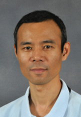 Qian Wang合作论文数Department of Chemistry and Biochemistry, University of South Carolina;McCausland College of Arts and Sciences, University of South Carolina9Roger Fourme合作论文数9clinton s potter合作论文数8Banerjee Manidipa合作论文数8
 Richard Kahn合作论文数Winship Cancer Institute, Emory University8
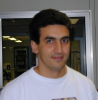 Mohamed Mezouar合作论文数Division of Experiments, European Synchrotron Radiation Facility8Roger W. Hendrix合作论文数8
 Jim Conway合作论文数Department of Structural Biology, University of Pittsburgh School of Medicine8
 Charles L. Brooks合作论文数College of Literature, Science, and the Arts, University of Michigan8
 Eric Girard合作论文数8
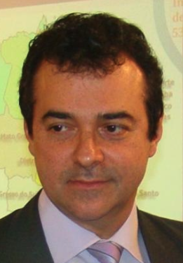 Jerson L. Silva合作论文数Instituto de Bioquímica Medica, Centro de Ciências da Saúde, Universidade Federal do Rio de Janeiro;National Institute of Science and Technology for Structural Biology and Bioimaging;Jiri Jonas National Center for Macromolecular Nuclear Magnetic Resonance, Universidade Federal do Rio de Janeiro8
 Mariana Tihova合作论文数7Tw Lin合作论文数7
 Routh Andrew合作论文数7Alasdair C. Steven合作论文数Laboratory of Structural Biology Research, National Institute of Arthritis and Musculoskeletal and Skin Diseases, National Institutes of Health7Matsui Tsutomu合作论文数7




