Background Same-day discharge after transcatheter aortic valve replacement (TAVR) may be feasible for selected patients if a low risk for adverse clinical events can be defined. We aimed to develop a clinical risk prediction model to facilitate same-day discharge planning. Methods A random forest machine learning algorithm was used to build a prediction model of adverse events occurring in-hospital after TAVR. Patients were categorized into low, moderate, or high-risk groups based on their estimated scores. Results Overall, 730 patients (median age, 81 years; 58.9% men) who had transfemoral TAVR performed with conscious sedation were examined. The risk score was built utilizing 9 clinical parameters. The prediction model had a median area under the receiver operating characteristic curve of 0.76. For determining the probability of events that would disallow same-day discharge, the model successfully identified 172 patients (23.6% of the population) as low-risk for same-day discharge, or for having an event rate of <3%, with all events occurring within 6 hours after TAVR. The low-risk group had no in-hospital events after a 6-hour observation, and no mortality at the 30-day follow-up. External testing in 158 patients showed 94% sensitivity in predicting overall adverse events and identified a low-risk group using the clinical risk score. Conclusions In this analysis, ∼1 in 4 patients may be candidates for same-day discharge after TAVR. This prediction model can identify such patients, with findings that may have implications for hospital resource allocation in those undergoing TAVR.
BACKGROUND:Tricuspid regurgitation (TR) progression following left-sided valvular heart disease (VHD) correction is a critical clinical concern. This study aimed to determine the incidence, predictors and outcomes of TR progression in a contemporary cohort. METHODS:We analysed 1644 patients (mean age 73 years, 62% men) without severe TR who underwent surgical or transcatheter treatment for aortic or mitral disease between 2014 and 2018. TR progression was defined as an increase in TR grade to moderate or severe on follow-up echocardiography. RESULTS:At 5 years, TR progression incidence was 12.0% (95% CI 10.5% to 13.7%). Baseline factors associated with TR progression included older age, female sex, atrial fibrillation, prior pacemaker implantation and larger tricuspid annular diameter (TAD). The relationship between TAD and TR progression was linear (HR 1.08; 95% CI 1.04 to 1.11; p<0.001), with sex differences mitigated by indexing TAD to body surface area. TR progression was associated with increased all-cause mortality (adjusted HR 2.77; 95% CI 2.16 to 3.56; p<0.001) and a combined endpoint of death or heart failure hospitalisation (adjusted HR 2.91; 95% CI 2.21 to 3.82; p<0.001). CONCLUSIONS:TR progression is common after left-sided VHD correction and is associated with adverse outcomes. Indexing TAD to body surface area mitigates sex differences in risk assessment. These findings suggest that lower thresholds for prophylactic tricuspid intervention may be warranted in high-risk patients.
Differentiating normal from minimally and mildly diseased coronary arteries on coronary computed tomographic angiography (CCTA) is crucial, impacting treatment decisions due to the extremely low coronary artery event risk associated with the former. Artificial intelligence quantitative computed tomographic (AI-QCT) can potentially identify subclinical atherosclerosis in cases deemed normal by reader interpretation. We aimed to evaluate AI-QCT’s ability to distinguish reader-determined normal coronary arteries from those with minimal and mild diseased on CCTA. We screened 849 consecutive patients without coronary artery stents or bypass grafts who underwent CCTA and AI-QCT for suspected coronary artery disease between October 2022 and February 2023. Clinical reads were blinded to AI-QCT results. 411 patients (mean age 60, 63
Coronary computed tomography angiography (CTA) analysis can help in the planning of percutaneous coronary intervention (PCI). Fractional flow reserve derived from coronary CTA (FFRCT), coronary CTA-derived regional myocardial mass, and FFRCT virtual PCI planner can facilitate decisions concerning sheath and guide catheter selection, stent lengths on the basis of predicted post-PCI FFRCT, optimal fluoroscopic angles, evaluation of provisional vs 2-stent bifurcation PCI techniques, and assessment of the magnitude of jeopardized myocardial mass in cases with side branch compromise. This case series illustrates the emerging opportunities for coronary CTA-based planning of bifurcation PCI.
Background Transcatheter aortic valve replacement (TAVR) is approved across all risk profiles, including patients with bicuspid aortic valves. These patients are generally younger, with a higher chance of reintervention. Objectives The aim of this study was to compare the feasibility of redo transcatheter aortic valve (TAV) between bicuspid and tricuspid aortic valves. Methods A computed tomographic (CT) simulation of redo-TAV was conducted using 913 post-TAVR CT studies from patients who underwent TAVR with SAPIEN 3 or SAPIEN 3 Ultra (S3; n = 623) or Evolut R, Evolut PRO, or Evolut PRO+ (Evolut; n = 290) valves. Fifty-nine cases were for bicuspid valves. Four redo-TAV scenarios—S3-in-S3, Evolut-in-S3, S3-in-Evolut, and Evolut-in-Evolut—were simulated. Second TAV size was determined from the averaged stent areas in the landing zone, except for Evolut-in-Evolut, for which same size was simulated. Redo-TAV feasibility (risk to coronary arteries) was estimated by the relationship between neoskirt plane and coronary risk plane or sinotubular junction and the narrowest valve-to-aorta distances. Results Patients with bicuspid valves, compared with those with tricuspid valves, were younger with lower surgical risk and had larger annular areas (522 mm2 [Q1-Q3: 461-597 mm2] vs 479 mm2 [Q1-Q3: 416-551 mm2]), sinus of Valsalva diameters (34 mm [Q1-Q3: 31-36 mm] vs 32 mm [Q1-Q3: 30-35 mm]), and sinotubular junction diameters (30 mm [Q1-Q3: 28-33 mm] vs 29 mm [Q1-Q3: 27-31 mm]) (P < 0.05 for all). Narrowest valve-to-aorta distances were longer in bicuspid cases, resulting in higher redo-TAV feasibility for S3-in-S3 and Evolut-in-S3 scenarios. Similar trends were observed for S3-in-Evolut and Evolut-in-Evolut, for which the feasibility tended to be higher in bicuspid cases. Conclusions Redo-TAV simulation using post-TAVR CT imaging performed at 30 days suggested higher redo-TAV feasibility in patients with bicuspid aortic valves compared with tricuspid aortic valves.
Coronary computed tomography angiography (CTA) analysis can help in the planning of percutaneous coronary intervention (PCI). Fractional flow reserve derived from coronary CTA (FFRCT), coronary CTA-derived regional myocardial mass, and FFRCT virtual PCI planner can facilitate decisions concerning sheath and guide catheter selection, stent lengths on the basis of predicted post-PCI FFRCT, optimal fluoroscopic angles, evaluation of provisional vs 2-stent bifurcation PCI techniques, and assessment of the magnitude of jeopardized myocardial mass in cases with side branch compromise. This case series illustrates the emerging opportunities for coronary CTA-based planning of bifurcation PCI.
BACKGROUND:Although pretranscatheter aortic valve replacement-computed tomography angiography (TAVR-CTA) has shown a good correlation with invasive coronary angiography (ICA) for ruling out obstructive coronary artery disease (CAD), its clinical effectiveness and safety as a gatekeeper for ICA pre-transcatheter aortic valve replacement (pre-TAVR) remain unclear. This study aims to determine whether routine TAVR-CTA, without premedication, could safely defer and guide the need for ICA pre-TAVR. METHODS:Patients who underwent TAVR evaluation with either TAVR-CTA or ICA to determine CAD between 2017 and 2022 were included. Patients with prior coronary artery bypass grafts were excluded, and the remaining patients were divided into CAD screening with TAVR-CTA or ICA groups. The primary outcome was symptom-driven revascularization at 1 year post-TAVR. RESULTS:Among 1165 patients (median age, 81 years; 46% women), 464 were in TAVR-CTA group and 701 were in the ICA group. Prevalence of CAD was similar (37% versus 41%; P=0.2). A total of 53% of patients were exempted from ICA after TAVR-CTA, given the absence of proximal obstructive CAD, whereas 17% had inconclusive TAVR-CTA interpretation, 15% desired for ICA despite CAD exclusion by TAVR-CTA, and 14% had obstructive CAD requiring further ICA. Elevated coronary artery calcium score and the presence of stents were associated with need for ICA after TAVR-CTA. TAVR-CTA revealed a per-patient sensitivity of 89%, specificity of 75%, positive predictive value of 69%, and negative predictive value of 91% for identifying obstructive CAD. Importantly, symptom-driven revascularization, acute coronary syndrome, and unplanned ICA at 1 year after TAVR were all low and not different between TAVR-CTA versus ICA (0.8% versus 1.8%, P=0.158; 1.6% versus 1.7%, P=0.846; 2.7% versus 2.8%, P=0.767; respectively). CONCLUSIONS:In comparison with routine ICA pre-TAVR, integration of TAVR-CTA in our program resulted in up to 53% of patients exempted from ICA, while maintaining patient safety. This approach could have important clinical implications to improve patient access, experience, value, and throughput.
HomeCirculationVol. 149, No. 2Highlights From the Circulation Family of Journals Free AccessIn BriefPDF/EPUBAboutView PDFView EPUBSections ToolsAdd to favoritesDownload citationsTrack citationsPermissions ShareShare onFacebookTwitterLinked InMendeleyReddit Jump toFree AccessIn BriefPDF/EPUBHighlights From the Circulation Family of Journals Originally published8 Jan 2024https://doi.org/10.1161/CIRCULATIONAHA.123.068221Circulation. 2024;149:155–159The November highlights from the Circulation family of journals cover several fascinating topics in cardiology. Among young patients with hypertrophic cardiomyopathy, an association of storage disease and preexcitation is reported in Circulation Arrhythmia and Electrophysiology. The associations of coronary and cerebrovascular microvascular dysfunction, heart failure, and cognitive dysfunction are reported in Circulation: Heart Failure. The incorporation of cost-effectiveness evaluations in clinical cardiology guidelines is reported in Circulation: Cardiovascular Quality and Outcomes. Investigators report an association of pericardial enhancement on cardiac magnetic resonance imaging after cardiac surgery with low-grade inflammation in Circulation: Cardiovascular Imaging. Last, an analysis of cardiac computed tomography characteristics before redo transcatheter aortic valve replacement for the risk of coronary occlusion is reported in Circulation: Cardiovascular Interventions.Circulation: Arrhythmia and ElectrophysiologyHypertrophic Cardiomyopathy and Ventricular Preexcitation in the Young: Cause and Accessory Pathway CharacteristicsRobert Przybylski, MD; Sakethram Saravu Vijayashankar, MD, MRCPCH; Edward T. O’Leary, MD; Robyn J. Hylind, MS, CGC; Jennifer Noon, MSN, CPNP-PC; Audrey Dionne, MD; Elizabeth S. DeWitt, MD; Vassilios J. Bezzerides, MD, PhD; Dominic J. Abrams, MD, MRCP, MBCorrespondence to: Robert Przybylski, MD, Department of Pediatrics, Inova Fairfax Hospital, 3600 Gallows Rd Falls Church, VA. Email robert.przybylski@inova.orgBACKGROUND: The cause of hypertrophic cardiomyopathy (HCM) in the young is highly varied. Ventricular preexcitation (preexcitation) is well recognized, yet little is known about the specificity for any cause and the characteristics of the responsible accessory pathways (AP).METHODS: Retrospective cohort study of patients <21 years of age with HCM/preexcitation from 2000 to 2022. The cause of HCM was defined as isolated HCM, storage disorder, metabolic disease, or genetic syndrome. Atrioventricular AP (true AP) were distinguished from fasciculoventricular fibers (FVF) using standard invasive electrophysiology study criteria. AP were defined as high risk if any of the following were <250 ms: shortest preexcited RR interval in atrial fibrillation, shortest paced preexcited cycle length, or anterograde AP effective refractory period.RESULTS: We identified 345 patients with HCM and 28 (8%) had preexcitation (isolated HCM, 10/220; storage disorder, 8/17; metabolic disease, 5/19; and genetic syndrome, 5/89). Six (21%) patients had clinical atrial fibrillation (1 with shortest preexcited RR interval <250 ms). Twenty-two patients underwent electrophysiology study which identified 23 true AP and 16 FVF. Preexcitation was exclusively FVF mediated in 8 (36%) patients. Five (23%) patients had AP with high-risk conduction properties (including ≥1 patient in each etiologic group). Multiple AP were seen in 8 (36%) and AP plus FVF in 10 (45%) patients. Ablation was acutely successful in 13 of 14 patients with recurrence in 3. One procedure was complicated by complete heart block after ablation of a high-risk midseptal AP. There were significant differences in QRS amplitude and delta wave amplitude between groups. There were no surface ECG features that differentiated AP from FVF.CONCLUSIONS: Young patients with HCM and preexcitation have a high likelihood of underlying storage disease or metabolic disease. Nonisolated HCM should be suspected in young patients with large QRS and delta wave amplitudes. Surface ECG is not adequate to discriminate preexcitation from a benign FVF from that secondary to potentially life-threatening AP.Circ Arrhythm Electrophysiol. 2023;16:e012191. doi: 10.1161/CIRCEP.123.012191Circulation: Heart FailureMicrovascular Dysfunction as a Possible Link Between Heart Failure and Cognitive DysfunctionElizabeth Hillier, PhD; Jason Covone, MSc; Kady Fischer, PhD; Hao Yu Chen, PhD; Tarik Hafyane, PhD; Matthias G. Friedrich, MDCorrespondence to: Matthias G. Friedrich, MD, Department of Medicine and Diagnostic Radiology, McGill University, 1001 Decarie Blvd, Montreal, QC H4A 3J1, Canada. Email matthias.friedrich@mcgill.caBACKGROUND: Microvascular function in the brain and heart may play an important role in the course of patients with heart failure (HF), but its relationship with ventricular and cognitive function is not well understood. We hypothesized that microvascular function in HF is closely related to both, cardiac and cognitive function.METHODS: In healthy controls and symptomatic patients with HF (New York Heart Association functional class II or III), we used oxygenation-sensitive magnetic resonance imaging during a standardized breathing maneuver to determine the cerebral oxygenation reserve and the myocardial oxygenation reserve (MORE) as markers for microvascular function. A stepwise multivariable linear regression was performed to determine the variables that best predict changes in cerebral oxygenation reserve and MORE. We also measured cognitive function using the Montreal Cognitive Assessment test.RESULTS: Twenty patients with HF (age 64.4±8.3 years; 50% female sex), and 21 healthy controls (age 55.0±5.1 years; 62% female sex) were included in the analysis. In patients with HF, cerebral oxygenation reserve and MORE were lower than in healthy controls (MORE, −0.1±3.3 versus 5.0±4.2, cerebral oxygenation reserve: 0.43±0.47 versus 1.21±0.60, respectively) as were Montreal Cognitive Assessment score results (HF, 23.9±3.7; healthy, 27.8±1.5; P=0.002). The Montreal Cognitive Assessment score in patients was correlated with cardiac output (r=0.55, P=0.011) and MORE (r=0.46, P=0.040). In addition to the presence of HF, significant predictors of cerebral and myocardial oxygenation reserve were cardiac output and end-diastolic volume, respectively.CONCLUSIONS: Our results indicate that heart failure is an independent predictor of coronary and cerebral microvascular dysfunction as defined by a reduced response to a vasodilatory breathing maneuver. This impaired response was associated with reduced cognitive function.Circ Heart Fail. 2023;16:e010117. doi: 10.1161/CIRCHEARTFAILURE.122.010117Circulation: Cardiovascular Quality and OutcomesEvolution of Value in American College of Cardiology/American Heart Association Clinical Practice GuidelinesAndrea Luviano, MD, MPH; Ankur Pandya, PhDCorrespondence to: Andrea Luviano, MD, MPH, Department of Health Policy, Harvard University, Center for Health Decision Science, Harvard T.H. Chan School of Public Health, 718 Huntington Ave, 2nd Floor, Boston, MA 02115. Email andrealuviano@g.harvard.eduBACKGROUND: In January 2014, the American College of Cardiology/American Heart Association released a policy statement arguing for the inclusion of cost-effectiveness analysis (CEA) and value assessments in clinical practice guidelines. It is unclear whether subsequent guidelines changed how they incorporated such concepts.METHODS: We analyzed guidelines of cardiovascular disease subconditions with a guideline released before and after 2014. We counted the words (total and per page) for 8 selected value- or CEA-related terms and compared counts and rates of terms per page in the guidelines before and after 2014. We counted the number of recommendations with at least 1 reference to a CEA or a CEA-related article to compare the ratios of such recommendations to all recommendations before and after 2014. We looked for the inclusion of the value assessment system recommended by the writing committee of the American College of Cardiology/American Heart Association policy statement of 2014.RESULTS: We analyzed 20 guidelines of 10 different cardiovascular disease subconditions. Seven of the 10 cardiovascular disease subconditions had guidelines with a greater term per page rate after 2014 than before 2014. Across all 20 guidelines, the proportion of recommendations with at least 1 reference to a CEA changed from 0.44% to 1.99% (P<0.01). The proportion of recommendations with at least 1 reference to a CEA-related article changed from 1.02% to 3.34% (P<0.01). Only 3 guidelines used a value assessment system.CONCLUSIONS: The proportion of recommendations with at least 1 reference to a CEA or CEA-related article was low before and after 2014 for most of the subconditions, however, with substantial variation in this finding across the guidelines included in our analysis. There is a need to organize existing CEA information better and produce more policy-relevant CEAs so guideline writers can more easily make recommendations that incentivize high-value care and caution against using low-value care.Circ Cardiovasc Qual Outcomes. 2023;16:e010086. doi: 10.1161/CIRCOUTCOMES.123.010086Circulation: Cardiovascular ImagingPrevalence of Pericardial Late Gadolinium Enhancement in Patients After Cardiac Surgery: Clinical and Histological CorrelationsAnna Giulia Pavon, MD; Ruben Martinez Fernandez, MS; Dimitri Arangalage, MD, PhD; Luca Bergamaschi, MD; Niccolò Maurizi, MD; Sebastien Colombier, MD; Samuel Rotman, MD; Anna Nowacka, MD; Judith Bouchardy, MD; Juerg Schwitter, MD; Matthias Kirsch, MD; Pierre Monney, MD; Tobias Rutz, MDCorrespondence to: Anna Giulia Pavon, MD, Division of Cardiology, Cardiocentro Ticino Institute, Ente Ospedaliero Cantonale, Via Tesserete, 48, 6900 Lugano, Switzerland. Email annagiulia.pavon@eoc.chBACKGROUND: Pericardial late gadolinium enhancement (LGE) is usually associated with active pericarditis, but it is not infrequently found in patients after cardiac surgery even a long time after the intervention. The clinical relevance of this finding and its histological correlates are unknown. We sought to determine the prevalence of chronic pericardial LGE in patients after cardiac surgery.METHODS: All consecutive patients with previous cardiac surgery, who were referred to cardiovascular magnetic resonance between January 2017 and December 2021 were enrolled in the study. Cardiovascular magnetic resonance examination protocol was adapted to clinical indication but always included standard LGE acquisitions. Two independent observers blinded to clinical data assessed the presence of pericardial enhancement on LGE sequences. Fifteen patients underwent cardiac reintervention and pericardial biopsies were obtained. The primary study end point was to assess the prevalence of pericardial enhancement after cardiac surgery and identify possible determinants. The secondary end point was to correlate pericardial enhancement with clinical symptoms and histopathology.RESULTS: Two hundred four patients were included in the study. The median time between surgery and cardiovascular magnetic resonance was 160 months (35–226 months). Pericardial LGE was observed in 90 patients (44%). All patients were asymptomatic, and no specific treatment for pericarditis was started. All patients remained asymptomatic at a 1-year clinical follow-up. Pericardial LGE was significantly correlated with the number of previous surgeries (P=0.03). Pericardial fibrosis was detected in all 15 pericardial biopsy specimens; pericardial LGE was present in 7 patients (47%) who underwent biopsy. Histological signs of low-grade inflammation were detected in 6 patients (40%) with severe, circumferential pericardial LGE but in no patient without pericardial enhancement.CONCLUSIONS: Pericardial LGE is a frequent finding even several years after cardiac surgery. Its histological correlate is a chronic subclinical post–pericardiotomy inflammation.Circ Cardiovasc Imaging. 2023;16:e015606. doi: 10.1161/CIRCIMAGING.123.015606Circulation: Cardiovascular InterventionsFeasibility of Redo-transcatheter Aortic Valve Replacement in Sapien Valves Based on In-vivo Computed Tomography AssessmentMiho Fukui, MD, PhD; Atsushi Okada, MD, PhD; Kiahltone R. Thao, BA; Marcus R. Burns, DNP; Hideki Koike, MD, PhD; Cheng Wang, MD; Asa Phichaphop, MD; John R. Lesser, MD; Paul Sorajja, MD; João L. Cavalcante, MD; Vinayak N. Bapat, MDCorrespondence to: Vinayak N. Bapat, MD, Minneapolis Heart Institute at Abbott Northwestern Hospital, 800 E 28th St, Ste 300, Minneapolis, MN 55407. Email vnbapat@yahoo.comBACKGROUND: Our aim was to assess the feasibility of repeat transcatheter aortic valve (TAV) replacement for degenerated Sapien3 (S3) prostheses by simulating subsequent implantation of S3 or Evolut, using in vivo computed tomography–based sizing and the impact on coronary and patient-prosthesis mismatch risks.METHODS: Computed tomography scans from 356 patients with prior S3 TAV replacement implantation were analyzed. The in vivo sizing for second TAV based on averaged area of 3 levels of outflow, mid (narrowest) and inflow, was compared with in vitro recommendations, that is, same size as index S3 for second S3 and 1 size larger for Evolut. Risks of coronary obstruction and patient-prosthesis mismatch were determined by valve-to-aorta distance and estimated effective orifice area, respectively.RESULTS: Overall, the majority of patients (n=328; 92.1%) had underexpanded index S3 with an expansion area of 94% (91%–97%), leading to significant differences in size selection of the second TAV between in vivo and in vitro sizing strategies. Expansion area <89% served as a threshold, resulting in 1 size smaller than the in vitro recommendations were selected in 45 patients (13%) for S3-in-S3 and 13 (4%) for Evolut-in-S3, while the remaining patients followed in vitro recommendations (P<0.01, in vivo versus in vitro sizing). Overall, 57% of total patients for S3-in-S3 simulation and 60% for Evolut-in-S3 were considered low risk for coronary complications. Deep index S3 implantation (odds ratio, 0.76 [interquartile range, 0.67–0.87]; P<0.001) and selecting Evolut as the second TAV (11% risk reduction in intermediate- or high-risk patients) reduced coronary risk. Estimated moderate or severe patient-prosthesis mismatch risk was 21% for S3-in-S3 and 1% for Evolut-in-S3, assuming optimal expansion of the second TAV.CONCLUSIONS: Redo-TAV replacement with S3-in-S3 and Evolut-in-S3 could be feasible with low risk to coronaries in ≈60% of patients, while the remaining 40% will be at intermediate or high risk. The feasibility of redo-TAV replacement is influenced by sizing strategy, type of second TAV, native annular anatomy, and implant depth.Circ Cardiovasc Interv. 2023;16:e013497. doi: 10.1161/CIRCINTERVENTIONS.123.013497FootnotesCirculation is available at www.ahajournals.org/journal/circ eLetters(0) eLetters should relate to an article recently published in the journal and are not a forum for providing unpublished data. Comments are reviewed for appropriate use of tone and language. Comments are not peer-reviewed. Acceptable comments are posted to the journal website only. Comments are not published in an issue and are not indexed in PubMed. Comments should be no longer than 500 words and will only be posted online. References are limited to 10. Authors of the article cited in the comment will be invited to reply, as appropriate. Comments and feedback on AHA/ASA Scientific Statements and Guidelines should be directed to the AHA/ASA Manuscript Oversight Committee via its Correspondence page. Sign In to Submit a Response to This Article Previous Back to top Next FiguresReferencesRelatedDetails January 9, 2024Vol 149, Issue 2 Advertisement Article Information Metrics © 2024 American Heart Association, Inc.https://doi.org/10.1161/CIRCULATIONAHA.123.068221PMID: 38190450 Originally publishedJanuary 8, 2024 PDF download Advertisement
Background Assessment of coronary artery disease (CAD) is critical in managing severe aortic stenosis. Unplanned coronary angiography after TAVR, with or without percutaneous coronary intervention, may present significant challenges. Objectives The aim of this study was to evaluate the incidence, predictors, and outcomes of unplanned coronary angiography after transcatheter aortic valve replacement (TAVR). Methods All TAVR procedures between July 2015 and December 2021 were examined for the occurrence of unplanned angiography and for procedural success with percutaneous coronary intervention if attempted, and a machine learning prediction model was created. Results Among 1,444 patients (median age 81 years, 59% men), 6.7% had unplanned post-TAVR angiography, 45% within the first year. The most common indication was acute coronary syndrome, which occurred in 3.3% overall. Patients with preprocedural CAD (50.1%) had a significantly higher incidence of unplanned angiography (10.5% vs 2.9%; P < 0.001) in comparison with others. In multivariable analysis, factors associated with unplanned angiography were age (>75 years; HR: 0.46; 95% CI: 0.30-0.71; P < 0.001), mean aortic valve gradient (HR: 0.82; 95% CI: 0.68-0.98; P = 0.031), dialysis (HR: 2.68; 95% CI: 1.07-6.74; P = 0.036), and CAD (HR: 2.96; 95% CI: 1.76-4.98; P < 0.001). In multivariate models, these same variables had areas under the curve of 0.71 to 0.77 for 5-year prediction of unplanned angiography. Conclusions Unplanned angiography post-TAVR occurs in about 1 in 15 patients, with about one-half occurring within the first year, about one-half due to acute coronary syndrome, and pre-existing CAD being the strongest predictor. For those considering TAVR and who have or are at risk for CAD, a comprehensive strategy to facilitate lifetime management is needed.
Coronary ischemia is uncommon in patients in their third decade of life. We present a 21-year-old woman with classic exertional angina secondary to a large cardiac paraganglioma. Cardiac paragangliomas are rare extra-adrenal neuroendocrine tumors that arise from chromaffin cells. Cardiac symptoms can be related to catecholamine excess or anatomical compression.
BACKGROUND Selection of transcatheter valve size using preprocedural computed tomography (CT) is standardized and well established. However, valve sizing for surgical aortic valve replacement (SAVR) is currently performed intraoperatively by using sizers, which may result in variation among operators and risk for prosthesis-patient mismatch. This study evaluated the usefulness of CT annulus measurement for SAVR valve sizing. METHODS This study included patients who underwent SAVR using Inspiris or Magna Ease and received preoperative electrocardiogram-gated CT imaging. Starting from June 2022, study investigators applied a CT sizing algorithm using CT-derived annulus size to guide minimum SAVR label size. The final decision of valve selection was left to the operating surgeon during SAVR. The study compared the appropriateness of valve selection (comparing implanted size with CTpredicted size) and prosthesis-patient mismatch rates without aortic root enlargement between 2 cohorts: 102 cases since June 2022 (CT sizing cohort) and 180 cases from 2020 to 2021 (conventional sizing cohort). RESULTS Implanted size smaller than CT predicted size and severe prosthesis-patient mismatch were significantly lower by CT sizing than by conventional sizing (12% vs 31% [ P = .001] and 0% vs 6% [ P = .039], respectively). Interoperator variability was a factor associated with implanted size smaller than CT predicted with conventional sizing, whereas it became nonsignificant with CT sizing. CONCLUSIONS Applying CT sizing to SAVR led to improved valve size selection, less prosthesis-patient mismatch, and less interoperator variability. CT sizing for SAVR could also be used to predict prosthesis-patient mismatch before SAVR and identify patients who need aortic root enlargement. (Ann Thorac Surg 2024;117:1154-63) (c) 2024 by The Society of Thoracic Surgeons. Published by Elsevier Inc.
BACKGROUND:SAPIEN3 (S3) is a ubiquitous redo-transcatheter aortic valve (TAV) replacement alternative for degenerated Evolut valves, but S3 sizing for S3-in-Evolut remains unclear. We sought to compare the impact of in vivo computed tomography (CT)-sizing on redo-TAV feasibility for S3-in-Evolut with traditional bench-sizing. METHODS:CT scans of 290 patients treated using Evolut R/PRO/PRO+ between July 2015 and December 2021 were analyzed. S3-in-Evolut was simulated using S3 outflow/neoskirt plane (NSP) at node-6, -5, and -4. CT-sizing for S3 was determined by averaging 4 areas of the Evolut stent frame at NSP level and 3 nodes below. Redo-TAV was deemed feasible if the NSP was below the coronaries, or the narrowest valve (virtual S3)-to-aorta distance was >4 mm. Risk of prosthesis-patient mismatch was estimated using predicted indexed-effective orifice area. RESULTS:Compared with bench-sizing, CT-sizing yielded smaller S3 size in 82% at node-6, 81% at node-5, and 84% at node-4. Factors associated with CT-sizing less than bench-sizing were larger index Evolut size, underexpansion of index Evolut, and shallower implant depth (all P<0.05). CT-sizing increased redo-TAV feasibility by +8% at node-6, +10% at node-5, and +4% at node-4. Redo-TAV feasibility increased with annulus size, sinotubular junction dimensions, coronary heights, index Evolut size, deeper Evolut implant depth, and lower NSP levels (all P<0.05). CT-sizing had a slightly higher estimated risk of severe prosthesis-patient mismatch (9% at node-6, 7% at node-5, and 6% at node-4), which could be mitigated by changing the NSP. CONCLUSIONS:CT-sizing for S3-in-Evolut is associated with higher feasibility of redo-TAV compared with bench-sizing, potentially reducing the risk of excessive oversizing and S3 underexpansion. Further validation using real-world clinical data is necessary.
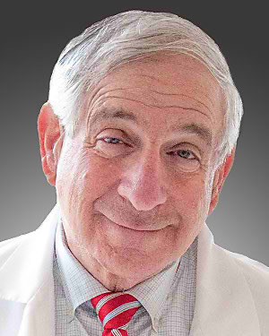 Barry J. Maron合作论文数Department of Cardiology, Lahey Hospital & Medical Center;Hypertrophic Cardiomyopathy Center, Lahey Hospital & Medical Center;Mayo Clinic83
 Robert S. Schwartz合作论文数Minneapolis Heart Institute76Fridolin Dittus合作论文数European Organization for Nuclear Research74
 Martin S. Maron合作论文数Department of Cardiology, Lahey Hospital & Medical Center;Tufts University School of Medicine74Sorajja Paul合作论文数73A. Catinaccio合作论文数72
 Mogens Dam合作论文数Niels Bohr Institute, University of Copenhagen72
 Stefan Koperny合作论文数Faculty of Physics and Applied Computer Science, AGH University of Science and Technology72
 Serhat Istin合作论文数Physics Department, Yeditepe University;CERN71V. A. Kantserov合作论文数71A. Lucotte合作论文数71
 Bruce Barnett合作论文数William H. Miller III Department of Physics and Astronomy, Krieger School of Arts and Sciences, Johns Hopkins University71
 Elżbieta Banaś合作论文数Division of Particle and Astroparticle Physics, Institute of Nuclear Physics, Polish Academy of Sciences;Department of Diffractive Processes, Institute of Nuclear Physics, Polish Academy of Sciences;CERN71K. Einsweiler合作论文数70O. L. Fedin合作论文数70
 Amir Farbin合作论文数Department of Physics, College of Science, The University of Texas at Arlington70Didier Ferrere合作论文数Département De Physique Nucléaire Et Corpusculaire, Faculty of Science, Université de Genève70
 Frederick C. Luehring合作论文数Department of Physics, College of Arts & Sciences, Indiana University Bloomington;European Organization for Nuclear Research70
 Alison James合作论文数Division of the Humanities, The University of Chicago;Department of Romance Languages and Literatures, The University of Chicago70G. Iacobucci合作论文数Argonne National Laboratory70
 Calin Alexa合作论文数Horia Hulubei National Institute of Physics and Nuclear Engineering70P. Kodys合作论文数69K. Lantzsch合作论文数69Tancredi Carli合作论文数CERN69Farkhad Khalil-Zada合作论文数Institute of Physics of ANAS69
 Maurice Garcia-Sciveres合作论文数Physics Division, Lawrence Berkeley National Laboratory;Berkeley Experimental Particle Physics Center, Lawrence Berkeley National Laboratory69F. Huegging合作论文数69
 Harutyun Hakobyan合作论文数TeamViewer Armenia69Michael Begel合作论文数Department of Physics, Brookhaven National Laboratory69Ivana Hristova合作论文数Rutherford69M. Cavalli-Sforza合作论文数69Z. Hubacek合作论文数69
 Davide Costanzo合作论文数School of Mathematical and Physical Sciences, University of Sheffield69
 Yuri A. Kulchitsky合作论文数CERN;Joint Institute for Nuclear Research69
 Tomáš Davídek合作论文数Institute of Particle and Nuclear Physics, Faculty of Mathematics and Physics, Charles University69
 A. Ereditato合作论文数Yale University69
 S. Giagu合作论文数INFN Sezione di Roma, Sapienza University69
 Markus Elsing合作论文数Experimental Physics Department, European Organization for Nuclear Research69
 Sergio Grancagnolo合作论文数Dipartimento di Matematica e Fisica 'Ennio De Giorgi', Università del Salento;INFN Sezione di Lecce69
 M. Cobal合作论文数Dipartimento di Chimica, Fisica e Ambiente, Università di Udine69
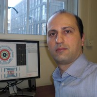 Nikolaos Konstantinidis合作论文数Department of Physics and Astronomy, Faculty of Mathematical and Physical Sciences, University College London;STFC Centre for Doctoral Training in Data Intensive Science, University College London69
 Bob Kowalewski合作论文数Victoria Subatomic Physics & Accelerator Research Centre, Department of Physics and Astronomy, Faculty of Science, University of Victoria69
 Remi Lafaye合作论文数Centre National De La Recherche Scientifique69Tristan Beau合作论文数Laboratoire de Physique Nucléaire et de Hautes Energies69
 Sergei Chekanov合作论文数High Energy Physics Division, Argonne National Laboratory69Kepka O合作论文数69Y. Ikegami合作论文数69
 Hans Peter Beck合作论文数Departement für Physik, Universität Fribourg;Physikalischen Institut, Universität Bern69
 Attilio Andreazza合作论文数Dipartimento di Fisica, Università degli studi di Milano;Laboratorio di Trattamento Numerico dei Dati Sperimentali, Università degli studi di Milano;Istituto Nazionale di Fisica Nucleare69




