BACKGROUND:Symptom burden of skull base tumor (SBT) patients, especially those undergoing multimodality treatment, is poorly understood. We aim to understand symptom burden in this patient group using the core module the MD Anderson Symptom Inventory (MDASI). METHODS:Twenty-nine patients with benign and malignant SBT underwent qualitative interviews to capture symptoms at diagnosis, during, and after treatment. An expert panel of 47 members (patients, caregivers, and providers) rated identified symptoms for relevance. RESULTS:Fifty-six individual symptoms were identified. Twenty-four symptoms met criteria for inclusion; 10 were already present in the MDASI core. Fourteen new SBT-specific symptoms were selected, adding to 13 core MDASI symptoms and 6 interference items to form the provisional MDASI-Skull Base. CONCLUSIONS:SBT patients experience wide ranging symptoms. A core cluster of symptoms is frequently reported across multiple SBT anatomical subsites. Further evaluation of the provisional MDASI-SB through psychometric testing in a larger group of patients is needed.
Purpose: Sleep-related symptom prevalence is not well-characterized and likely under-recognized in head and neck cancer. We investigated longitudinal symptom severity trajectory, prevalence of moderate/severe symptoms, and association between severe sleep-related patient-reported symptoms and oncologic endpoints in oropharyngeal cancer (OPC) patients receiving radiotherapy (RT). Methods: A longitudinal cohort of 372 patients with OPC treated with curative-intent RT with/without chemotherapy at a single-site tertiary center was queried at RT start, end of RT, and six months post-RT. All completed baseline MD Anderson Symptom Inventory-Head and Neck (MDASI-HN) at start of RT. Sleep-related symptom items included "fatigue", "disturbed sleep", and "drowsiness" (mild, moderate, severe). Clinical data were collected, and overall survival (OS) was evaluated using Kaplan-Meier/log-rank tests and multivariable Cox proportional hazards models with covariate adjustment. Results: Severe fatigue, disturbed sleep, and drowsiness were infrequent at baseline and at RT start, but peaked at RT end (26%, 15% and 18%) and returned to near-baseline prevalence by six months after RT (6%, 4%, 2%). In Cox proportional hazard models, severe longitudinal fatigue (HR 5.05, 95% CI 1.25-17.07, p = 0.013), sleep (HR 3.86, 95% CI 1.23 to 10.16, p = 0.0104), and drowsiness burden (HR 8.24, 95% CI 1.22 to 32.62, p = 0.008) were all independently associated with worse OS. Additionally, severe fatigue (HR = 3.58, 95% CI: 1.09 to 10.11, p = 0.0375), sleep disturbance (HR 5.08, 95% CI 1.60-13.51, p = 0.0081), and drowsiness (HR=7.76, 95% CI 1.45-33.47, p=0.0193) reported at RT start were also independently associated with significantly worse OS. Aside from age, severe average daily symptom scores, and severe symptoms reported at RT start, were independently associated with worse OS. Conclusion: Severe sleep related symptoms were uncommon at baseline or RT start, but a substantial proportion of OPC patients experienced clinically meaningful sleep disturbances by the end of RT. At an average follow up of 84 months, severe longitudinal fatigue, sleep, and drowsiness burden were all independently associated with worse OS in OPC. Severe fatigue, drowsiness and sleep disturbance reported at RT start were also independently associated with worse OS. Assessment of sleep disturbances along with potential physiologic correlates are an important part of comprehensive care. Keywords: survival, oropharyngeal cancer, radiotherapy, sleep disturbance, fatigue, drowsiness ### Competing Interest Statement All authors have completed the ICMJE uniform disclosure form at www.icmje.org/coi_disclosure.pdf and declare: KH is supported by OPC SURVIVOR: Optimizing OroPharyngeal Cancer SURVivorship Project Number: 1P01CA285249-01A1 and has received travel, speaker honoraria, and/or registration fee waivers unrelated to this project from Atos Medical, MedBridge, Inc., Gap Education, UCSF, OSU, NRG, American Board of Swallowing and Disorders. GEM is supported by: U.S. NSF CNS-2320261, U.S. NIH NCI-R01CA258827, U.S. NIH UG3-TR004501, UIC Institute for Health Data Science Research. CDF has received travel, speaker honoraria, and/or registration fee waivers unrelated to this project from Siemens Healthineers/Varian, Elekta AB, Philips Medical Systems, The American Association for Physicists in Medicine, The American Society for Clinical Oncology, The Royal Australian and New Zealand College of Radiologists, Australian & New Zealand Head and Neck Society, The American Society for Radiation Oncology, The Radiological Society of North America, and The European Society for Radiation Oncology. ### Funding Statement This manuscript is the result of funding in whole or in part by the National Institutes of Health (NIH) ### Author Declarations I confirm all relevant ethical guidelines have been followed, and any necessary IRB and/or ethics committee approvals have been obtained. Yes The details of the IRB/oversight body that provided approval or exemption for the research described are given below: After institutional review board approval by the University of Texas MD Anderson Cancer Centers IRB (RCR03-0800), data from a philanthropically funded observational cohort at The University of Texas MD Anderson Cancer Center (Stiefel Oropharynx Cancer Cohort, PA14-0947) were extracted for retrospective acquisition. I confirm that all necessary patient/participant consent has been obtained and the appropriate institutional forms have been archived, and that any patient/participant/sample identifiers included were not known to anyone (e.g., hospital staff, patients or participants themselves) outside the research group so cannot be used to identify individuals. Yes I understand that all clinical trials and any other prospective interventional studies must be registered with an ICMJE-approved registry, such as ClinicalTrials.gov. I confirm that any such study reported in the manuscript has been registered and the trial registration ID is provided (note: if posting a prospective study registered retrospectively, please provide a statement in the trial ID field explaining why the study was not registered in advance). Yes I have followed all appropriate research reporting guidelines, such as any relevant EQUATOR Network research reporting checklist(s) and other pertinent material, if applicable. Yes In accordance with NOT-OD-21-013, Final NIH Policy for Data Management and Sharing, anonymized/de-identified data that support the findings of this study are openly available in an NIH-supported generalist scientific data repository (figshare) at https://doi.org/10.6084/m9.figshare.29971432.v1 no later than the time of an associated publication.
LRF by KRAS and assigned treatment. Rates of local failure based on treatment and KRAS-variant status, with KRAS-variant patients being depicted by blue lines and nonvariant patients by red lines and dashed lines representing cetuximab-treated patients.
Grade 3 to 4 treatment-related AEs over time by KRAS and assigned treatment. Rates of treatment-related AEs with 95% CI by treatment and KRAS-variant status, with KRAS-variant patients being depicted by blue (cisplatin-treated) and red (cetuximab-treated) and nonvariant patients depicted by green (cisplatin-treated) and brown (cetuximab-treated).
Background: Sinonasal undifferentiated carcinoma (SNUC) is an extremely rare, high-grade, and aggressive tumor of the sinonasal tract. Due to the rarity of this malignancy, current treatment guidelines are based on small and often/mainly single-center retrospective datasets. In the absence of a universally accepted standard of care for SNUC, treatment approaches vary across countries and institutions, reflecting real-world clinical practice. The primary aim of this study was to describe real-world treatment and outcomes for patients with confirmed SNUC. Methods: This was an international, multi-center, retrospective, observational cohort study that pooled patients into the largest SNUC dataset to date. Fifteen centers were enrolled to contribute data, including seven from Europe, four from the United States, three from the United Kingdom, and one from Canada. In the absence of a universally accepted standard of care for SNUC, treatment approaches varied across countries and institutions, reflecting real-world clinical practice. Patients included were those with histologically confirmed SNUC who were treated between 1997 and 2021. Results: This study yielded 485 patients treated for SNUC. The median age at diagnosis was 55.6 years (IQR: 44.5-67.6), and 63.7% were male. Most cases presented at advanced stages, with 70.8% as T4a or T4b. Overall survival (OS) outcomes were available for 412 patients, with a median follow-up of 26.0 months. The 5- and 10-year OS were 47.2% (95% CI: 40.8-53.3%) and 39.6% (95% CI: 32.5-46.6%), respectively. Advanced age, dichotomized T-stage (T4a/b vs. T1-3), M-stage, and orbital involvement were significant poor prognostic factors on univariable analysis (p's < 0.01). On multivariable analysis, orbital involvement (HR: 2.73, 95% CI: 1.42-5.27, p = 0.003) and distance metastasis stage (HR: 3.00, 95% CI: 1.25-7.21, p = 0.014) were both independently associated with worse OS. Conclusions: This observational study presents the largest multi-center cohort analysis of SNUC to date, providing new insights into prognostic factors for a rare cancer treated at global centers of excellence. Orbital involvement and the presence of metastases are candidate independent risk factors associated with poorer OS.
OBJECTIVE:Hearing loss from immune checkpoint inhibitors (ICIs) has been documented in case reports and case series. We present the largest retrospective study investigating the rate of ICI-related ototoxicity in a monitored cohort of platinum-naïve patients. STUDY DESIGN:Retrospective cohort study. SETTING:Tertiary-care center. METHODS:Patients treated with ICI between January 1, 2017, and December 31, 2022, with baseline and post-treatment audiograms, were included. Patients with a history of platinum-based chemotherapy were excluded. Demographics, oncologic diagnosis, ICI treatment details, and temporal bone irradiation (TBRT) were recorded. Audiometric thresholds were compared before and after ICI therapy. The primary outcome measure was a change in hearing as defined by the National Cancer Institute Common Terminology Criteria for Adverse Events (CTCAE). Secondary outcome measures included changes in hearing using the American Speech-Language-Hearing Association (ASHA) and TUNE criteria. RESULTS:Among 15,390 ICI recipients, 29 platinum-naïve patients met the inclusion criteria. Six of 29 patients (20.7%) experienced a CTCAE grade 1 or higher hearing loss. The proportions of hearing loss as defined by ASHA and TUNE criteria were 44.8% and 27.6%, respectively. The length of time between baseline and follow-up audiograms was significantly longer among patients with hearing loss (mean difference [days]: CTCAE: 590.3, P < .01; ASHA: 280.9, P = .05; and TUNE: 122.1, P = .46). No other potential covariates believed to be confounders were significantly associated with the outcome. CONCLUSION:A significant proportion of our monitored platinum-naïve ICI patients met hearing-loss criteria. Prospective studies with standardized audiologic surveillance are needed to further quantify the true incidence of ICI ototoxicity.
Multivariable Cox Models for KRAS as a Predictive Biomarker for Local-Regional Failure
Purpose: NRG/RTOG 1016 was a phase III noninferiority trial comparing IMRT + cisplatin versus IMRT + cetuximab for human papillomavirus-positive oropharyngeal squamous cell cancer (HPV+ OPSCC). A germline mutation (the KRAS-variant) previously identified patients with improved outcomes to radiation + cetuximab + cisplatin; thus, we investigated whether there may be similar benefits for IMRT + cetuximab.Patients and Methods: 562 patients were tested, with 16% positive for the KRAS-variant. Clinical endpoints were per NRG/RTOG 1016, and hazard ratios (HR) were estimated by Cox models. Negative binomial regression modeled treatment-related acute and late grade 3 to 5 adverse events (AE). All models included KRAS, assigned treatment, their interaction, and other prognostic variables tested at two-sided 0.05.Results: There was no association between the KRAS-variant and clinical outcomes. There were higher local-regional failure rates (LRF) in nonvariant patients treated with cetuximab [HR = 1.83 (1.19-2.82)] versus KRAS-variant patients [HR (95% CI), 0.90 (0.34-2.41)]. Grade 3 to 4 acute mucositis was higher in nonvariant patients treated with cetuximab [OR = 1.41 (95% CI, 0.98-2.03)] and lower in KRAS-variant patients [OR = 0.60 (95% CI, 0.25-1.41)]. However, the mean late toxicity ratio was lower in nonvariant patients [0.55 (95% CI, 0.35-0.87)] versus KRAS-variant patients [1.62 (95% CI, 0.57-4.62)]. KRAS by treatment interaction for acute and late AEs was not significant (P = 0.0717 and 0.0659, respectively).Conclusions: Possible lower LRF and less acute toxicity with cetuximab versus cisplatin for KRAS-variant patients seem to be offset by potentially increased late toxicity. Further evaluation of this class of biomarkers is warranted.Significance: Germline microRNA-disrupting biomarkers are functional biomarkers being studied to help guide treatment personalization in cancer. The work in this study is a hypothesis-generating evaluation of the KRAS-variant as a potential biomarker to help predict outcomes and toxicity in patients with head and neck cancer treated with radiation and cetuximab. Findings from this work suggest that these biomarkers may have the potential to differentiate patients at risk of acute versus late toxicity, supporting further study of these biomarkers in radiation studies.
Advanced imaging is the cornerstone of modern radiation oncology, contributing to each phase of patient care, from diagnosis and treatment planning to delivery and follow-up. It has evolved from providing purely geometric guidance to enabling biological and dynamic precision, capturing detailed spatial and functional information about tumors and surrounding tissues. This progress has also generated vast amounts of complex data that remain largely underexplored. AI-based methods have shown promises to unlock the potential of these data, ensuring quality and standardization while extracting previously inaccessible insights. AI-driven tools can enhance accuracy, efficiency, and personalization of radiation oncology through precision diagnosis, automated segmentation, adaptive treatment planning, real-time image guidance, and predictive response assessment. In this review, we conducted a systematic bibliometric analysis of relevant literature published in the last decade and explored current advancements in AI and radiomics applications across radiation oncology. We also addressed ongoing challenges, such as data heterogeneity, model interpretability, and clinical implementation, and discussed future directions for integrating AI-powered imaging solutions into routine practice to advance precision cancer care.
Univariate and Multivariable Cause-Specific Cox Models for KRAS as a Prognostic Biomarker for Distant Metastasis
BACKGROUND:The role of SABR in head and neck cancer remains uncertain. Historically, it has been used mainly for reirradiation, and its application in other clinical scenarios has expanded with recent advances. OBJECTIVE:To develop an international expert consensus on indications, technical parameters, and clinical application of head and neck SABR using a modified Delphi process. METHODS:A three-round modified Delphi process was conducted between July 2021 and January 2023. Radiation oncologists with experience in head and neck SABR were invited to participate. Round 1 consisted of open-ended questions. Rounds 2 and 3 involved rating agreement with 103 statements addressing indications, contraindications, planning, dose and fractionation, toxicity mitigation, systemic therapy integration, and follow-up. Consensus was defined as ≥75% agreement, and major agreement as 65% to 74%. RESULTS:Seventeen of 26 invited experts (65.4%) completed all rounds, with representation from North America, Europe, and Asia-Oceania. Of the 103 statements, 56 (54.3%) reached consensus, and 12 (11.7%) achieved major agreement. Consensus supported SABR for reirradiation of small isolated recurrences, unresectable tumors, or second primaries in previously irradiated fields, and oligometastatic lesions in untreated patients. Use in local palliation and in patients unfit for standard fractionation within a clinical trial also met consensus. No consensus was reached for postoperative SABR, including its use for positive margins or extranodal extension, nor for SABR boost in first-course treatment. Technically, consensus favored volumetric modulated arc therapy, tight planning target volume margins of 2 to 3 mm, multimodality imaging (thin-slice computed tomography, magnetic resonance imaging, and positron emission tomography/computed tomography), and daily volumetric imaging with physician verification. Clinical target volume expansion was discouraged, and elective nodal irradiation was not recommended. Every-other-day fractionation and dose reduction near critical structures were endorsed. Consensus emphasized counseling on key toxicities and supported imaging at 2 to 3 months post-SABR. No consensus was reached on systemic therapy integration. CONCLUSION:This multi-institutional Delphi study provides expert-derived recommendations reflecting the current state of head and neck SABR practice and identifies priorities for future research.
BACKGROUND:Adenoid cystic carcinoma of the anterior craniofacial region (ACF-ACC) is challenging to treat due to extensive subclinical spread and proximity to critical structures. Although surgery followed by radiotherapy (RT) is the current standard, real-world outcomes with modern photon and particle therapy remain insufficiently characterized. METHODS:We retrospectively analyzed 578 patients with ACF-ACC treated at eight international centers (1984-2023). Clinicopathologic features, treatment patterns, and outcomes were assessed. Anatomical extension was classified using hierarchical clustering. Comparative analyses of gross total resection (GTR) and non-surgical treatment (NST) were adjusted using propensity score matching and multivariable Cox and Fine-Gray models. Primary endpoints were local recurrence-free survival (LRFS) and cumulative incidence of local recurrence (LRCI). RESULTS:Most tumors arose in the sinonasal tract (75.8%) and were low/intermediate grade (68.6%). Long-term outcomes showed high local and distant recurrence (20-year LRCI: 74.1%; cumulative incidence of distant metastasis: 55.6%). GTR followed by adjuvant RT, especially with proton therapy (PT), achieved the best local control. R2 resections provided no advantage over NST. Within the NST cohort (n = 110), PT yielded higher complete response rates than photon RT, while responders demonstrated local control comparable to surgically treated patients. Ten-year ≥G3 toxicity incidence was 36%. CONCLUSIONS:For ACF-ACC, GTR plus modern RT provides the strongest local control, and R2 surgery should be avoided. PT is an effective definitive option for selected patients, supporting future response-guided treatment strategies.
e18143 Background: Myoepithelial carcinoma (MEC) is a neoplasm derived from myoepithelial cells and characterized by an infiltrative growth pattern. These are very rare malignancies with limited published data describing outcomes. We sought to define the natural history of MEC and to identify best available treatments. Methods: In this retrospective series, we identified 70 patients with MEC seen at our institution from 1997-2025. Electronic medical records were reviewed to determine the patient and tumor characteristics, treatment regimens and response, and outcomes. The Kaplan Meier method was used to estimate survival, and log-rank tests were used to compare groups. Results: In our institutional cohort of 70 patients, MEC was most common in middle-aged individuals (median age = 52 years), with a predilection for head and neck sites (54/70, 77%). There was a slight predilection for male sex (40/70, 57%). The median overall survival (mOS) in our cohort was 87.2 months. Of 24 patients who developed metastatic disease, the predominant location was lung (n = 15, 62.5%) followed by bone (n = 11, 45%). For patients with localized disease (n = 66), the median recurrence-free survival (mRFS) was 55.4 months. The mRFS was not significantly different in patients who had resection alone (n = 39, mRFS = 60.0 months) compared with those treated with adjuvant radiation and/or chemotherapy (n = 27, mRFS = 51.1 months, p = 0.70). The primary systemic therapy utilized was platinum-based with few patients receiving doxorubicin or gemcitabine-based regimens. There were no significant clinical responses noted with systemic therapy. The median progression-free survival (mPFS) in patients with metastatic disease who received any systemic therapy was 3.1 months (n = 16). No significant difference in mOS was observed based on age, though there was a trend toward worse outcomes in younger patients (mOS for <30 years = 62.5 months, mOS for >30 years = 93.5 months, p = 0.88). The sample size of patients in the younger age group (n = 11) was small and only three patients in that subset were free of disease for >15 years. Conclusions: In our institutional experience with MEC, most patients were middle-aged with head and neck as the most common primary site. Outcomes were generally worse for younger patients. The lack of significant difference between the mOS between these age groups is attributed to a small sample size of patients in the younger age group with a wide confidence interval. Outcomes with currently available systemic therapy for metastatic disease were uniformly poor, emphasizing the need to develop novel treatment regimens moving forward. Future work is needed to discern the impact of specific genomic alterations (e.g. EWSR1-KLF15 fusion) on tumor biology and response to therapy.
Importance:Imaging-detected extranodal extension (iENE) has been included in the American Joint Committee on Cancer (AJCC) and Union for International Cancer Control (UICC) protocol tumor-node-metastasis 9th edition staging system (TNM9) for human papillomavirus (HPV)-mediated (p16-positive) oropharyngeal cancer (HPV-positive OPC). Objective:To confirm the new TNM9 staging system on a prospective dataset. Design, Setting, and Participants:This prognostic study included patients with HPV-positive OPC with clinically node-positive disease treated at 182 health care centers in the US and Canada and enrolled in the NRG Radiation Therapy Oncology Group (RTOG) 1016 trial between 2011 and 2014 were included. Deidentified images were reviewed between September and December 2021. Data were analyzed between May and September 2023. Exposure:Computed tomography simulation scans were reviewed by 2 expert head and neck radiologists documenting the presence of iENE. Main Outcomes and Measures:Kaplan-Meier analysis was used to estimate overall survival (OS) and progression-free survival (PFS). Cumulative incidence curves were generated for locoregional failure (LRF) and distant metastasis. Univariate (UVA) and multivariate analyses (MVA) were performed incorporating iENE with other known prognostic factors. The TNM9 clinical nodal schema incorporating iENE was compared with the previous TNM8. Model goodness of fit and comparison of models was done using Akaike information criterion (AIC) and Harrell concordance statistic (C index). Results:Of the 987 patients enrolled in NRG RTOG 1016, 586 met inclusion criteria (median age, 58 years [range, 33-83 years]; 534 male [91.1%]), 350 (60%) of whom had iENE. Median (IQR) follow-up was 4.2 (3.3-5.0) years. Patients with iENE had inferior 5-year OS (79.2% vs 87.0%; difference, 7.8% [95% CI, 1.2%-14.4%]) and 5-year PFS (68.8% vs 80.4%; difference, 11.6% [95% CI, 4.1%-19.2%]) compared with those without iENE. On MVA, iENE was independently associated with inferior OS (HR, 1.63; 95% CI, 1.04-2.56) and PFS (HR, 1.78; 95% CI, 1.24-2.57). TNM9 and TNM8 clinical nodal schemas had similar performance in terms of C index and AIC (TNM8: C, 0.73 [95% CI, 0.68-0.78]; AIC = 1109.34 vs TNM9: C, 0.72 [95% CI, 0.67-0.77]; AIC = 1112.70). Conclusions and Relevance:This study showed that the updated TNM9 clinical nodal schema, in which patients with iENE-positive disease are upstaged 1 stratum from their TNM8 clinical N category, discriminates better than the TNM8 category between patients. Trial Registration:ClinicalTrials.gov Identifier: NCT01302834.
 Adam S. Garden合作论文数Department of Radiation Oncology, Division of Radiation Oncology, The University of Texas MD Anderson Cancer Center232
 G. Brandon Gunn合作论文数Department of Radiation Oncology, Division of Radiation Oncology, The University of Texas MD Anderson Cancer Center213
 Clifton David Fuller合作论文数Department of Radiation Oncology, Division of Radiation Oncology, The University of Texas MD Anderson Cancer Center, Houston196
 Steven Frank合作论文数Department of Anesthesiology and Critical Care Medicine, School of Medicine, Johns Hopkins University138
 David N. Rosenthal合作论文数Pediatrics Department, Stanford University118
 William Robert Morrison (Rob)合作论文数Center for Grain and Animal Health Research, Agricultural Research Service, United States Department of Agriculture100
 Renata Ferrarotto合作论文数85
 D.I. Rosenthal合作论文数76Reddy Jay P合作论文数Department of Molecular &Cellular Biology, Baylor College of Medicine;Department of Molecular & Cellular Biology, Baylor College of Medicine75William H Morrison合作论文数61
 Beth Beadle合作论文数Department of Radiation Oncology, Stanford University59Ehab Hanna合作论文数57
 Shirley Y. Su合作论文数50
 Mohamed Abdallah合作论文数49
 Erich M. Sturgis合作论文数Department of Head and Neck Surgery, The University of Texas M.D. Anderson Cancer Center/Population Science & Cancer Control, UT Southwestern Simmons Comprehensive Cancer Center/Head and Neck Surgery, Baylor College of Medicine48
 Katherine A. Hutcheson合作论文数Department of Communication Sciences and Disorders, Division of Surgery, The University of Houston College of Liberal Arts and Social Sciences;Section of Speech Pathology and Audiology, Department of Head and Neck Surgery, The University of Texas MD Anderson Cancer Center48
 Abdallah Sherif Radwan Mohamed合作论文数Department of Radiation Oncology, Baylor College of Medicine45
 Stephen Y. Lai合作论文数Department of Head and Neck Surgery, Division of Surgery, The University of Texas MD Anderson Cancer Center;Department of Radiation Oncology, Division of Radiation Oncology, The University of Texas MD Anderson Cancer Center;Department of Molecular and Cellular Oncology, Division of Basic Science Research, The University of Texas MD Anderson Cancer Center44
 Steven J. Frank合作论文数Department of Radiation Oncology, Division of Radiation Oncology, The University of Texas MD Anderson Cancer Center43
 Amy Moreno合作论文数Department of Surgical Oncology42
 Houda Bahig合作论文数Radiation Oncology Department, Centre Hospitalier de l'Université de Montréal;Department of Radiology, Radiation Oncology and Nuclear Medicine, Université de Montréal41
 Heath D. Skinner合作论文数Department of Radiation Oncology, School of Medicine, University of Pittsburgh;Hillman Cancer Center, University of Pittsburgh Medical Center34
 Anna Lee合作论文数33
 Sweet Ping Ng合作论文数33
 Franco DeMonte合作论文数Department of Neurosurgery, Division of Surgery, The University of Texas MD Anderson Cancer Center29
 Neil D. Gross合作论文数Department of Head and Neck Surgery, Division of Surgery, The University of Texas MD Anderson Cancer Center28C. Pollard合作论文数23Michael T. Spiotto合作论文数Department of Radiation Oncology, Stanford University School of Medicine23Shaan M. Raza合作论文数22Michael E Kupferman合作论文数21
 Baher Elgohari合作论文数Faculty of Medicine, University of Almansoura20
 Randal S. Weber合作论文数Department of Individual Giving, Division of Philanthropy, The University of Texas MD Anderson Cancer Center;Department of Head and Neck Surgery, Division of Surgery, The University of Texas MD Anderson Cancer Center20
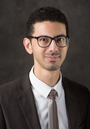 Hesham Elhalawani合作论文数Department of Radiation Oncology, Brigham and Women's Hospital;Harvard Medical School;Dana-Farber Brigham Cancer Center20
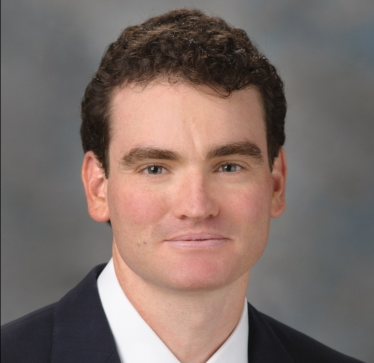 Mark E. Zafereo合作论文数Department of Head and Neck Surgery, Division of Surgery, The University of Texas MD Anderson Cancer Center20Vinita Takiar合作论文数20
 Bonnie S. Glisson合作论文数University of Texas M.D. Anderson Cancer Center20
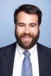 C. Cardenas合作论文数Dept Radiat Oncol, Univ Alabama Birmingham19
 Quynh-Thu Le合作论文数Department of Radiation Oncology, School of Medicine, Stanford University19
 Diana M. Bell合作论文数Division of Anatomic Pathology, Department of Pathology, School of Medicine, University of Pittsburgh19
 Maura L. Gillison合作论文数Department of Thoracic/Head and Neck Medical Oncology, Division of Cancer Medicine, The University of Texas MD Anderson Cancer Center19Karen Reue合作论文数Department of Human Genetics, David Geffen School Of Medicine, University of California, Los Angeles18A.C. Moreno合作论文数17Galloway Thomas J合作论文数Department of Radiation Oncology, Fox Chase Cancer Center17
 Adel K. El-Naggar合作论文数Department of Pathology, Division of Pathology, MD Anderson Cancer Center, The University of Texas17H. Wang合作论文数17
 Jinzhong Yang合作论文数Department of Radiation Physics, Division of Radiation Oncology, The University of Texas MD Anderson Cancer Center;The University of Texas Graduate School of Biomedical Sciences at Houston17
 He Wang (王鹤)合作论文数Computer Science Department, School of Computing, Faculty of Engineering and Physical Sciences, University of Leeds17Goepfert Ryan P合作论文数16
 Sue S. Yom合作论文数Department of Radiation Oncology, University of California, San Francisco16




