Introduction:Magnetic resonance-guided radiotherapy enables repeated on-treatment imaging during chemoradiotherapy for glioblastoma (GBM), providing an opportunity to study dynamic tumor changes. We evaluated longitudinal volumetric change and spatial migration of T2/FLAIR signal abnormalities during treatment using a 1.5 T MR-Linac and examined their association with early disease progression. Methods:GBM patients suitable for chemoradiation on a 1.5 T MR-Linac were prospectively enrolled. Regions of T2/FLAIR signal abnormalities were contoured at baseline (F0), during selected treatment fractions, and at one-month post-radiotherapy (PM1). Tumor dynamics were quantified using percentage change in volume relative to F0 (Vrel) and migration distance (dmigrate), defined as the maximum linear displacement relative to F0. Patients were classified as early progressors (disease progression within 6 months after chemoradiation) or non-early progressors. Linear mixed-effects models evaluated longitudinal differences by early progression status, methylation, and extent of resection. Results:Thirty-three patients were included; 28 had ≥ 6 months follow-up and 12 were classified as early progressors. In adjusted linear mixed-effects models, Vrel was higher in early progressors than in non-early progressors from F7 onward (all p < 0.025), with larger differences later in treatment (F15: 38%, p = 0.004; F30: 104%, p = 0.019). Median Vrel was 2% at F1, 2% at F15, and 18% at F30. Median dmigrate increased from 5.0 mm at F1 to 12.6 mm at F30 and was higher in early progressors from F19 onward (difference 5.9 mm, p = 0.02) and at F30 (8.5 mm, p = 0.01). At end of treatment, Vrel ≥ 10% occurred in 12/28 patients (43%) and dmigrate ≥ 10 mm in 13/28 (46%). Among patients with Vrel ≥ 10%, 10/12 (83%) were early progressors; among those with dmigrate ≥ 10 mm, 9/13 (69%) were early progressors. Conclusion:Serial 1.5 T MR-Linac imaging enables longitudinal assessment of T2/FLAIR abnormality volume (Vrel) and spatial migration (dmigrate) during chemoradiotherapy for glioblastoma. Greater increases in Vrel and dmigrate were associated with early progression, supporting the potential role of on-treatment imaging in informing risk-adapted MR-guided radiotherapy strategies.
BACKGROUND:In patients with locally advanced, non-metastatic oropharyngeal squamous cell carcinoma (OPSCC), surgery-based and nonsurgical treatment approaches have not been adequately compared in randomized Phase III clinical trials. Current clinical guidelines recommend various surgical and nonsurgical strategies across disease stages for both p16-negative and p16-positive OPSCC, without clearly favoring a single approach. OBJECTIVE:This review summarizes the available evidence relevant to clinical decision-making between primary surgical and primary nonsurgical treatment approaches in patients with non-metastatic OPSCC. METHODS:We reviewed studies evaluating tumor control across different treatment modalities, with emphasis on the highest-quality available evidence and post-treatment quality of life (QoL). Outcomes were discussed in relation to disease extent (early-stage vs. locally advanced) and treatment approaches. Emerging therapeutic strategies with the potential to improve the efficacy-to-toxicity ratio were also reviewed. RESULTS:In the absence of Phase III randomized trials in non-metastatic OPSCC, the most informative comparative data derive from studies employing propensity score matching to reduce selection bias. These analyses suggest that survival outcomes are largely comparable between surgery-based and nonsurgical approaches in both p16-positive and p16-negative OPSCC. With respect to post-treatment QoL, surgery alone is generally associated with the lowest toxicity burden, whereas surgery followed by adjuvant (chemo)radiotherapy and definitive (chemo)radiotherapy demonstrate broadly comparable, albeit modality-specific, toxicity and QoL profiles. p16-positive status is consistently associated with more favorable post-treatment QoL. De-escalation strategies in p16/HPV-associated OPSCC, as well as emerging modalities such as immune checkpoint inhibitors, proton therapy, and adaptive radiotherapy, may influence future treatment paradigms. CONCLUSIONS:In the absence of clear differences in oncologic efficacy between primary surgical and nonsurgical approaches, treatment selection should be guided primarily by anticipated toxicity and its impact on QoL, within the context of multidisciplinary assessment and patient preferences. The influence of treatment costs in the decision-making process varies across healthcare systems, reflecting differences in the valuation and financing of health interventions. The role of emerging treatment strategies in OPSCC will need to be defined through prospective, disease-specific clinical trials.
Purpose:The magnetic resonance linear accelerator (MRL) enables daily adaptation of radiation therapy plans. This study presents our institutional experience with the clinical outcomes and quality of life (QoL) of glioblastoma patients treated using a 1.5 Tesla (T) MRL. Materials and Methods:Eligible patients with glioblastoma were enrolled in a study and treated with MRL between August 2021 and May 2024. Acute toxicity was assessed. QoL was evaluated using the EORTC QLQ-C30 and QLQ-BN20 questionnaires. Cumulative overall survival (OS) and progression-free survival (PFS) were estimated using Kaplan-Meier analysis. Results:27 patients were included, with a median follow-up of 12.2 months (range 0.5-30.6 months). The mean age was 63 years; 14 were male and had an ECOG performance status of 0-1. Eleven patients underwent gross total resection, 9 had subtotal resection, and 7 had biopsy only. 11 were MGMT-methylated, 15 nonmethylated, and 1 had unknown status. Twenty-five received their first radiotherapy course on the MRL. The mean interval from CT/MRI simulation to radiotherapy start was 12.6 days. 26 patients completed all prescribed fractions on the MRL. The median on-table treatment time per fraction was 28.2 (range: 11.7-66.5) minutes. Eleven patients (39.3%) required Adapt-To-Shape or re-planning within the first week due to tumor progression, indicated by increased T2/FLAIR signal beyond the initial CTV. No Grade 3-4 toxicities were reported. At 3 months, treatment was associated with declines in QoL domains at 3 months with recovery by 6 months. The median OS and PFS were 14.1 and 11.2 months. Conclusion:Treatment of glioblastoma with MR-guided adaptive radiotherapy is feasible, with manageable acute toxicities and adaptive capabilities to address tumor changes.
INTRODUCTION:Liver metastases are common in several malignancies, with only 15-20% of patients eligible for surgical resection. For non-surgical candidates, stereotactic body radiation therapy (SBRT) is an effective alternative with acceptable local control (LC) rates. Higher biologically effective doses (BED10 > 100 Gy10) are associated with improved LC, and single-fraction ultra-high dose SBRT (35-40 Gy) has demonstrated excellent LC with minimal toxicity. MR-guided adaptive SBRT enhances precision and may optimizing single-fraction SBRT for liver metastases. METHODS AND ANALYSIS:ULTRAS is a multi-center, phase III randomized trial evaluating whether ultra-high dose MR-guided single-fraction SBRT (38 Gy, BED10 ≈ 180 Gy10) improves LC of liver oligometastases/oligoprogression compared to high-dose MR-guided single-fraction SBRT (27 Gy, BED10 ≈ 100 Gy10). Primary endpoint is LC of treated intrahepatic target lesion(s). Secondary endpoints include overall survival (OS), progression-free survival (PFS), intrahepatic and extrahepatic progression, toxicity (CTCAE v5.0 and PRO-CTCAE v1.0), and quality of life (QOL) (EORTC QLQ-C15-PAL, EORTC QLQ-LM21). Patients will be randomized 1:1 to receive either 27 Gy or 38 Gy, stratified by primary tumor site/histology, lesion characteristics, and prior systemic therapy. A total of 114 patients (57 per arm) will be enrolled over three years, with two years of follow-up. Assessments at predefined intervals will monitor response and toxicity. LC will be analyzed using cumulative incidence function. Widespread and intrahepatic progression will be assessed using Fine & Gray regression; OS and PFS via Cox models; toxicity and QOL via Chi-squared test and mixed-effects model. ETHICS AND DISSEMINATION:The ULTRAS trial obtained ethical approval from the University Health Network and will follow ICMJE reporting standards. TRIAL REGISTRATION:ClinicalTrials.gov ID: NCT06362395.
Oesophageal cancer remains a significant global health burden with poor survival outcomes despite multimodal treatment. Recent advances in magnetic resonance imaging (MRI) have opened opportunities to improve radiotherapy delivery. This review examines the role of MRI and MR-guided radiotherapy (MRgRT) in oesophageal cancer, focusing on applications in staging, treatment planning, and response assessment, with particular emphasis on magnetic resonance linear accelerator (MR-Linac)-based delivery. Compared to computed tomography (CT), MRI offers superior soft-tissue contrast, enabling more accurate tumour delineation and the potential for reduced treatment margins. Real-time MR imaging during treatment can facilitate motion management, while daily adaptive planning can accommodate anatomical changes throughout the treatment course. Functional MRI sequences, including diffusion-weighted and dynamic contrast-enhanced imaging, offer quantitative data for treatment response monitoring. Early clinical and dosimetric studies demonstrate that MRgRT can significantly reduce radiation dose to critical organs while maintaining target coverage. However, clinical evidence for MRgRT in oesophageal cancer is limited to small early-phase studies, with no phase II/III trials demonstrating improvements in survival, toxicity, or patient-reported outcomes. Long-term clinical benefits and cost-effectiveness remain unproven, highlighting the need for prospective outcome-focused studies to define the role for MRgRT within multimodality treatment pathways.
BACKGROUND AND OBJECTIVE:Differentiating glioma recurrence from radiation necrosis remains a critical diagnostic challenge in neuro-oncology due to overlapping imaging features. Positron emission tomography (PET) and magnetic resonance imaging (MRI) offer complementary metabolic and structural information that may enhance diagnostic accuracy. This narrative review evaluates the performance of PET, advanced MRI techniques, and their integration in distinguishing tumour recurrence from post-treatment effects. METHODS:A comprehensive search of PubMed, Medline, and Embase databases was conducted to identify studies published after April 2020. Inclusion criteria comprised studies on histologically diagnosed gliomas using PET and MRI, reporting sensitivity, specificity, or accuracy. KEY CONTENT AND FINDINGS:Eight cohort studies met the inclusion criteria, examining PET tracers (18F-FDG, 18F-FET, 11C-MET, 18F-DOPA) and advanced MRI techniques [diffusion kurtosis imaging (DKI), dynamic susceptibility contrast (DSC) perfusion, magnetic resonance spectroscopy (MRS)]. The integration of PET and MRI outperformed standalone modalities in diagnostic accuracy in all but one study. 18F-FET PET demonstrated high diagnostic accuracy, with sensitivity up to 95% and no diagnostic improvement when combined with MRI in high-grade glioma, though its availability remains limited. MRI alone had variable sensitivity (33-77%) and specificity (50-100%), depending on the technique and tracer used. DKI showed promise in enhancing sensitivity (72-100%). Heterogeneity in protocols and patient cohorts limited direct comparisons across studies. CONCLUSIONS:The combination of PET and MRI provides a superior diagnostic approach for differentiating glioma recurrence from radiation necrosis, with 18F-DOPA PET and advanced MRI sequences emerging as the most effective pairing. Further multicenter studies with standardized protocols are required to validate these findings and address challenges related to tracer accessibility and protocol variability. These advancements have the potential to optimize patient management in neuro-oncology.
INTRODUCTION:Stabilised hyaluronic acid (sHA) gel can be used as a breast cancer tumour bed marker. This study aimed to assess the gel on post-treatment surveillance mammography (MMG) and breast ultrasound (USS). METHODS:Patients who underwent breast-conserving surgery and adjuvant radiotherapy, with sHA gel markers (0.3-0.5 mL drops) in breast tissue had their routine 1- and 2-year post-treatment MMG and USS undergo central review by one experienced radiologist. The central review questionnaire included whether the gel was visible, description if visible, and whether it impacted diagnostic assessment of the images using a 5-point Likert scale. The images were also standardly reported by radiologists who were blinded to the gel history. RESULTS:Of the 33 patients eligible for evaluation, sHA gel was visible on the 1-year post-treatment MMG in 3% of patients, described as well circumscribed nodules adjacent to the surgical scar. sHA gel was visible on the 1-year post-treatment USS in 38% of patients, described as simple round cystic lesions near the surgical bed. sHA gel was still visible on the same patient's 2-year post-treatment MMG (3%) and was visible on the 2-year post-treatment USS in 19% of patients. In all cases, the gel was rated 'unlikely' or 'very unlikely' to impair diagnostic assessment of the images and did not lead to any additional investigations. The blinded radiologists reported the gel as non-suspicious appearing cysts without clinical concern. CONCLUSION:sHA gel used as breast tumour bed marker was not visible in most patients' post-treatment MMG and USS and did not affect diagnostic assessment.
HPV-positive OPSCC shows a favourable prognosis, prompting evaluation of de-escalated and adaptive strategies. Quantitative imaging may provide scalable biomarkers to individualise care. Quantitative imaging can support baseline risk stratification, early on-treatment decision-making, and posttreatment surveillance in HPV-positive OPSCC. Real-world translation requires standardised reporting, calibration/harmonisation across centres, rigorous model validation, and workflow integration with radiotherapy planning. Quantitative MRI, CT, and PET, augmented by radiomics and AI, show convergent promise as non-invasive biomarkers to enable safe individualisation of therapy in HPV-positive OPSCC, contingent on methodological rigour and prospective, externally validated studies. Despite this promise, clinical translation faces substantial barriers, including limited external validation, heterogeneous methodologies, and the need for standardised, prospectively validated pipelines.
Abstract Increasing numbers of clinical practice guidelines have been published by international/national groups for nasopharyngeal carcinoma (NPC), providing valuable references for clinicians in making evidence‐based decisions on treatment. However, there are substantial discrepancies in various recommendations, leading to uncertainties in choosing the optimal strategies. The authors systematically searched databases and organizational websites for NPC guidelines published between January 2000 and November 2025. All identified guidelines underwent quality appraisal; in total, 26 clinical practice guidelines rated recommended for use were included. The recommendations covering all management aspects (diagnosis, staging, radiotherapy, systemic therapy, follow‐up surveillance, biomarkers, and salvage of recurrent/metastatic diseases) were summarized and comparatively analyzed for consistency and disparities. Strong consensus exists for diagnostic workup, staging systems, and induction chemotherapy plus concurrent chemoradiotherapy for advanced disease, whereas marked disparities exist on radiotherapy details, particularly target volume delineation, elective coverage extent, and dose specifications. Although systemic therapy strategies for different stage groups were mostly consistent, substantial disparities exist in alternative options and treatment details. This first comprehensive systematic synthesis of international NPC guidelines provides a practical reference for clinicians to understand all recommendations and select optimal options based on local resources and expertise while identifying current controversies that demand future research for further standardization and harmonization.
BACKGROUND:The role of SABR in head and neck cancer remains uncertain. Historically, it has been used mainly for reirradiation, and its application in other clinical scenarios has expanded with recent advances. OBJECTIVE:To develop an international expert consensus on indications, technical parameters, and clinical application of head and neck SABR using a modified Delphi process. METHODS:A three-round modified Delphi process was conducted between July 2021 and January 2023. Radiation oncologists with experience in head and neck SABR were invited to participate. Round 1 consisted of open-ended questions. Rounds 2 and 3 involved rating agreement with 103 statements addressing indications, contraindications, planning, dose and fractionation, toxicity mitigation, systemic therapy integration, and follow-up. Consensus was defined as ≥75% agreement, and major agreement as 65% to 74%. RESULTS:Seventeen of 26 invited experts (65.4%) completed all rounds, with representation from North America, Europe, and Asia-Oceania. Of the 103 statements, 56 (54.3%) reached consensus, and 12 (11.7%) achieved major agreement. Consensus supported SABR for reirradiation of small isolated recurrences, unresectable tumors, or second primaries in previously irradiated fields, and oligometastatic lesions in untreated patients. Use in local palliation and in patients unfit for standard fractionation within a clinical trial also met consensus. No consensus was reached for postoperative SABR, including its use for positive margins or extranodal extension, nor for SABR boost in first-course treatment. Technically, consensus favored volumetric modulated arc therapy, tight planning target volume margins of 2 to 3 mm, multimodality imaging (thin-slice computed tomography, magnetic resonance imaging, and positron emission tomography/computed tomography), and daily volumetric imaging with physician verification. Clinical target volume expansion was discouraged, and elective nodal irradiation was not recommended. Every-other-day fractionation and dose reduction near critical structures were endorsed. Consensus emphasized counseling on key toxicities and supported imaging at 2 to 3 months post-SABR. No consensus was reached on systemic therapy integration. CONCLUSION:This multi-institutional Delphi study provides expert-derived recommendations reflecting the current state of head and neck SABR practice and identifies priorities for future research.
BACKGROUND AND PURPOSEL:Anatomical changes over the many weeks of radiotherapy for Head and Neck cancer patients can compromise the original treatment plan, potentially increase the risk of side effects and impact outcomes. It is difficult to decide, based on daily imaging alone, which patients will most benefit from adapted treatment plans. This work presents an automated approach to evidence-based assessment of treatment plan robustness to anatomy changes. METHODS:Directed Adaptive Radiotherapy (DART) generates a synthetic CT for calculating dose changes in the treatment planning system (TPS). The process first enhances the CBCT, then uses a double fusion to insert the smaller field-of-view CBCT into the larger CT. The double fusion captures both neck tilt and weight loss changes, to accurately capture the position of the head and related critical organs. The deformed CT and contours are sent to TPS for recalculation of the original plan and quantify dose changes. A retrospective study was undertaken using 12 H&N patients who had a replanning CT during their treatment. The quality of the dose distributions calculated on synthetic CT and deformed contours were evaluated, along with the sensitivity and specificity of the synthetic CT as a predictor for adaptation. RESULTS:The contour and dose comparison between synthetic CT and clinical replan CT showed good agreement. The average Dice Similarity Coefficient (DSC) was > 80% for all contours, except for the sub-mandibular (> 75%) and excluding cochlea. The average mean distance-to-agreement (MDA) was < 2 mm, and all MDAs were < 3 mm. The average DVH differences were within 1%, and all dose differences were within 3%. The total gamma passing rates were > 95%. Both the sensitivity and specificity of the synthetic CT for predicting the need for adaptation were 92%, based on a tolerance of 2 Gy change for both target coverage and OAR dose constraint limits. CONCLUSIONS:By quantifying the impact of anatomical changes on dose distributions, the DART process helps determine whether a patient would benefit from adaptive replanning. The DART method improves efficiency by reducing unnecessary imaging and replanning when the treatment remains robust against anatomy changes.
Objective: To evaluate the impact of postoperative radiotherapy (PORT) on oncological outcomes in node-negative early-stage oral squamous cell carcinoma (OSCC) with perineural invasion (PNI). Methods: A systematic review and meta-analysis was conducted using the PubMed, EMBASE, and Scopus databases for the period from 2000 to 2024. Studies comparing PORT versus observation in pN0 early-stage OSCC with PNI were included. Oncological outcomes assessed included overall survival (OS), disease-free survival (DFS), and local control (LC). A random-effects model was used to calculate log odds ratios, and heterogeneity was assessed using tau2, chi2, and I2 statistics. Results: Seven retrospective studies comprising 522 patients (281 PORT, 241 no-PORT) were included. The 3-year overall survival (OS) was 86.3% in the PORT group compared to 71.1% in the no-PORT group (logOR = −1.03, p = 0.0012), while the 5-year OS was 88.1% versus 77.3% (logOR = −0.97, p = 0.0061). Disease-free survival (DFS) also favored PORT, with 3-year DFS at 86.3% versus 58.1% (logOR = −1.19, p < 0.001) and 5-year DFS at 86.3% versus 55% (logOR = −0.78, p = 0.003). Local control (LC) was higher in the PORT group, with 3-year LC rates of 89% compared to 72.2% in the no-PORT group (logOR = −1.13, p = 0.025). Conclusions: PORT significantly improves OS, DFS, and LC in node-negative early-stage OSCC with PNI as the sole adverse feature.
Abstract Introduction Locoregional recurrence (LR) is common in locally advanced head and neck cancer (HNSCC), posing challenges for treatment. We analysed outcome parameters and toxicities for patients being treated with radiotherapy (RT) for LR-HNSCC and investigated patient and disease related prognostic factors in this prognostically unfavourable group. Methods This analysis includes 101 LR-HNSCC patients treated with RT, radio-chemotherapy (RCT) or radio-immunotherapy (RIT) between 2010 and 2018 at a high-volume tertiary centre. Patient characteristics, tumour and treatment details were retrospectively collected. Overall survival (OS), progression-free survival (PFS) and toxicities according to Common Terminology Criteria for Adverse Events (CTCAE) v5.0 were assessed. Results 62% of patients were radiotherapy-naïve (initial RT group) while 38% were re-irradiated at site of LR (re-RT group). Median OS for initial RT was 24 months, for re-RT 12 months (p < 0.01). In the RCT subgroup, patients with initial RT had significantly longer OS with 35 months compared to re-RT 12 months (p < 0.05). Patients with UICC grade IV tumours and percutaneous endoscopic gastrostomy (PEG) tube had significantly shorter OS in multivariate analysis: initial RT 13 vs. re-RT 32 months and initial RT 12 vs. re-RT 32 months respectively. Salvage surgery before RT at recurrence was a positive prognostic factor for OS (initial RT 35 vs. re-RT 12 months). Other significant factors for longer OS in univariate analysis included low inflammatory status (Glasgow Prognostic Score 0) and radiation doses ≥ 50 Gy. We detected 37 (15%) ≥ CTCAE Grade 3 events for initial RT and 19 (15%) for re-RT patients. Conclusion In this analysis, we identified key prognostic factors including PEG tube and inflammation status that could guide treatment decision. Our findings suggest salvage surgery as preferred treatment option with postoperative RT at LR. Adverse events due to re-RT were acceptable. A radiation dose of ≥ 50 Gy should be administered to achieve better outcomes.
Introduction: This is a radiobiological modelling study aimed at comparing stereotactic ablative body radiotherapy (SABR) with conventional palliative radiotherapy (CPRT) and curative-dose volumetric-modulated arc therapy (CD-VMAT) in the palliation of patients with previously untreated head and neck cancer. Methods: Three radiotherapy plans were generated for 8 patients with head and neck cancer: SABR, prescribed to 45 Gy in 5 fractions; CPRT, prescribed to 30 Gy in 5 fractions and CD-VMAT, prescribed to 70 Gy in 35 fractions. The tumour control probability (TCP) and normal tissue complication probability for salivary and swallowing function (NTCPsaliva and NTCPswallow, respectively) were determined. From those values, the therapeutic ratio, as measured by the uncomplicated tumour control probability (UTCP), was determined. Results: Dosimetric objectives were achieved in all treatment plans. SABR had a higher mean TCP compared to CPRT and CD-VMAT (100% vs 81% vs 93%, p = 0.003). There were no statistically significant differences in the mean NTCPs for salivary or swallowing function (mean NTCPsaliva 27% vs 41% vs 36%, p = 0.093 and mean NTCPswallow 9% vs 12% vs 23%, p = 0.093). This resulted in the mean UTCP being statistically significantly higher for SABR plans compared to CPRT and CD-VMAT (66% vs 42% vs 49%, p = 0.004). Conclusion: It is feasible to create SABR plans that satisfy the dosimetric objectives in this study. Based on radiobiological modelling, SABR has superior TCP and similar NTCP, leading to a better therapeutic ratio than CPRT and CD-VMAT.
BACKGROUND AND PURPOSE:Compare breast cancer tumour bed (TB) delineation using stabilised hyaluronic acid (sHA) gel and MRI-simulation versus surgical clips and CT-simulation within same patient cohort. MATERIALS AND METHODS:Prospective single arm study of patients undergoing breast conserving surgery. Patients had both clips (≥5) and sHA gel markers inserted to define the TB and underwent MRI and CT simulation scans. Six observers delineated the TB aided by gel on MRI, and clips on CT. The primary measure was the inter-observer variability (inter-OV) of the delineated TB using the overlap difference of using clips and CT versus sHA gel and MRI, measured according to the general definition of the conformity index (CIgen). RESULTS:Of 35 patients recruited, 30 underwent inter-OV analysis of contours and 5 required further breast surgery for positive margins. There was no significant difference in mean CIgen between TB delineated using gel and MRI versus clips and CT (0.463 vs 0.434, p = 0.235). Observers reported higher usefulness of gel in patients who underwent oncoplastic surgery (median utility score 8.2 vs 6.6, p = 0.024), and higher visibility of gel in patients who had their scans within 6 weeks than beyond post-op (median visibility score 8.1 vs 6.1, p = 0.013). When the cavity visualisation score was higher (4-5), the median utility score of gel was lower (5.54 vs 8.0, p < 0.001), and the mean CIgen of clips and CT delineated TB was higher (0.64 vs 0.37, p < 0.001). CONCLUSION:sHA gel has similar inter-OV of TB delineation compared to ≥5 clips, hence is a reliable alternative to clips when MRI-simulation is used.
 Clifton David Fuller合作论文数Department of Radiation Oncology, Division of Radiation Oncology, The University of Texas MD Anderson Cancer Center, Houston40
 Jack Phan合作论文数33
 G. Brandon Gunn合作论文数Department of Radiation Oncology, Division of Radiation Oncology, The University of Texas MD Anderson Cancer Center32
 Adam S. Garden合作论文数Department of Radiation Oncology, Division of Radiation Oncology, The University of Texas MD Anderson Cancer Center30
 Houda Bahig合作论文数Radiation Oncology Department, Centre Hospitalier de l'Université de Montréal;Department of Radiology, Radiation Oncology and Nuclear Medicine, Université de Montréal29
 Richard Khor合作论文数Peter MacCallum Cancer Centre24
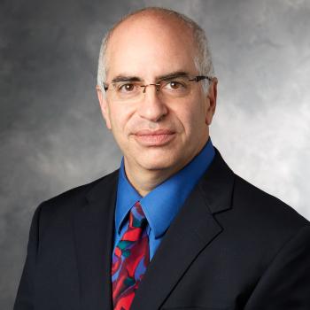 David N. Rosenthal合作论文数Pediatrics Department, Stanford University24
 Steven Frank合作论文数Department of Anesthesiology and Critical Care Medicine, School of Medicine, Johns Hopkins University21
 Eddie Lau合作论文数18
 William Robert Morrison (Rob)合作论文数Center for Grain and Animal Health Research, Agricultural Research Service, United States Department of Agriculture17
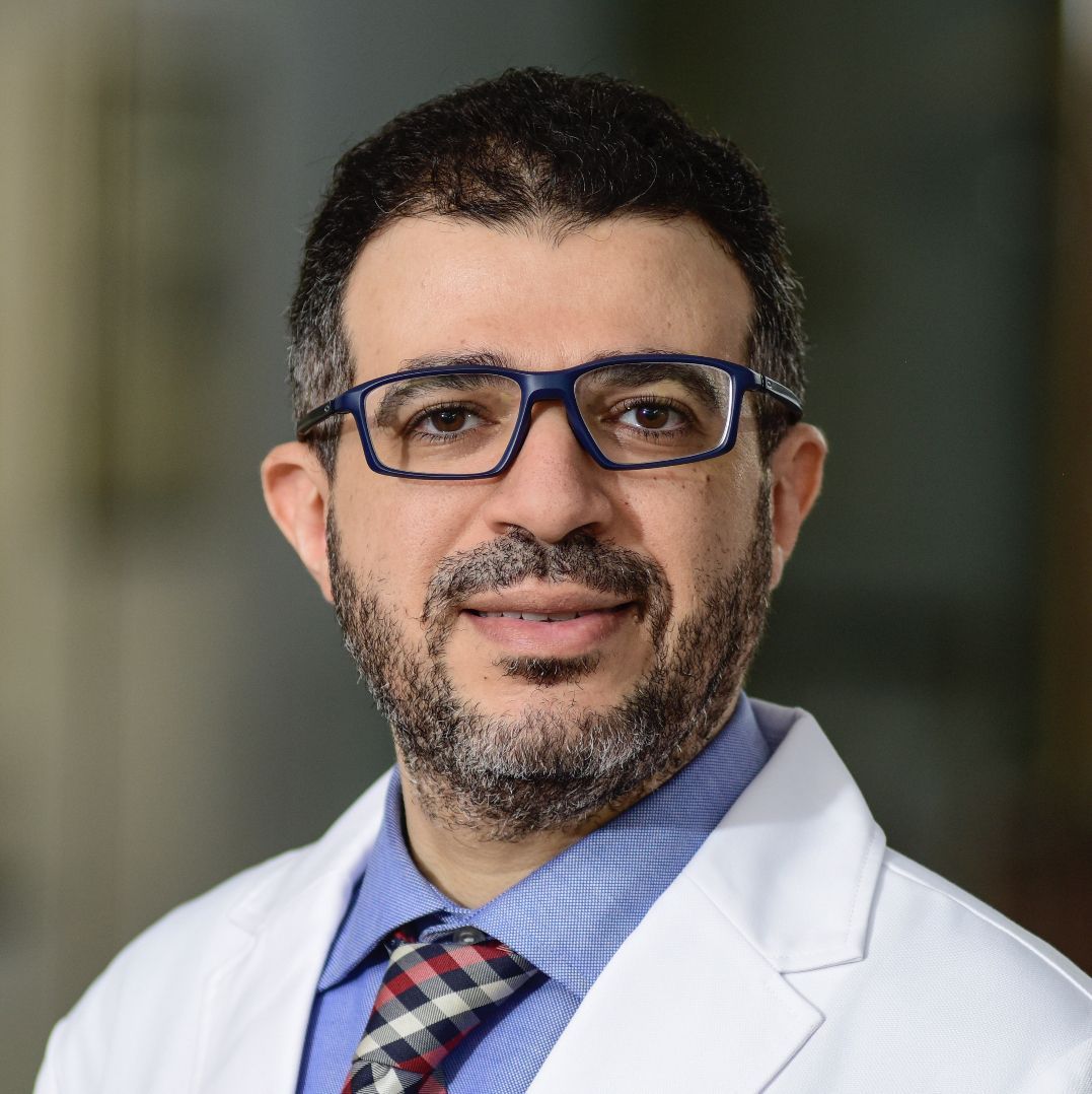 Abdallah Sherif Radwan Mohamed合作论文数Department of Radiation Oncology, Baylor College of Medicine16
 Baher Elgohari合作论文数Faculty of Medicine, University of Almansoura14
 Alfio Ferlito合作论文数ENT Clinic, University of Udine School of Medicine/International Head and Neck Scientific Group14
 C. Cardenas合作论文数Dept Radiat Oncol, Univ Alabama Birmingham13
 Renata Ferrarotto合作论文数13
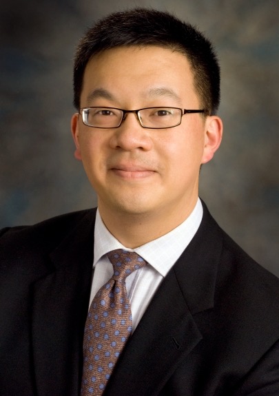 Stephen Y. Lai合作论文数Department of Head and Neck Surgery, Division of Surgery, The University of Texas MD Anderson Cancer Center;Department of Radiation Oncology, Division of Radiation Oncology, The University of Texas MD Anderson Cancer Center;Department of Molecular and Cellular Oncology, Division of Basic Science Research, The University of Texas MD Anderson Cancer Center12
 Katherine A. Hutcheson合作论文数Department of Communication Sciences and Disorders, Division of Surgery, The University of Houston College of Liberal Arts and Social Sciences;Section of Speech Pathology and Audiology, Department of Head and Neck Surgery, The University of Texas MD Anderson Cancer Center12
 Sandra Nuyts合作论文数Department of Radiation Oncology, UZ Leuven;Department of Oncology, KU Leuven, University of Leuven12
 Farshad Foroudi合作论文数Division of Radiation Oncology, Peter MacCallum Cancer Centre11
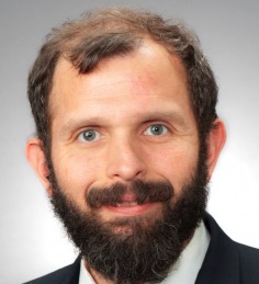 Heath D. Skinner合作论文数Department of Radiation Oncology, School of Medicine, University of Pittsburgh;Hillman Cancer Center, University of Pittsburgh Medical Center10
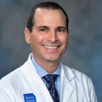 Erich M. Sturgis合作论文数Department of Head and Neck Surgery, The University of Texas M.D. Anderson Cancer Center/Population Science & Cancer Control, UT Southwestern Simmons Comprehensive Cancer Center/Head and Neck Surgery, Baylor College of Medicine10
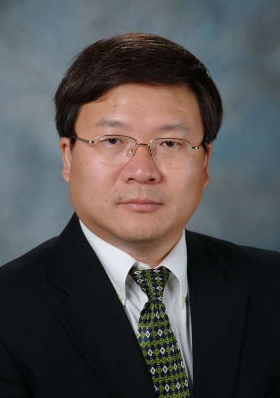 Jihong Wang合作论文数Department of Radiation Physics, Division of Radiation Oncology, MD Anderson Cancer Center, The University of Texas10
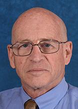 Avraham Eisbruch合作论文数Department of Radiation Oncology, University of Michigan10C. Pollard合作论文数9
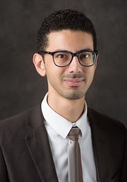 Hesham Elhalawani合作论文数Department of Radiation Oncology, Brigham and Women's Hospital;Harvard Medical School;Dana-Farber Brigham Cancer Center9Yao Ding合作论文数8Primož Strojan合作论文数8
 Hui Gan合作论文数Tumour Targeting Laboratory, Olivia Newton-John Cancer Research Institute;Austin Health8
 Joseph M Herman合作论文数7
 Mohamed Abdallah合作论文数7Reddy Jay P合作论文数Department of Molecular &Cellular Biology, Baylor College of Medicine;Department of Molecular & Cellular Biology, Baylor College of Medicine7
 Mona Kamal合作论文数7
 June Corry合作论文数Division of Radiation Oncology Locked Bag No 1, Peter MacCallum Cancer Centre7Morikatsu Wada合作论文数6
 Jason Michael Johnson合作论文数6June Yap合作论文数6Alisha Moore合作论文数6Yuan Ying合作论文数6Roslyn Francis合作论文数6
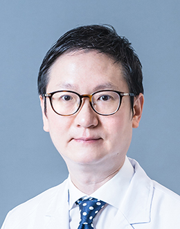 Wai Tong Ng (吴伟棠)合作论文数Li Ka Shing Faculty of Medicine, The University of Hong Kong;Department of Clinical Oncology, School of Clinical Medicine, The University of Hong Kong6
 Eugene J. Koay合作论文数Department of Radiation Oncology, Division of Radiation Oncology, The University of Texas MD Anderson Cancer Center;Department of Gastrointestinal Radiation Oncology, Division of Radiation Oncology, The University of Texas MD Anderson Cancer Center;Department of Nanomedicine, Houston Methodist Research Institute6
 Mark Blayne Pinkham合作论文数Department of Clinical Oncology, The Christie NHS Foundation Trust;School of Medicine, University of Queensland;School of Medicine, University of Queensland6Lin Peter合作论文数6
 Shirley Y. Su合作论文数6
 Amy Moreno合作论文数Department of Surgical Oncology6
 Antti Mäkitie合作论文数Department of Ophthalmology and Otorhinolaryngology, Faculty of Medicine, University of Helsinki;Department of Clinical Science, Intervention and Technology, Karolinska Institutet;Helsinki University Hospital6Ehab Hanna合作论文数6Trevor Leong合作论文数6S. T. Lee合作论文数6




