Whole Genome Sequencing (WGS) provides comprehensive genomic profiling of brain tumours. In Wessex, the integration of WGS into clinical workflow has been adopted rapidly to enable this emerging clinical tool. This requires multi-speciality working and the support of a dedicated Genomics Medicine team. This project reviews the current WGS pathway infrastructure within a large tertiary UK neuro-oncology unit. Patients with primary brain tumours eligible for WGS as per NHS/GLH criteria were identified from theatre lists between January-December 2024. Electronic patient records, histopathological reports and WGS reports were analysed. Rates of adequate sample collection and completed WGS reports were recorded. Uncompleted WGS reports were analysed for root cause. Pre-operative clinical assessment of patients undergoing surgical intervention for a primary brain tumour in- cludes discussion of WGS testing. 104 patients were identified as eligible for WGS. Of these, 70 (67.3%) had ade- quate frozen tissue taken at time of surgery to facilitate WGS. The Genomics Medicine team consent patients to WGS and track samples from the point of collection to ensure all samples proceed. From the samples collected, all have progressed to one of three categories: WGS report, low DNA content, clinical reason to abandon test- ing. The weekly GTAB meeting led by the Genomics Medicine team and clinical scientists works closely with clinicians to interpret results and identify potential therapeutic targets/clinical trials. A review to investigate barriers to adequate sample collection/uncompleted WGS is ongoing. BNOS and Darzi reports separately recommend the routine use of WGS for all patients with primary brain tumours. This audit demonstrates the rapid adoption of the this tool in clinical practice with a robust multi- disciplinary clinical pathway. The Genomics Medicine team facilitate sample processing to a conclusion in all instances and are imperative to WGS interpretation.
Previous evidence has demonstrated feasibility of day-case biopsy in selected neuro-oncology cohorts, but its widespread application has not yet occurred. There is potential for rationalising care pathways and finite healthcare resources. We present our 11-year experience of day-case frameless stereotactic biopsy without intra-operative frozen section in an adult neuro-oncology practice. Single centre retrospective study of stereotactic biopsies in adult neuro-oncology patients between January 2014-December 2024. Patient demographics, performance status (PS), radiological, operative, histological and outcome data were collected. Univariate analysis was performed to identify predictors of post-procedural intracranial haemorrhage (ICH) and unsuccessful same day discharge. Inpatient biopsy was performed if time-sensitive and non-dischargeable. Of 715 stereotactic biopsies, 559 (78.2%) were planned day-case procedures. Of the day-case cohort, median age was 65 years, with a male preponderance (329/559; 58.9%) and PS 0-1 (345; 79.6%). Majority of tumours were multifocal (359/559; 64.2%), of which 37.6% (135/359) were multicentric. Midline involvement was observed in 237/559 (42.4%). Median operative time was 30 minutes and 525/559 (93.9%) were performed under conscious sedation. Same day discharge was achieved in 92% (514/559) and a non-diagnostic biopsy in 2.7% (15/559). Post-procedure ICH was observed in 13.1% (73/559), of which 5 were symptomatic (0.89%). All cause 30-day readmission and mortality rates were 9.6% (54/559) and 0.89% (5/559) respectively. Univariate logistic regression was performed to identify factors associated with higher odds of unsuccessful same day discharge or ICH. No statistically significant factors were identified related to demographic, radiological or histological variables. Increased operative time was associated with higher odds of ICH (OR=1.02, p=0.045). Awake day-case biopsy across a wide range of neuro-oncology patients is feasible with an acceptable safety profile and diagnostic yield. We demonstrate that day-case awake frameless biopsy has treatment equipoise to inpatient pathways. Early discharge has additional economic and quality of life considerations in life limiting condition.
Multifocal glioblastoma multiforme (GBM) is associated with a poor prognosis, with a median survival of approximately six months. We present the case of a 51-year-old female diagnosed with multifocal WHO Grade 4 IDH-wildtype GBM, involving bilateral frontal infiltrating tumours with extension into the left centrum semiovale and corpus callosum, and exhibiting MGMT promoter methylation (25–50%). Management employed an individualised, multimodal approach incorporating surgery, chemotherapy, radiotherapy, and genomic profiling. A stereotactic burr hole biopsy on 15/09/2024 confirmed the diagnosis, undertaken due to eloquent location, fixed neurological deficit despite dexamethasone, and multifocal disease. Genomic profiling identified pathogenic mutations in TP53, RB1, KMT2D, TERT, BCOR, and PTEN, along with variants of uncertain significance in FGFR3 and PIK3CG. Tumour mutation burden was low, microsatellite instability was not detected, PD-L1 was negative, and mismatch repair proficient. Temozolomide chemotherapy was initiated on 23/09/2024. Hypofractionated radiotherapy (40.05 Gy in 15 fractions) commenced on 14/10/2024, targeting the dominant enhancing, biopsied left frontal lesion, concurrent with the second chemotherapy cycle. Following treatment initiation, the patient’s Karnofsky Performance Status improved from 70% to 90–100%, facilitating independent ambulation and enhanced quality of life. After the seventh cycle of temozolomide, oligoprogression was observed with enlargement of a right frontal lesion. The patient underwent craniotomy for resection on 04/05/2025, with plans for further radiotherapy and continued chemotherapy targeting this site. This case underscores that, despite the typically poor prognosis of multifocal GBM, a personalised, stepwise multimodal approach—guided by molecular insights and functional status—can yield meaningful clinical benefit and extended survival. At the time of reporting, the patient maintains excellent performance status and quality of life, emphasising the value of adaptive, individualised care in this aggressive disease.
Maximal safe cytoreduction remains the optimal surgical strategy in GBM. Despite this, a significant proportion of patients are not amenable to resection. Stereotactic biopsy remains essential to provide an integrated diagnosis and guide further treatment. There is a paucity of recent literature examining survival variables of this cohort post the WHO 2021 classification. Single centre retrospective study of stereotactic biopsies in IDHwt-GBM patients between January 2014-December 2024. Patient demographic, performance status (PS), radiological, histological and outcome data were collected. MGMT-hypermethylated tumours were stratified into 5-10% vs >10% cohorts. Univariate and Kaplan-Meier survival analyses were performed. Basal ganglia, thalamus, brainstem and corpus callosum defined midline structures. 394 cases were identified. Median age was 66 with a male:female ratio=1.77:1, 285 (72.5%) cases were PS0-1. Multifocal tumours accounted for 65.9% (259) of cases, of which 99 were multicentric. 184(46.7%) patients had midline involvement. Median survival(mOS) with further treatment and best supportive care was 7.5 months (95% CI: 6.47-8.23) and 2.1 months (95% CI: 1.60-2.53). Kaplan-Meier and univariate survival analysis demonstrated significant improved survival distributions for age less than 65 (Logrank-Mantel-Cox, p=0.0004) and pre-biopsy PS 0-1 (Logrank-mantel-cox, p<0.0001). These were also predictive of undergoing further treatment. Involvement of midline structures had reduced mOS compared to non-midline lesions (Logrank Mantel-Cox, 0.0005). MGMT hypermethylation was associated with improved mOS in patient undergoing adjuvant treatment (8.57 vs 7.63months, Logrank Mantel-Cox p=0.0145). Whilst degree of MGMT-hypermethylation did not demonstrate significance in mOS for the adjuvant treatment cohort (8.2 vs 8.6 months, p=0.1828), >10% MGMT-hypermethylation cohort demonstrated improved 2-year OS (17.5% vs 6.3%). Despite recent advances in therapeutics, inoperable GBM remains a challenging entity to treat. Here we quantify pertinent peri-operative factors which may be utilised to council patients and manage prognostic expectations. Poorer prognostic cohorts may be better served with enrolment in clinical trials or best supportive care.
Purpose The role of repeat resection for recurrent glioblastoma (rGB) remains equivocal. This study aims to assess the overall survival and complications rates of single or repeat resection for rGB. Methods A single-centre retrospective review of all patients with IDH-wildtype glioblastoma managed surgically, between January 2014 and January 2022, was carried out. Patient survival and factors influencing prognosis were analysed, using Kaplan–Meier and Cox regression methods. Results Four hundred thirty-two patients were included, of whom 329 underwent single resection, 83 had two resections and 20 patients underwent three resections. Median OS (mOS) in the cohort who underwent a single operation was 13.7 months (95% CI: 12.7–14.7 months). The mOS was observed to be extended in patients who underwent second or third-time resection, at 22.9 months and 44.7 months respectively ( p < 0.001). On second operation achieving > 95% resection or residual tumour volume of < 2.25 cc was significantly associated with prolonged survival. There was no significant difference in overall complication rates between primary versus second ( p = 0.973) or third-time resections ( p = 0.312). The use of diffusion tensor imaging (DTI) guided resection was associated with reduced post-operative neurological deficit (RR 0.37, p = 0.002), as was use of intraoperative ultrasound (iUSS) (RR 0.45, p = 0.04). Conclusions This study demonstrates potential prolongation of survival for rGB patients undergoing repeat resection, without significant increase in complication rates with repeat resections. Achieving a more complete repeat resection improved survival. Moreover, the use of intraoperative imaging adjuncts can maximise tumour resection, whilst minimising the risk of neurological deficit.
BACKGROUND: Hemangioblastomas are benign, often cystic tumors occurring most commonly in the cerebellum. We describe resection of cystic cerebellar hemangio-blastomas using an endoscope-assisted technique.METHODS: We present a series of 9 cases and discuss the endoscopic technique for resection of cystic heman-gioblastoma. Frameless neuronavigation was used in cases. A small 1-to 1.5-cm corticotomy lined with Surgicel and patties allows access to the peritumoral cyst. Using bimanual microsurgical techniques and the 30-degree rigid endoscope to provide visualization, the mural nodule can be resected and the cyst cavity inspected.RESULTS: Complete resection of the mural nodule was achieved in all cases. One patient had a postsurgery pseudomeningocele requiring ventriculoperitoneal shunt. Hospital length of stay was short for all the patients.CONCLUSIONS: Endoscope-assisted resection is a safe and effective technique for surgical resection of cystic hemangioblastoma.
Abstract AIMS Despite evidence of correspondence with intraoperative stimulation, there remains limited data on MRI diffusion tractography (DT)’s sensitivity to predict morbidity after brain tumour resection. Our aims were: (1) evaluate DT against subcortical stimulation mapping and performance changes during and following awake surgery; (2) evaluate the use of early post-operative DT to predict recovery from post-surgical deficits. METHOD We performed a retrospective review our first 100 awake neurosurgery procedures using DTneuronavigation. Intra-operative stimulation and performance outcomes were assessed to classify DT predictions for sensitivity and specificity calculations. RESULTS Between 2014 and 2019 a total of 91 adult brain tumour patients (mean age 49.2 years) underwent 100 awake surgeries with subcortical stimulation. The sensitivity and specificity of pre-operative DT predictions were 92.2% and 69.2%, varying among tracts. Post-operative deficits occurred after 41 procedures (39%), but were prolonged (>3 months) in only 4 patients (4%). Post-operative DT was available in 51 patients and confirmed surgical preservation of tracts. Post-operative DT anticipated complete recovery in a patient with supplementary motor area syndrome, and indicated infarct-related damage to corticospinal fibers associated with delayed, partial recovery in a second patient. CONCLUSION Pre-operative DT provided very accurate predictions of the spatial location of tracts in relation to a tumour. As expected, however, the presence of a tract did not provide information on its functional status, resulting in variable DT specificity among individual tracts. While prolonged deficits were rare in our series, DT in the immediate post-operative period offered additional information to monitor neurological deficits and anticipate recovery potential.
Purpose Gliomas are the most commonly occurring brain tumour in adults and there remains no cure for these tumours with treatment strategies being based on tumour grade. All treatment options aim to prolong survival, maintain quality of life and slow the inevitable progression from low-grade to high-grade. Despite imaging advancements, the only reliable method to grade a glioma is to perform a biopsy, and even this is fraught with errors associated with under grading. Positron emission tomography (PET) imaging with amino acid tracers such as [ 18 F]fluorodopa ( 18 F-FDOPA), [ 11 C]methionine ( 11 C-MET), [ 18 F]fluoroethyltyrosine ( 18 F-FET), and 18 F-FDOPA are being increasingly used in the diagnosis and management of gliomas. Methods In this review we discuss the literature available on the ability of 18 F-FDOPA-PET to distinguish low- from high-grade in newly diagnosed gliomas. Results In 2016 the Response Assessment in Neuro-Oncology (RANO) and European Association for Neuro-Oncology (EANO) published recommendations on the clinical use of PET imaging in gliomas. However, since these recommendations there have been a number of studies performed looking at whether 18 F-FDOPA-PET can identify areas of high-grade transformation before the typical radiological features of transformation such as contrast enhancement are visible on standard magnetic resonance imaging (MRI). Conclusion Larger studies are needed to validate 18 F-FDOPA-PET as a non-invasive marker of glioma grade and prediction of tumour molecular characteristics which could guide decisions surrounding surgical resection.
Introduction Awake craniotomy is widely used for surgery in eloquent brain in order to facilitate maximal safe resection of brain tumours. There has been huge progress in both surgical and anaesthetic techniques used during awake craniotomy. This study reports a single surgeon experience of awake craniotomy for tumour resection over a 14 year period focusing on the safety and feasibility of a simple technique. Methods Patients who underwent awake craniotomy for tumour resection between 2006 and 2019 (inclusive) were identified retrospectively from theatre logbooks in an NHS neurosciences centre. Case note review was performed to collect data on demographics, histology, intra-operative mapping and complications. Results Four hundred and sixty-nine patients were included with a mean age of 52.0 ± 14.3 years. Three hundred and seventy-seven (80.2%) of the tumours were primary tumours of which WHO grade IV were the most common (n = 204, 54.1%). Ninety-two (19.6%) of the tumours were metastases with breast (n = 23, 25.0%), skin (n = 22, 23.91%) and lung (n = 22, 23.9%) being the most common primary malignancies. The frontal lobe was the most common location (n = 221, 47.1%). The median length of stay was 1 day. One hundred and seven (22.8%) patients had complications with neurological deficits (n = 73, 15.6%), being the most common however only 8 patients (1.7%) had permanent neurological deficits. Discussion This is the largest published UK series of awake craniotomy for tumour resection. It demonstrates the safety and feasibility of a simple and easily reproducible technique with a low incidence of permanent neurological deficits and short durations of hospital admission.
OBJECTIVES:Masson tumor or intravascular papillary endothelial cell proliferation was first described in 1923. Only a few cases of intracranial Masson tumor have been reported following stereotactic radiosurgery (SRS). We report a series of 6 cases, age range 28-56 years, with intracranial Masson tumor following SRS for treatment of an intracranial arteriovenous malformation (AVM).METHODS:We performed a retrospective case note review, reviewed the imaging, SRS records, and neuropathology specimens following surgical excision.RESULTS:In our series all patients received Leksell SRS with the periphery of the AVM receiving doses ranging from 22-25 Gy. The time lapse from SRS to a clear enhancing mass appearing on imaging ranged from 5-10 years. Four patients underwent craniotomy and excision of the enhancing lesion for persistent edema and an enlarging cyst resulting in a resolution of symptoms.CONCLUSIONS:SRS is an effective treatment for obliteration of intracranial AVMs.
Background. In recent years an increasing number of patients with cerebral metastasis (CM) have been referred to the neuro-oncology multidisciplinary team (NMDT). Our aim was to obtain a national picture of CM referrals to assess referral volume and quality and factors affecting NMDT decision making. Methods. A prospective multicenter cohort study including all adult patients referred to NMDT with 1 or more CM was conducted. Data were collected in neurosurgical units from November 2017 to February 2018. Demographics, primary disease, KPS, imaging, and treatment recommendation were entered into an online database. Results. A total of 1048 patients were analyzed from 24 neurosurgical units. Median age was 65 years (range, 21-93 years) with a median number of 3 referrals (range, 1-17 referrals) per NMDT. The most common primary malignancies were lung (36.5%, n = 383), breast (18.4%, n = 193), and melanoma (12.0%, n = 126). A total of 51.6% (n = 541) of the referrals were for a solitary metastasis and resulted in specialist intervention being offered in 67.5% (n = 365) of cases. A total of 38.2% (n = 186) of patients being referred with multiple CMs were offered specialist treatment. NMDT decision making was associated with number of CMs, age, KPS, primary disease status, and extent of extracranial disease (univariate logistic regression, P < .001) as well as sentinel location and tumor histology (P < .05). A delay in reaching an NMDT decision was identified in 18.6% (n = 195) of cases. Conclusions. This study demonstrates a changing landscape of metastasis management in the United Kingdom and Ireland, including a trend away from adjuvant whole-brain radiotherapy and specialist intervention being offered to a significant proportion of patients with multiple CMs. Poor quality or incomplete referrals cause delay in NMDT decision making.
Abstract Background Over the recent years an increasing number of patients with brain metastasis are being referred to the neuro-oncology multi-disciplinary team (NMDT). Our aim was to determine if referrals of this group of patients to the NMDT in the UK & Ireland comply with NICE guidelines and to assess referral volume, quality of information provided and its impact on NMDT decision-making. Methods Prospective multicentre oberservational study including all adult patients referred with ≥1 cerebral metastasis. Data was collected in neurosurgical units from 11/2017 to 02/2018. Demographics, primary disease, Karnofsky performance status (KPS), imaging and treatment recommendation were entered into an online database. Results 1049 patients were analysed from 24 neurosurgical units. Median age was 63[range 21–93] years with a median number of 3[range 1–17] referrals per NMDT. The most common primary malignancies were lung (36.5%, n=383), breast (18.5%, n=194) and melanoma (12.0%, n=126). 51.6% (n=541) of the referrals to the NMDT were within the NICE 2006 guidelines, and resulted in specialist intervention being offered in 68.8%. 41.2% (n=197) of patients being referred outside of the NICE 2006 guidelines were offered specialist treatment. NMDT decision-making was influenced by number of metastases, age, KPS, primary disease status and extent of extracranial disease (univariate logistic regression, p<0.0001) as well as metastasis location/histology (p<0.05). Conclusions This study confirmed a national change in culture of referral patterns. We identified a delay in NMDT decision-making in ~20%, contributing to increased NMDT workload. New stratification tools may be needed to reflect advancements in diagnostics and treatment modalities.
Abstract In the United Kingdom widespread adoption of day-case image-guided biopsy (DIB) for brain tumour has yet to develop. We review a decade of experience of DIB for suspected supra-tentorial brain tumour and recommend discharge criteria for patients post-operatively. 30-day complications, post-operative admissions into hospital and patient satisfaction are examined. METHODS Published protocols and procedures for DIB of brain tumours were used. Tissue samples were sent for fixed histological preparation without intraoperative neuropathological assessment. All patients undergoing an image-guided biopsy for tumour were retrospectively identified from operative logbooks (01/10/2006–30/09/2016), and information recorded from online records. Patients completed satisfaction questionnaires. RESULTS 706 image-guided biopsies for supratentorial tumour were performed of which approximately 60% were identified pre-operatively as candidates for DIB. 92% of DIB patients were successfully discharged 6 hours postoperatively. 4.5% of DIB patients were admitted directly from the day-case unit following identification of a postoperative haematoma >2 cm diameter on CT head performed at 4 hours. 3/401 DIB patients (< 1%) required surgical evacuation for the haematoma, all made a full recovery, with no delay to commencing their oncological management. A diagnostic accuracy of 98% was achieved. Overall 90% of patients were satisfied about the timing of their discharge from hospital and 92.5 % felt they had enough medical support following discharge. DISCUSSION Conscious sedation with enhanced recovery techniques, lack of intra-operative neuropathological analysis and a stream lined service with robust communication between patients, carers/families and the oncology allied medical professionals has ensured that DIB for diagnosis of tumour is safe, reliable and feasible for most patients and is not associated with increased morbidity or mortality. The procedure is well tolerated with good patient satisfaction. We recommend that patients with a good pre-operative functional baseline, requiring a supra-tentorial biopsy are offered day-case surgery in a dedicated unit specialising in this procedure.
OBJECTIVE:External ventricular drain (EVD) insertion is one of the most common emergency neurosurgical procedures. EVDs are traditionally inserted freehand (FH) in an emergency setting, but often result in suboptimal positioning. Image-guided surgery (IGS) is selectively used to assist placement. However, the accuracy and practicality of IGS use is yet to be reported. In this study, the authors set out to assess if IGS is practical and improves the accuracy of EVD placement. METHODS:Case notes and images obtained in patients who underwent frontal EVD placement were retrospectively reviewed. Ventriculomegaly was determined by the measurement of the Evans index. EVD location was classified as optimal (ipsilateral frontal horn) or suboptimal (any other location). Propensity score matching of the two groups (IGS vs FH) for the Evans index was performed. Data were analyzed for patient age, diagnosis, number of EVDs, and complications. Those without postoperative CT scans were excluded. RESULTS:A total of 607 patients with 760 EVDs placed were identified; 331 met inclusion criteria. Of these, 287 were inserted FH, and 44 were placed with IGS; 60.6% of all unmatched FH EVDs were optimal compared with 75% of the IGS group (p = 0.067). The IGS group had a significantly smaller Evans index (p < 0.0001). Propensity score matching demonstrated improved optimal position in the IGS group when compared with the matched FH group (75% vs 43.2%, OR 4.6 [1.5-14.6]; p = 0.002). Patients with an Evans index of ≥ 0.36 derived less benefit (75% in IGS vs 66% in FH, p = 0.5), and those with an Evans index < 0.36 derived more benefit (75% in IGS vs 53% in FH, p = 0.024). The overall EVD complication rate was 36% in the FH group versus 18% in the IGS group (p = 0.056). Revision rates were higher in the FH group (p = 0.035), and the operative times were similar (p = 0.69). Long intracranial EVD catheters were associated with tip malposition irrespective of the group. CONCLUSIONS:Image guidance is practical and improves the accuracy of EVD placement in patients with small ventricles; thus, it should be considered for these patients.
OBJECTIVES:External ventricular drain (EVD) placement is required frequently in neurosurgical patients to divert cerebrospinal fluid and monitor intracranial pressure. The usual practice is the tunneled EVD technique performed in operating theaters. EVD insertion through a bolt in intensive care also is described. We employ both practices in our institute. Herein, we compare the indications, accuracy, safety, and costs of the 2 techniques. METHODS:This was a retrospective cohort study of a prospectively maintained EVD database of all patients undergoing first frontal EVD placement between January 2010 and December 2015. Those patients with preceding cerebrospinal fluid infection were excluded. We compared bolt EVD with tunneled EVD techniques in terms of accuracy of EVD tip location by analyzing computed tomography scans to grade catheter tip location as optimal (ipsilateral frontal horn) or otherwise suboptimal, and complications that include infection and revision rates. RESULTS:In total, 579 eligible patients aged 3 months to 84 years were identified; 430 had tunneled EVDs and 149 bolt EVDs. The most frequent diagnosis was intracranial hemorrhage (73% bolt vs. 50.4% tunneled group; P < 0.001). Other diagnoses included tumor (4.7% bolt vs. 19.1% tunneled; P < 0.001) and traumatic brain injury (17.5% bolt vs. 17.4% tunneled). In the bolt EVD group 66.4% of EVD tips were optimal, compared with 61.0% in the tunneled group (P = 0.33). Infection was confirmed in 15 (10.0%) bolt EVDs compared with 61 (14.2%) tunneled EVDs (P = 0.2). Each bolt EVD kit costs £260, whereas placing a tunneled one in the theater costs £1316. CONCLUSIONS:Bedside bolt EVD placement is safe, accurate, and cost effective in selective patients with hemorrhage-related hydrocephalus.
Image-guided biopsy of intrinsic brain tumours is a common neurosurgical procedure. In 2008 we reported the first UK series of day-case craniotomy and biopsy. In 2017 our unit presented a series of 645 biopsies over 10 years, of which 355 were discharged the same day. We now report the results of the first UK patient satisfaction questionnaire of day-case brain biopsy. Consecutive patients undergoing image-guided biopsies were sent questionnaires. All patients received sedation for their procedure and had a CT brain scan 6 hours post operatively before discharge home. Over nine months a total of 60 brain biopsies were carried out. 43 of these were identified as being suitable for day-case surgery and 39/43 were discharged 6 hours postoperatively. The remainder were thought to be unsuitable for day-case surgery. 32 questionnaires were sent out and 26 responses received. 24/26 patients felt involved in the decision making process to undergo a biopsy. Of the operation 8/26 remember nothing, 8/26 most of, and 10/26 remembered little of the procedure. 22/26 patients felt the timing of discharge was acceptable, only one thought they should remain in hospital for longer. All of the patients received adequate discharge information and felt supported once home. Previous articles have demonstrated the safety of day-case image guided brain biopsies yet enhanced recovery programmes remain scarce in neurosurgery. Sustainability and transformation projects for the NHS require services to look at ways of reducing length of stay to reduce unnecessary admissions. Day-case neurosurgery is a practical option with clear significant cost saving implications. In the last 10 -12 years our unit has challenged accepted practice and undertaken day-case and short stay neuro-oncology surgery. This satisfaction survey of day-case brain biopsies demonstrates that it is well tolerated and accepted by patients and imparts confidence to continue this practice.
ObjectivesExternal ventricular drain (EVD) insertion is a common neurosurgical procedure. EVD-related infection (ERI) is a major complication that can lead to morbidity and mortality. In this study, we aimed to establish a national ERI rate in the UK and Ireland and determine key factors influencing the infection risk.MethodsA prospective multicentre cohort study of EVD insertions in 21 neurosurgical units was performed over 6 months. The primary outcome measure was 30-day ERI. A Cox regression model was used for multivariate analysis to calculate HR.ResultsA total of 495 EVD catheters were inserted into 452 patients with EVDs remaining in situ for 4700 days (median 8 days; IQR 4–13). Of the catheters inserted, 188 (38%) were antibiotic-impregnated, 161 (32.5%) were plain and 146 (29.5%) were silver-bearing. A total of 46 ERIs occurred giving an infection risk of 9.3%. Cox regression analysis demonstrated that factors independently associated with increased infection risk included duration of EVD placement for ≥8 days (HR=2.47 (1.12–5.45); p=0.03), regular sampling (daily sampling (HR=4.73 (1.28–17.42), p=0.02) and alternate day sampling (HR=5.28 (2.25–12.38); p<0.01). There was no association between catheter type or tunnelling distance and ERI.ConclusionsIn the UK and Ireland, the ERI rate was 9.3% during the study period. The study demonstrated that EVDs left in situ for ≥8 days and those sampled more frequently were associated with a higher risk of infection. Importantly, the study showed no significant difference in ERI risk between different catheter types.
Citation for published version: Jamjoom, A, Joannides, A, Poon, M, Chari, A, Zaben, M, Abdulla, MAH, Roach, J, Glancz, LJ, Solth, A, Duddy, J, Brennan, P, Bayston, R, Bulters, DO, Mallucci, CL, Jenkinson, MD, Gray, WP, Kandamsay, J, Hutchinson, PJ, Kolias, AG, Ahmed, AI & Loan, J 2017, 'Prospective, Multi-Centre Study of External Ventricular Drainage-Related Infections in the United Kingdom and Ireland' Journal of Neurology, Neurosurgery & Psychiatry. DOI: 10.1136/jnnp-2017-316415
 Paul Grundy合作论文数9
 Crispin Weidmann合作论文数5
 Diederik O. Bulters合作论文数Department of Neurosurgery, Addenbrooke’s Hospital5Laurence J Glancz合作论文数4
 Paul Brennan合作论文数Centre for Clinical Brain Sciences, Edinburgh Medical School, The University of Edinburgh4
 Aswin Chari合作论文数John Radcliffe Hospital, University of Oxford Medical School4
 Michael Jenkinson合作论文数Department of Pharmacology and Therapeutics, Institute of Systems, Molecular and Integrative Biology, Faculty of Health and Life Sciences, University of Liverpool4
 Puneet Plaha合作论文数Nuffield Department of Clinical Neurosciences, University of Oxford4
 Andrew Durnford合作论文数Department of Neurosurgery, University Hospital Southampton NHS Foundation Trust3
 Laurent James Livermore合作论文数3
 Vasileios Apostolopoulos合作论文数Imperial Healthcare NHS Trust and BTRC Research Group, Imperial College3
 Victoria Wykes合作论文数Institute of Cancer and Genomic Sciences, College of Medical and Dental Sciences, University of Birmingham3Jonathan Duffill合作论文数2Benjamin Gaastra合作论文数2Anna Solth合作论文数2James Clerkin合作论文数2Emma Dalton合作论文数2Jeng Ching合作论文数2Dr Richard Chapman合作论文数2
 Kenny Yu合作论文数2Mark Fabian合作论文数2Susruta Manivannan合作论文数2
 Josephine Jung合作论文数2
 Aminul Islam Ahmed合作论文数Kings Coll London, Kings Coll Hosp2
 Roger Bayston合作论文数2
 Keyoumars Ashkan合作论文数Department of Basic & Clinical Neuroscience, School of Neuroscience, Institute of Psychiatry, Psychology & Neuroscience, King’s College London2
 Francesco Vergani合作论文数2
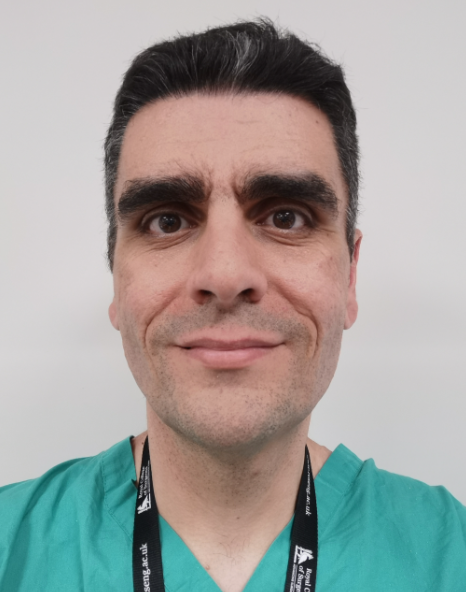 Angelos Kolias合作论文数Department of Clinical Neuroscience, Addenbrooke's Hospital;CRUK Cambridge Centre, Cancer Research UK Cambridge Institute, University of Cambridge2
 Budohoski Karol P合作论文数Department of Neurosurgery, University of Cambridge2
 Zakaria Rasheed合作论文数Department of Neurosurgery, The Walton Centre NHS Foundation Trust2Lucy Brazil合作论文数2
 Peter J A Hutchinson合作论文数Department of Clinical Neurosciences, Robinson College, University of Cambridge2John Duddy合作论文数2
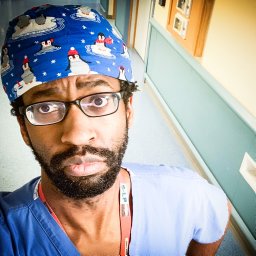 Simon Lammy合作论文数Department of Neurosurgery, Aberdeen Royal Infirmary2Richard Stacey合作论文数2Patrick Gray合作论文数Pascal Biosciences2Catey Bunce合作论文数2Enrico Clarke合作论文数Sant’ Andrea Hospital, University “La Sapienza”,2Jothy Kandasamy合作论文数2
 Malik Zaben合作论文数2Alexis Joannides合作论文数Division of Neurosurgery, University of Cambridge2
 Conor Mallucci合作论文数Department of Neurosurgery, Royal Liverpool Children’s Hospital2
 Anan B Y Shtaya合作论文数2
 Ahmed-Ramadan Sadek合作论文数2
 Jignesh Tailor合作论文数Department of Paediatric Surgery, John Radcliffe Hospital2Mutwakil Abdulla合作论文数1Simon McElligott合作论文数1B Jamjoom合作论文数1Alexander Dando合作论文数1




