BACKGROUND AND OBJECTIVES:Contemporary skull base surgeons must understand transcranial and endoscopic endonasal approaches to the petrous apex (PA). We provide an anatomic overview and comparison of main approaches to the PA through illustrative anatomic dissections. METHODS:On 10 sides of 5 specimens, transcranial approaches to the PA including the anterior petrosal, transcochlear, and retrosigmoid with suprameatal extension were performed. For endoscopic endonasal approaches, the transclival approach was performed in the midline of 5 specimens and its contralateral transmaxillary extension was performed on 10 sides. RESULTS:The anterior petrosal approach offers an anterolateral view of the PA bounded by petrous ridge posteromedially, internal auditory canal posterolaterally, greater superficial petrosal nerve anterolaterally, and lateral boundary of cranial nerve V, the Gasserian ganglion, and V3 anteromedially; it exposes the middle fossa and, once the PA is removed, the superomedial cerebellopontine angle. The transcochlear approach affords a lateral view of the PA defined medially and inferiorly by inferior petrosal sinus, posteriorly by posterior fossa dura, anteriorly by the petrous internal carotid artery, and superiorly by the superior petrosal sinus and middle fossa dura. Through a retrosigmoid approach, the PA is bounded by the superior petrosal sinus superiorly, the sagittal plane of cranial nerve VI medially, and the axial and sagittal plane of the internal auditory canal porus inferiorly and laterally, respectively. It affords a panoramic view of the posterior fossa and access to Meckel's cave after PA drilling. Endoscopic endonasal approaches target the anteromedial PA, and it is demarcated by a triangle consisting of cranial nerve VI posterolaterally, the paraclival segment of the internal carotid artery anteriorly, and the petroclival synchondrosis inferiorly and medially. The addition of the contralateral transmaxillary approach enhances lateral access up to the internal auditory canal. CONCLUSION:We provide a comprehensive overview of the main approaches to the PA through illustrative anatomic dissections and representative cases.
Biparietal osteodystrophy, also known as bilateral parietal calvarium bone thinning, is a rare calvarial condition characterized by loss of diploic bone within the parietal region. Although historically described in anthropologic and radiologic literature, current understanding of its epidemiology, pathogenesis, clinical relevance, and management remains fragmented. This review synthesizes current evidence and highlights emerging studies on biparietal osteodystrophy. Epidemiologic reports indicate that biparietal osteodystrophy affects a broad age range but appears most frequently in older adults, with several series suggesting a slight female predominance. Prevalence is difficult to estimate due to incidental detection, though modern imaging has increased recognition of the condition. Histologically and anatomically, biparietal osteodystrophy is marked by thinning of the outer table and loss of the diploic space, with little osteoblastic or osteoclastic remodeling. Proposed pathogenic mechanisms include chronic degenerative change, impaired vascular supply to the diploë, and other theories, although no single explanation fully accounts for its complete presentation. Clinically, most patients remain asymptomatic, with complications such as calvarial fractures and traumatic brain injury being rare but highlighting the structural vulnerability associated with severe thinning. Management strategies are not standardized, and the use of anti-osteoporotic drugs and cranioplasty has been reported to offer benefit in select cases, although no treatment has been formally validated for biparietal osteodystrophy. As the understanding of biparietal osteodystrophy continues to evolve, future research is needed to clarify its natural history, identify potential etiologic pathways, and establish evidence-based management guidelines.
Understanding motor representation in the human brain requires mapping beyond the primary motor cortex, into the distributed networks that coordinate complex movements. The insular cortex, a multifunctional hub buried within the Sylvian fissure, has been implicated in motor control through clinical observations and neuroimaging. Yet its precise relation to primary sensorimotor processing remains one of the least understood aspects of motor neurophysiology. To address this gap, we quantified electrophysiological changes from implanted depth electrodes in patients performing simple movement tasks combined with single-pulse electrical stimulation (SPES) to map effective connectivity. The movement data reveal somatotopically specific representation bilaterally, as well as intereffector regions that are active for different movement types. Hand representation is centered along the contralateral ventral aspect of the middle and posterior short gyri bilaterally, while tongue/mouth tuned sites cluster in the dorsal posterior short gyrus and the dorsal long gyri. Insular activity temporally follows the primary motor cortex (M1) and precedes movement onset. SPES revealed somatotopically specific connectivity between corresponding sites in M1 and insula (hand-to-hand, tongue-to-tongue) and between bilateral insulae. These observations establish that somatotopy is a conserved property of distributed motor control incorporating the insular representations and connectivity.
BACKGROUND AND OBJECTIVES:Endoscopic transnasal surgery offers a minimally invasive alternative to transcranial approaches for accessing midline skull base lesions involving the petrous apex and petroclival region. However, lateral access, particularly behind the petrous internal carotid artery (ICA), remains limited. The contralateral transmaxillary (CTM) approach provides a more direct surgical trajectory, more parallel to the petrous ICA. Yet, lateral access through this approach can be restricted by anatomic barriers, including the pterygoid process and lateral buttress of the maxillary sinus. We aimed to evaluate the contralateral transmaxillary-transpterygoid (CTMP) approach, with and without the lateral buttress removal (CTMP-LBR), to determine whether these modifications could enhance surgical access to the petroclival region. METHODS:Ten sides from 5 formalin-fixed, latex-injected cadaveric specimens were dissected to compare CTM, CTMP, and CTMP-LBR approaches. Petrous bone drilling was directed posterolaterally behind the petrous ICA toward the region medial to the cochlea. In addition, 30 sides from computed tomography angiography scans were analyzed to obtain radiographic measurements for each approach. RESULTS:Compared with CTM, both CTMP and CTMP-LBR significantly increased access for retrocarotid petrous bone drilling (P < .001) and yielded a wider surgical angle relative to midline (P < .001), in both cadaveric and radiographic analyses. In dissections, 20% of sides required CTM only, 50% benefited from CTMP, and 30% required CTMP-LBR to achieve adequate lateral access. Radiographic analysis revealed that CTMP required a median of 4 mm of pterygoid drilling, whereas CTMP-LBR required 9 mm and enabled a trajectory parallel to the petrous ICA with only 11 mm of soft tissue displacement. Patients with smaller angles between the petrous ICA and the horizontal plane were significantly more likely to benefit from CTMP over CTM (P = .005). CONCLUSION:Modification of the CTM approach with pterygoid drilling and optional LBR improves retrocarotid access. Preoperative evaluation of the petrous ICA angle may help optimize patient-specific planning in endoscopic petroclival surgery.
OBJECTIVE:Early discharge following uncomplicated endonasal resection of pituitary adenoma has been described as safe and feasible. Discharge on postoperative day (POD) 1 is not a widely implemented practice in the United States. This study aimed to evaluate the safety of routine POD1 discharges by assessing postoperative unplanned medical care rates compared with those of patients discharged on POD 2 or later (POD2+). The secondary endpoint was to identify predictors of delayed discharge in the POD2+ cohort. METHODS:A retrospective database review of the medical records of 534 patients who underwent endonasal resection of pituitary adenoma at the Mayo Clinic was performed. Relevant demographics, tumor characteristics, and perioperative metrics were abstracted from the medical record, including intraoperative CSF leakage, postoperative complications, emergency department visits, or inpatient readmissions within 30 days. Descriptive, univariate, and multivariate analyses were performed. RESULTS:In comparison to 422 (79%) patients discharged on POD1, 112 (21%) patients discharged on POD2+ had a higher unplanned medical care rate (13% vs 5.5%, p = 0.019). There were significantly more men in the POD1 group (48%) than in the POD2+ group (37%) (p < 0.04). The most common reasons for POD1 unplanned care were endocrine dysfunction (2.4%), split evenly between syndrome of inappropriate antidiuretic hormone and adrenal insufficiency, whereas the most frequent reason in the POD2+ cohort was medical (0.06%). Univariate analyses indicated that female sex, higher BMI, intensive care unit (ICU) admission, obstructive sleep apnea, continuous positive airway pressure dependence, intraoperative CSF leakage, and diabetes insipidus (DI) increased the likelihood of POD2+ discharge. The reduced logistic regression model demonstrated a higher likelihood of POD2+ discharge with older age, ICU admission, intraoperative CSF leakage, and DI (p < 0.001). Common factors contributing to discharge after POD1 included endocrine (n = 66, 35%) and/or medical (n = 65, 34%) reasons. CONCLUSIONS:Routine patient discharge on POD1 following uncomplicated endonasal resection of pituitary adenoma is a safe practice, as evidenced by the significantly lower rates of unplanned medical care and complications compared with those of patients discharged on POD2 or later. Delayed discharge was associated with older age, ICU admission, intraoperative CSF leakage, and DI. The most frequent reasons for POD1 readmission were endocrine versus medical reasons for POD2+. Preoperative identification of risk factors for delayed discharge and preemptive management of endocrine and medical complications may reduce the length of stay and return to medical care.
Abstract Chondrosarcoma and chordoma represent two profoundly different and challenging central skull base pathologies. Both are largely surgical diseases with varying outcomes based on the completeness of resection. Adjuvant therapy is controversial, though radiation therapy is commonly employed postoperatively.
BACKGROUND:Olfactory neuroblastoma (ONB) exhibits variable recurrence patterns, with significant late recurrences occurring years after treatment. We investigated cutoff periods to define late recurrence and identified predictors that distinguish patients at risk of late recurrence from those considered cured. METHODS:From a multi-institutional cohort of 219 ONB patients treated at nine centers, we analyzed 77 patients with documented recurrence. Using a 48-month cutoff to define early (< 48 months) versus late (≥ 48 months) recurrence, we evaluated 35 clinical, anatomic, and treatment variables using Firth penalized logistic regression across three comparisons: (1) late versus early recurrence timing, (2) late recurrence versus no recurrence, and (3) early recurrence versus no recurrence. RESULTS:Thirty-four of the 77 (44.2%) recurrences were classified as late. Among the 77 patients who recurred, smaller tumor volume (OR 0.243, p = 0.035) and lower novel Dulguerov staging (OR 0.802, p = 0.038) were associated with late, rather than early recurrence. When patients with late recurrence were compared to those who remained disease-free, radiographic skull base bone involvement was the strongest independent predictor (OR 2.90, 95% CI 1.14-8.64, p = 0.025), and this effect strengthened after adjusting for tumor volume (OR 3.80, p = 0.029). Pathologic dural invasion (OR 3.29, p = 0.013), Kadish stage (OR 2.02, p = 0.026), and T-stage (OR 1.32, p = 0.040) were also significant. CONCLUSIONS:A 48-month cutoff effectively stratifies ONB recurrence risk. Skull base bone involvement independently predicts recurrence when compared to nonrecurrent cases and uniquely persists as a risk factor for late recurrence. These findings establish skull base bone involvement as a novel biomarker for risk-stratified surveillance protocols.
OBJECTIVE:Deep brain stimulation (DBS) is emerging as an additional surgical option for refractory, multifocal epilepsy. Four-lead DBS systems can be used when seizure onset is poorly localized, generalized, or involves multiple targetable circuits. The goal of this study was to assess preliminary outcomes and complication rates in 4-lead DBS systems for treating epilepsy. METHODS:A consecutive series of 32 patients with 4-lead DBS implants who had at least 6 months of follow-up was reviewed, and demographics, outcomes, and complications were abstracted. RESULTS:Thirty-two patients implanted with 4 DBS leads (including off-label use) were included. The median age at surgery was 28.5 years, the median age at epilepsy diagnosis was 7.5 years, and 18 patients (56%) were women. The median epilepsy duration prior to implantation was 21 years. The most common seizure localization was frontotemporal (53%), followed by generalized (28%) and parietoccipital (13%). Prior vagus nerve stimulation had been trialed in 20 patients (63%), and an invasive stereotactic EEG recording was performed in 17 patients (59%). Eight patients had prior epilepsy surgery (25%). DBS targets included the anterior thalamic nucleus in combination with the centromedian thalamic nucleus, hippocampus, pulvinar nucleus, cingulate, insula, and globus pallidus. The median surgical duration was 5 hours 22 minutes, with a median operating room time of 8 hours 21 minutes. The median postoperative follow-up was 27 (interquartile range 17-41) months. Three patients experienced transient neurological deficits and 2 patients required intraoperative lead repositioning. There were no infections or hemorrhages. Most patients benefited from stimulation (22% Engel class I, 22% class II, 28% class III, and 28% class IV). The median patient-reported seizure reduction rate was 70%, with a responder rate of 72%. One patient elected to have the system explanted. CONCLUSIONS:Four-lead DBS is safe and can achieve meaningful improvement in challenging cases of multifocal epilepsy with a low complication rate.
This multicenter, multi-surgeon retrospective study aimed to identify risk factors for postoperative cerebrospinal fluid (CSF) leak following endonasal resection of craniopharyngioma, while evaluating the “perception gap” through a surgeon survey. A retrospective review was conducted on 416 patients who underwent endoscopic transsphenoidal surgery (ETS) for craniopharyngioma from 20 institutions. Factors were compared between patients with and without postoperative CSF leak, and between Early (2007–2015) and Late (2016–2025) Epochs. Complementing the clinical data, a survey of 19 neurosurgeons captured expert perspectives on risk stratification and management strategies. Overall postoperative CSF leak rate was 13.5
Targeted therapy for BRAF V600E mutant papillary craniopharyngioma (PCP) has been rapidly accepted, though given the rarity of the tumor, there is limited guidance for appropriate use. Here, we retrospectively evaluated current practice patterns for the implementation of targeted therapeutics and their outcomes in the treatment of papillary craniopharyngioma (PCP). Practice patterns at the institutions in the Registry for Adenomas of the Pituitary and Related Disorders (RAPID) for treatment of BRAF V600E mutated PCPs using targeted therapy were evaluated. Baseline clinical and demographic variables were retrospectively recorded. Imaging response was evaluated. Dosing, treatment course, adverse events (AEs) and other therapeutic strategies employing BRAF/MEK inhibitors were studied. Adjuvant and/or salvage therapies, including radiation and surgery were recorded. Most patients demonstrated at least partial response to BRAF and/or BRAF/MEK inhibition (13/19, 72.2
OBJECTIVE:Communicating hydrocephalus may occur following stereotactic radiosurgery (SRS) for vestibular schwannomas (VSs), yet identifying individual patient risk factors associated with this post-SRS complication remains a challenge. This study examined predictors of nonobstructive ventricular enlargement and symptomatic communicating hydrocephalus following primary SRS treatment for VS via a single-center institutional cohort review and meta-analysis of the literature. METHODS:A retrospective single-institution cohort study and systematic literature review and meta-analysis examining post-SRS communicating hydrocephalus in VS was performed. RESULTS:The institutional cohort consisted of 634 patients who received primary SRS as treatment for VS. The cohort was 51.6% female, with a median age of 64 (range 18-89) years. Following SRS treatment, 364 patients (57.4%) experienced tumor shrinkage, 218 (34.4%) had no change in the size of their lesion, and 52 (8.2%) experienced tumor growth. Nonobstructive ventricular enlargement was observed in 23 patients (3.6%) following SRS treatment, of whom 9 (39.1%) remained asymptomatic and 14 (60.9%) required placement of a ventriculoperitoneal (VP) shunt, with a median time to shunt placement of 8 months. In the multivariate analysis, patients ≥ 65 years old (p = 0.038), SRS target volume ≥ 5 cm3 (p < 0.001), maximum SRS dose ≥ 26 Gy (p = 0.015), and tumor growth at the most recent follow-up (p = 0.002) were associated with an increased risk of post-SRS ventricular enlargement. Similarly, patients with older age (p = 0.049), increased SRS target volume (p = 0.002), and tumor growth (p = 0.016) were at an increased risk of symptomatic communicating hydrocephalus requiring VP shunt placement. Twenty-nine studies, including the cohort in this study, met inclusion criteria in the meta-analysis. Of the pooled 7825 patients, the overall incidence of hydrocephalus following SRS was 5%, and a subanalysis of 7081 patients demonstrated the incidence of symptomatic hydrocephalus requiring a VP shunt to be 4%. In this subanalysis, the overall shunting rate in patients who experienced post-SRS ventriculomegaly was 92%. Among individual studies in the literature, increased tumor size was most commonly found to be a statistically significant risk factor for post-SRS hydrocephalus. CONCLUSIONS:Approximately 5% of patients may experience nonobstructive ventricular enlargement following primary SRS treatment for VS. However, not all patients may be symptomatic and require shunting. Patients who are older (≥ 65 years), those with larger tumor volumes, and those with post-SRS tumor growth may be at increased risk of communicating hydrocephalus and may benefit from closer clinical monitoring.
To characterize invasive neuromodulation for insular drug-resistant epilepsy, we retrospectively evaluated 22 patients treated with responsive neurostimulation (RNS; n = 14) or chronic subthreshold stimulation (CSS; n = 8). Stereoelectroencephalography was performed in 20 cases (91%). Median baseline seizure frequency was 10.5/month. Forty-one percent had prior epilepsy surgery. At year 3, median seizure reduction (MSR) was 83% (p = .01, Wilcoxon signed-rank test), with a responder rate (RR) of 77%; MSR was 92% for CSS and 75% for RNS. There were no significant differences in MSR (p ≥ .4) or RR (p > .9) when comparing RNS versus CSS. Three patients achieved seizure freedom by year 3. Patients undergoing combined insular-thalamic stimulation (n = 5) demonstrated an MSR of 84%, 86%, and 88% at 1, 2, and 3 years; seizure reduction in thalamocortical versus cortical-only stimulation was not different (p ≥ .1). Parasagittal lead trajectories achieved closer insular proximity than transopercular trajectories (p < .0001, Mann-Whitney U-test). Median charge density at last follow-up was 1.3 μC/cm2. There were no acute surgical complications and all stimulation-related side effects resolved with programming adjustments. This study expands the limited literature on insular neuromodulation by demonstrating effectiveness of RNS and CSS, suggesting potential benefit from combined thalamocortical targeting, and defining parasagittal trajectories as a surgical approach for optimal lead proximity.
Although complications of autologous bone flap cranioplasty are well described, data on long-term bone flap survival and the comorbidities associated with secondary reconstruction are limited. This study aimed to quantify the need for secondary alloplastic cranioplasty within a standardized patient cohort. Patients who underwent delayed cranioplasty following craniectomy with banked, frozen autologous bone between 2012 and 2025 were identified and analyzed using Cox regression, spline analysis, and Kaplan-Meier survival analysis. A total of 367 patients with an average age of 39.2±19.7 years (range: 0.3-84.4 years) at the time of cranioplasty were included. A secondary cranioplasty was required in 13.6% of patients (n=50 of 367), with resorption (52%) and infection (42%) as the most common indications. In adjusted analyses, patients who underwent cranioplasty following a traumatic injury (HR: 2.37; 95% CI: 1.07-5.26; P=0.03) and patients younger than 18 years (HR: 2.99; 95% CI: 1.35-6.62; P=0.01) were more likely to require a secondary cranioplasty due to bone flap resorption. Increasing defect size was also associated with a higher risk of secondary cranioplasty due to bone flap resorption, becoming significant at roughly 95 cm² (HR: 2.54; 95% CI: 1.04-6.20; P=0.04). The estimated cumulative incidence of secondary cranioplasty was 37% at ten years after autologous reconstruction. These results indicate that although autologous bone flap cranioplasty is largely effective, select patients face a substantially higher risk of secondary cranioplasty, and alternative reconstructive options may be warranted.
Importance:Adjuvant radiotherapy can improve locoregional control and survival in patients with olfactory neuroblastoma (ONB), particularly with advanced-stage and histologic-grade disease. Standard radiotherapy treatment is with intensity-modulated radiotherapy (IMRT). Proton beam radiotherapy (PBRT) provides theoretical advantages in greater sparing of dose to uninvolved organs at risk. Objective:To investigate if there are differences in the effectiveness and radiation treatment-related adverse events (RTAEs) between adjuvant IMRT and PBRT for patients with ONB. Design, Setting, and Participants:This propensity score-matched cohort study included patients with ONB treated between February 2005 and April 2021 with either IMRT or PBRT at 9 academic tertiary care centers in North America. Patients were matched 1:2 based on age, modified Kadish stage, and Hyams grade. Data were analyzed from July 2024 to January 2025. Exposure:Adjuvant IMRT or adjuvant PBRT. Main Outcomes and Measures:Local recurrence-free survival (RFS), any RFS, and overall survival (OS). RTAEs, ie, grade 2 events or higher based on Common Terminology Criteria for Adverse Events, were recorded for both modalities. Results:Of 54 included patients, 27 (50%) were female, and the mean (SD) age was 46.2 (15.4) years. A total of 18 were treated with PBRT and 36 were treated with IMRT. Most patients had modified Kadish stage C disease (33 of 54 [61%]), and 24 patients (44%) had Hyams grade III or IV disease. The RTAE rate was 20% (8 of 40); IMRT had a rate of 21% (6 of 29), and PBRT had a rate of 18% (2 of 11). The difference in the point estimates for 10-year RFS showed a potential clinical benefit favoring IMRT, although the wide confidence interval indicates uncertainty (10-year RFS: IMRT, 63.3%; 95% CI, 44.6-89.8; PBRT, 37.8%; 95% CI, 14.2-100; difference, 25.5 percentage points; 95% CI, -17.6 to 68.6). There were no clinically meaningful differences in 10-year local RFS (IMRT, 75.6%; 95% CI, 59.8-95.4; PBRT, 72.7%; 95% CI, 45.2-100; difference, 2.9 percentage points; 95% CI, -35.9 to 41.7) or 10-year OS (IMRT, 61.8%; 95% CI, 42.8-89.1; PBRT, 57.1%; 95% CI, 24.3-100; difference, 4.7 percentage points; 95% CI, -49.2 to 58.6), although wide confidence intervals indicate considerable uncertainty. Conclusions and Relevance:Due to the imprecision of estimates, no definitive conclusions can be made regarding the comparative effectiveness of IMRT vs PBRT for patients with ONB. These preliminary data may inform the design of appropriately powered prospective studies evaluating the efficacy of PBRT vs IMRT in this population.
OBJECTIVE:Frailty is a known predictor of adverse outcomes in pituitary surgery, yet its impact regarding craniopharyngioma remains unclear. The authors assessed preoperative frailty, measured by the 11-factor modified frailty index (mFI-11), and its association with perioperative morbidity among adult patients undergoing craniopharyngioma surgery. METHODS:Using the Registry for Adenomas of the Pituitary and Related Disorders (RAPID) database, the authors performed a retrospective cohort study of the records of patients ≥ 18 years of age who underwent craniopharyngioma surgery between June 2007 and November 2024. Patients were stratified into the following categories according to their numeric mFI-11 score: fit (scores 0 and 1), managing well (scores 2 and 3), and mildly frail (scores 4-6). Primary outcomes included hospital length of stay (LOS) and discharge disposition. Secondary outcomes included 90-day readmission, surgical outcomes, and complications. Multivariable analyses were performed to identify independent predictors of hospital LOS and non-home discharge, adjusting for age, surgeon experience, American Society of Anesthesiologists class, complications, and surgical approach. RESULTS:Among 278 patients (221 fit, 43 managing well, 14 mildly frail), higher-frailty patients were older (mean age 47.8 ± 16.1 years in fit vs 60.1 ± 12.5 years in managing well vs 67.6 ± 8 years in mildly frail, p < 0.001) and more likely to present urgently (24.2% vs 36.4% vs 78.6%, p = 0.001). Gross-total resection was achieved less frequently in frail patients (44.8% vs 24.3% vs 23.1%, p = 0.030). Overall complication rates were similar; however, increasing frailty was associated with higher rates of postoperative mortality (0% vs 2.8% [1/36] vs 7.7% [1/13], p = 0.018), pneumonia (0.5% [1/195] vs 2.8% [1/36] vs 15.4% [2/13], p = 0.006), and reintubation (2.6% [5/195] vs 2.8% [1/36] vs 30.8% [4/13], p = 0.002). Higher frailty was associated with a stepwise rise in median [IQR] LOS (5 [3-8] days vs 7 [4-12] days vs 13 [6.5-21.8] days, p < 0.001) and remained an independent predictor of extended LOS on multivariable analysis (β = 1.729 per point, p = 0.006). Rates of non-home discharge showed a similar pattern (9.2% vs 30.0% vs 64.3%, p < 0.001), with frailty independently predicting non-home discharge (OR 2.352, 95% CI 1.527-3.822). Unplanned 90-day readmission rates were significantly higher in the mildly frail cohorts (22.5% vs 45.7% vs 53.8%, p = 0.002). CONCLUSIONS:The mFI-11 scores independently predicted perioperative morbidity following craniopharyngioma resection. Incorporating frailty screening into preoperative screening may improve risk stratification, family and patient counseling, and discharge planning.
Bidirectional interactions between sleep, seizures, and epilepsy remain incompletely understood. Evidence from animal models and people with focal epilepsy suggest that seizures may engage mechanisms of memory consolidation during post-ictal sleep to reinforce and strengthen synaptic connections within the pathological networks that generates seizures, termed seizure-related consolidation (SRC). Human studies of post-ictal sleep changes supportive of SRC, however, are limited by small sample size and restricted observations of post-ictal sleep. We investigated the interplay between seizures and sleep by analyzing sleep-wake and seizure catalogs derived from continuous local field potential (LFP) recordings in 11 people (6 males and 5 females) with drug-resistant focal epilepsy implanted with novel investigational devices and living in their natural environments. Our findings demonstrate that post-ictal rapid-eye-movement sleep duration is reduced, whereas slow-wave sleep duration, slow-wave LFP spectral power, and waveform slope are increased compared with inter-ictal nights without preceding seizures. The most significant changes localize to the epileptogenic networks generating the participants' habitual seizures. These results reveal parallels between SRC and physiological memory consolidation, providing novel insights into the potential role of post-ictal sleep in strengthening epileptic neural engrams and may have implications for targeted disruption of post-ictal sleep and SRC in focal epilepsy.
 Michael J. Link合作论文数Department of Neurologic Surgery, Mayo Clinic143
 Gregory A. Worrell合作论文数Department of Neurology, Mayo Clinic;Department of Physiology & Biomedical Engineering, Mayo Clinic103
 Matthew L Carlson合作论文数84Choby Garret W合作论文数68Maria Peris Celda合作论文数65
 Brian Nils Lundstrom合作论文数Department of Physiology and Biophysics, University of Washington56
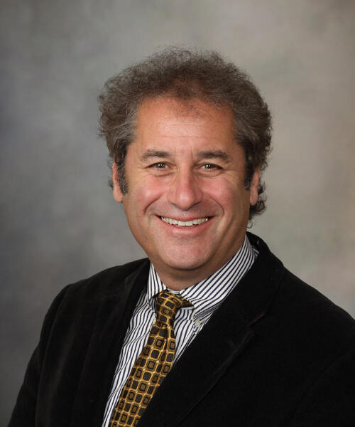 Fredric B. Meyer合作论文数Departments of Neurosurgery, Mayo Clinic;Center for Regenerative Biotherapeutics, Mayo Clinic;Mayo Clinic Comprehensive Cancer Center52
 Kai J. Miller合作论文数Department of Neurosurgery, University of Washington49
 Benjamin H. Brinkmann合作论文数Section of Nuclear Medicine, Mayo Clinic and Mayo Foundation49
 Colin L W Driscoll合作论文数Dept Otorhinolaryngol, Mayo Clin44Brian A. Neff合作论文数Department of Otolaryngology, Mayo Clinic41John L. D. Atkinson合作论文数39
 Irina Bancos合作论文数Division of Endocrinology, Diabetes, Metabolism, & Nutrition, Department of Internal Medicine, Mayo Clinic38
 Nicholas Gregg合作论文数Department of Neurology, Mayo Clinic37Dana Erickson合作论文数37
 Christopher S Graffeo合作论文数Department of Neurologic Surgery, Mayo Clinic36
 Vaclav Kremen合作论文数Faculty of Electrical Engineering, Czech Technical University in Prague36
 Matt Stead合作论文数36
 Jeffrey R Janus合作论文数35Stokken Janalee合作论文数34
 Yuki Shinya合作论文数Department of Neurosurgery, The University of Tokyo Hospital33
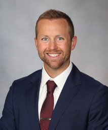 Carlstrom Lucas P合作论文数Mayo Graduate School, Mayo Clinic: College of Medicine33
 Panagiotis Kerezoudis合作论文数Dept Neurol Surg, Montefiore Med Ctr30W. Richard Marsh合作论文数29Eric J. Moore合作论文数25
 Aditya Raghunathan合作论文数24Avital Perry合作论文数23
 Caterina Giannini合作论文数Comprehensive Cancer Center, Mayo Clinic;Department of Laboratory Medicine & Pathology, Mayo Clinic;Dipartimento di Scienze Biomediche e Neuromotorie, University of Bologna23
 Jeffrey W Britton合作论文数23Grewal Sanjeet S合作论文数Department of Surgery, University of Cincinnati Medical Center21Paul A. Gardner合作论文数Department of Neurological Surgery, University of Pittsburgh School of Medicine21Vladimir Sladky合作论文数20
 John P Marinelli合作论文数Brooke Army Medical Center20Christine Lohse合作论文数Mayo Clinic20
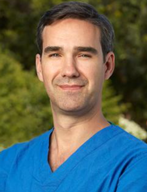 Juan Carlos Fernandez-Miranda合作论文数Department of Neurosurgery, Stanford University School of Medicine;Skull Base and Pituitary Surgery Division, Stanford University School of Medicine20Megan M J Bauman合作论文数19
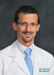 Siviero Agazzi合作论文数Department of Neurosurgery, University of South Florida19
 Erin K O'Brien合作论文数Department of Otolaryngology – Head & Neck Surgery, Mayo Clinic College of Medicine18
 Carlos Pinheiro-Neto合作论文数Department of surgery, Albany Medical Center18
 Zwagerman Nathan T合作论文数Children's Hospital of Pittsburgh, University of Pittsburgh Medical Center18
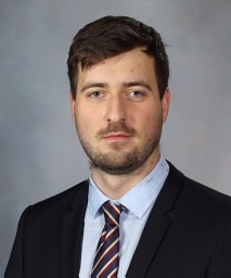 Filip Mivalt合作论文数Faculty of Electrical Engineering and Communication, Brno University of Technology17
 Gregory D. Cascino合作论文数Department of Neurology, Mayo Clinic17
 Lily Wong-Kisiel合作论文数17Carlos D Pinheiro-Neto合作论文数16
 Kendall H Lee合作论文数1Department of Neurologic Surgery, Mayo Clinic16
 Harry R. Van Loveren合作论文数Department of Neurosurgery and Brain Repair, College of Medicine, University of South Florida 16
 John Ignatius Lane合作论文数16
 Keith Starnes合作论文数15
 Inyong Kim合作论文数Bioelect Neurophysiol & Engn Lab, Mayo Clin15




