The instruction course program will again include some interactive courses. These courses use an interactive keypad
Background and Methods. The use of corticosteroids to treat optic neuritis is controversial. At 15 clinical centers, we randomly assigned 457 patients with acute optic neuritis to receive oral prednisone (1 mg per kilogram of body weight per day) for 14 days; intravenous methylprednisolone (1 g per day) for 3 days, followed by oral prednisone (1 mg per kilogram per day) for 11 days; or oral placebo for 14 days. Visual function was assessed over a six-month follow-up period.Results. Visual function recovered faster in the group receiving intravenous methylprednisolone than in the placebo group; this was particularly true for the reversal of visual-field defects (P = 0.0001). Although the differences between the groups decreased with time, at six months the group that received intravenous methylprednisolone still had slightly better visual fields (P = 0.054), contrast sensitivity (P = 0.026), and color vision (P = 0.033) but not better visual acuity (P = 0.66).The outcome in the oral-prednisone group did not differ from that in the placebo group. In addition, the rate of new episodes of optic neuritis in either eye was higher in the group receiving oral prednisone, but not the group receiving intravenous methylprednisolone, than in the placebo group (relative risk for oral prednisone vs. placebo, 1.79; 95 percent confidence interval, 1.08 to 2.95).Conclusions. Intravenous methylprednisolone followed by oral prednisone speeds the recovery of visual loss due to optic neuritis and results in slightly better vision at six months. Oral prednisone alone, as prescribed in this study, is an ineffective treatment and increases the risk of new episodes of optic neuritis.
Venous stasis retinopathy is an uncommon sign of carotid artery obstruction. It is found in those patients who have very severe stenosis, or complete occlusion, of the arterial lumen. Affected patients usually have advanced multivessel disease with compromise of both carotid arteries. The retinopathy is ipsilateral to the more severely compromised artery; it is caused by progressive, chronic hypoxic injury to retinal tissues, which causes an ophthalmoscopic picture of venous dilation and tortuosity, retinal hemorrhages, macular edema, and eventual neovascular proliferation. The retinal findings are quite similar to those found in diabetic retinopathy, except for unilaterality corresponding to the more obstructed artery and early onset in the retinal midzone rather than the posterior pole. The primary treatment is panretinal photocoagulation to ischemic areas, followed by either medical or surgical management of obstructive carotid disease or carotid endarterectomy alone.
Enlargement of the optic nerve and chiasm as demonstrated by high-resolution CT scanning or magnetic resonance imaging is often assumed to indicate tumor. However, this is not always true. The mode of clinical presentation of the disease process must be correctly applied to the interpretation of the radiographic studies. In the case illustrated here, neuro-ophthalmologic evaluation prevented an unnecessary biopsy in a patient whose presentation was atypical for anterior visual pathway glioma and whose clinical course was otherwise compatible with optic neuritis.
Our website uses cookies to enhance your experience. By continuing to use our site, or clicking "Continue," you are agreeing to our Cookie Policy | Continue
A patient presented with an 8-month history of a progressive left homonymous visual field deficit, left hemiparesis, and a left thalamocortical sensory deficit that was not detectable by repeated conventional neurodiagnostic evaluations. Proton nuclear magnetic resonance (NMR) imaging revealed a right parietal lesion characterized by a prolonged T2 (spin-spin relaxation time). At surgery, the mass proved to be an anaplastic astrocytoma. NMR appears to be more sensitive than x-ray computerized tomography scanning in some patients with malignant gliomas and offers the clinician an additional probe with which to evaluate these patients.
Members of the Advisory Committee, in conjunction with the Program Committee and the Academy's Board of Directors, are continually exploring ways to improve the educational value of the instruction program and ways of encouraging the largest possible number to participate in our educational courses at the least expense.
Chlorpromazine is frequently administered to patients with hyperprolactinemia to stimulate an increase in the serum levels of prolactin. A patient with a prolactin secreting adenoma is described in whom pituitary apoplexy developed in association with a hypotensive episode following the administration of 25 mg of chlorpromazine. Prolactin levels fell from more than 2,000 ng/ml to 340 ng/ml following infarction of the pituitary tumor. Pituitary apoplexy should be considered as a rare complication of chlorpromazine stimulation in a patient with a pituitary tumor.
 Ana Berlin合作论文数2Robert B Kalina合作论文数2J. James Rowsey合作论文数2Carl Migliazzo合作论文数2
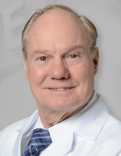 Jerry Shields合作论文数Wills Eye Hospital;Sidney Kimmel Medical College, Thomas Jefferson University2
 Michael Marmor合作论文数Department of Environmental Medicine, Grossman School of Medicine, New York University2
 Roy W. Beck合作论文数Jaeb Center for Health Research2Edward G. Buckley合作论文数Eye Center, Duke University2Jonathan D Trobe合作论文数2William Benson合作论文数2
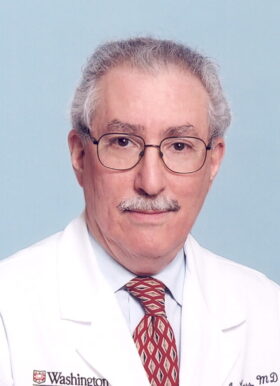 Michael Kass合作论文数Kass Lab, Department of Ophthalmology & Visual Sciences, School of Medicine in St.Louis, Washington University2Peter J. Savino合作论文数2
 Jack T Holladay合作论文数2David J. Schanzlin合作论文数2Joseph W. Sassani合作论文数2Melvin L. Rubin合作论文数2Milton Boniuk合作论文数2Alan B. Scott合作论文数2Herbert E. Kaufman合作论文数2
 Peter Robert Laibson合作论文数Wills Eye Hospital2W. Sanderson Grizzard合作论文数2San Francisco合作论文数1New Orleans合作论文数1Oklahoma City合作论文数1Myron Anoff合作论文数1Ronald S. Fishman合作论文数1Sheridan Lam合作论文数1Frederick Blodi合作论文数1Kenneth L. Cohen合作论文数1Malcolm Luxenberg合作论文数1Stuart Fourman合作论文数1Howard Barnebey合作论文数1New York合作论文数1R W DARRELL合作论文数1K Ossoinig合作论文数1Bradley F. Jost合作论文数1George Baerveldt合作论文数1Alan Kimura合作论文数1Stephen R Russell合作论文数1William H. Knobloch合作论文数1Gordon R. Douglas合作论文数1Marc F Lieberman合作论文数1Po Kohler合作论文数1Karla J. Johns合作论文数1Larry E. Magargal合作论文数1Bj Katz合作论文数1Jeffrey A. Jackson合作论文数1Alex E. Jalkh合作论文数1merrill grayson合作论文数1




