Study Design:Retrospective cohort.Summary of Background Data:Although fusion surgery is the established recommendation for degenerative lumbar spondylolisthesis (DLS) with instability, a decompression alone might be needed in some cases based on the patient's age, comorbidity burden, surgical fitness, and preference.Objective:To analyze the outcomes of minimally invasive decompression alone in patients with L4-5 DLS and translational motion >= 2 mm and compare with fusion over short term.Methods:Patients who underwent minimally invasive decompression or fusion for L4-5 DLS with translational motion >= 2 mm and had a minimum of 1-year follow-up (maximum follow-up of 2 y) were included. Postoperative improvement in patient-reported outcome measures (PROMs) was analyzed. The decompression and fusion groups were compared for improvement in PROMs, minimal clinically important difference (MCID), patient acceptable symptom state (PASS), and response on global rating change (GRC) scale.Results:Eighty-four patients were included, out of which 60 (71.4%) underwent fusion. The decompression group had a significantly higher average age compared with fusion (69.3 vs. 64.8 y, P=0.036). There was no significant difference between the groups in other demographic variables and preoperative PROMs. The decompression group showed significant improvement in PROM postoperatively. The decompression group had a comparable magnitude of improvement in PROMs and MCID and PASS achievement rates as fusion over short term follow-up. More than 80% of patients reported feeling better compared with preoperative at both the timepoints with no significant difference in the responses between the 2 groups.Conclusion:Minimally invasive decompression alone does lead to significant postoperative improvement over the short term and may be considered as an option in patients with unstable spondylolisthesis where fusion cannot be done. However, these are preliminary results and future research with a larger sample size and longer follow-up is required to further investigate this topic.Level of Evidence:Level III.
STUDY DESIGN:Retrospective cohort study. OBJECTIVE:To evaluate the impact of heterotopic ossification (HO) severity on segmental and global cervical range of motion (ROM) following cervical disc replacement (CDR). SUMMARY OF BACKGROUND DATA:Heterotopic ossification is the most commonly reported complication following CDR, with rates ranging from 40% to over 70% at mid- to long-term follow-up. While high-grade HO is known to restrict motion, the dose-response relationship between HO severity and ROM across multiple spinal levels remains incompletely characterized. METHODS:Patients who underwent single-level CDR with minimum 1-year follow-up and flexion-extension radiographs were identified from a single-institution database. HO was graded per McAfee (Grade 0-4) and patients stratified into Grade 0-2 (non-severe HO), Grade 3 (severe HO), and Grade 4 (severe HO with bridging ankylosis). ROM was measured at the operative disc space and C2-7 levels at final follow-up. Pairwise Mann-Whitney comparisons were Bonferroni-corrected across six planned tests (three disc space, three C2-7). Reported pairwise P values reflect adjustment. Kruskal-Wallis omnibus tests were not adjusted. RESULTS:A total of 146 patients met inclusion criteria (mean age 45.6±9.0 y, 63.7% male, mean follow-up 2.8±1.9 y). Grade 0-2 HO was present in 88 (60.3%), Grade 3 in 47 (32.2%), and Grade 4 in 11 (7.5%). Disc space ROM differed significantly across groups (Kruskal-Wallis P <0.001): Grade 0-2 mean 9.1°±5.4°, Grade 3 mean 6.2°±4.2° (P=0.009), Grade 4 mean 1.3°±2.0° (P <0.001). C2-7 ROM did not differ significantly across HO groups (Kruskal-Wallis P=0.090), with all pairwise comparisons non-significant. CONCLUSION:Higher-grade HO following CDR is associated with stepwise reduction in disc space ROM. Grade 3 HO significantly reduces segmental motion compared to Grade 0-2, and Grade 4 HO leads to near-complete segmental motion loss. Regardless of HO severity, C2-7 ROM is preserved, potentially suggesting transfer of motion to adjacent segments. LEVEL OF EVIDENCE:4.
STUDY DESIGN:Retrospective radiographic analysis of patients undergoing cervical disc replacement (CDR). OBJECTIVE:To assess the impact of anteroposterior endplate coverage on the development, location, and severity of heterotopic ossification (HO) following CDR, and to establish clinically actionable coverage thresholds. SUMMARY OF BACKGROUND DATA:Heterotopic ossification (HO) is a common complication following CDR that may limit the range of motion. While endplate coverage has been implicated as a risk factor, the direction-specific relationship between undercoverage location and HO location, as well as dose-response thresholds, remains poorly defined. METHODS:Patients undergoing CDR (2015-2024) with a minimum 1-year radiographic follow-up were retrospectively assessed (221 levels). Implant-to-endplate depth ratio (ID: ED) and exposed endplate ratio (EER) were measured on the first postoperative radiographs. HO was graded on final radiographs using the McAfee classification (0-4). Dose-response, direction-matched correlations, and ROC analyses were performed. RESULTS:Heterotopic ossification (HO) was present in 85.8% of levels, with 42.7% developing severe HO (grade 3-4). Posterior HO was more common than anterior (84.3% vs. 41.3%, P<0.001). ID: ED correlated inversely with HO grade at both endplates (P<0.001). Dose-response analysis demonstrated that severe HO rates increased from 22.2% (Q1) to 57.9% (Q4; RR=2.61, P<0.0001). Direction-matched analysis confirmed anterior undercoverage predicted anterior HO (r=0.422, P<0.0001) and posterior undercoverage predicted posterior HO (r=0.300, P<0.0001). ROC analysis identified an ID: ED ≤0.850 as the optimal threshold (AUC=0.668, P<0.001). CONCLUSIONS:Heterotopic ossification (HO) following CDR is strongly associated with inadequate endplate coverage. A direction-specific relationship exists whereby undercoverage location predicts HO location. Maintaining ID>85% and EER <15% may help limit severe HO risk. LEVEL OF EVIDENCE:Level III-prognostic study.
STUDY DESIGN:Retrospective cohort study. OBJECTIVES:To compare 90-day complication rates and clinical outcomes in patients undergoing minimally invasive lumbar fusion with and without inflammatory arthritis. SUMMARY OF BACKGROUND DATA:Patients with inflammatory arthritis are at special risk of developing spinal pathology. There is a paucity of literature investigating outcomes following minimally invasive lumbar fusion in patients with inflammatory arthritis. METHODS:Patients who had undergone minimally invasive fusion between 2017 and 2023 for the treatment of degenerative pathology of the lumbar spine with a minimum of 6 months follow-up were included and separated into inflammatory arthritis (IA) or noninflammatory arthritis (non-IA) cohorts. Patient demographics, perioperative variables, 90-day complications, patient-reported outcome measures, and rates of MCID achievement were compared between the IA cohort and the unmatched and 3:1 case-control matched non-IA cohort at early (<6 mo) and late (≥6 mo) follow-up. RESULTS:Six hundred sixty-nine patients were included (IA cohort, n=23; non-IA cohort, n=646). Patient demographics and perioperative characteristics did not differ significantly between the two cohorts except for IA patients reporting a higher age-adjusted Charlson comorbidity index (P=0.004). There were no significant differences in 90-day complications, reoperations, or readmissions. The IA cohort demonstrated significant improvements in most PROMs at the early and late postoperative time points. PROM scores, magnitudes of improvement in PROMs, MCID achievement rates, and time to achieve MCID for most PROMs were not significantly different between the 2 cohorts. After 3:1 case-control matching, there were no significant differences in complications, reoperations or admissions. In addition, there were no significant differences between the two cohorts in most PROM scores or rates of MCID achievement. Magnitudes of improvement and time to achieve MCID were comparable between the two cohorts for all PROMs. CONCLUSION:Patients with inflammatory arthritis undergoing minimally invasive lumbar fusion have similar clinical outcomes as compared with controls. LEVEL OF EVIDENCE:Level III.
STUDY DESIGN:Retrospective cohort study. OBJECTIVE:To determine whether coronal implant position and implant alignment on first postoperative radiographs predict flexion-extension range of motion (ROM) following cervical disc replacement (CDR). SUMMARY OF BACKGROUND DATA:CDR is a motion-preserving alternative to anterior cervical discectomy and fusion. While sagittal alignment factors have been studied, the impact of coronal implant positioning on ROM preservation remains poorly understood. METHODS:This retrospective radiographic study evaluated 236 CDR levels at a single academic institution. On first postoperative anteroposterior radiographs, we measured superior and inferior anatomic axis deviation (AAD), superior and inferior mechanical axis deviation (MAD), disc level deviation (DLD), and plumbline deviation (PD). Flexion-extension ROM was assessed at final follow-up at the disc space (DS ROM), functional spinal unit (FSU ROM), and C2-7 levels. Pearson correlations, multivariable linear regression adjusted for age, sex, and body mass index, dose-response quartile analysis, and receiver operating characteristic curve analysis were performed. RESULTS:The mean follow-up was 1.8±1.7 years. Superior MAD demonstrated a significant negative correlation with FSU ROM (r=-0.149, P=0.023), which persisted after multivariable adjustment (β=-0.36, P=0.032). Dose-response analysis revealed that patients in the best alignment quartile had significantly higher FSU ROM than those in the worst quartile (10.2 vs. 8.0 degrees, P=0.046). Every 3 degrees of MAD corresponded to ∼1 degree of FSU ROM loss. Superior MAD >3.8 degrees was associated with a 2.27-fold increased odds of poor FSU ROM (P=0.024). CONCLUSIONS:Coronal MAD significantly impacts FSU ROM following CDR. Surgeons should target MAD <3 degrees to optimize motion preservation. LEVEL OF EVIDENCE:Level III-therapeutic study.
STUDY DESIGN:Retrospective review of electronic health records system. OBJECTIVE:To compare the indications and complication rates between rhBMP-2 and non-rhBMP-2 use in spine fusion at a high-volume tertiary orthopedic institution. SUMMARY OF BACKGROUND DATA:Bone morphogenetic proteins are growth factors that play a critical role in bone development and proliferation. rhBMP-2 is a biological approved for select spinal fusion procedures to improve fusion rates. It is used in off-label indications. There are limited studies describing the use of rhBMP-2 in current practice. MATERIALS AND METHODS:Patients who underwent spinal fusion from February 2016 to January 2022 were included. Surgery types were categorized by the approach listed as the primary procedure. A subgroup analysis was performed to assess differences in rhBMP-2 use for short and long fusions. Complications present within one year as ICD-10 codes were assessed. RESULTS:A total of 9809 patients were assessed. rhBMP-2 was used in 0.3% of ACF and 64.5% of PCF surgeries. Patients who underwent PCF with rhBMP-2 had higher rates of osteoporosis. rhBMP-2 was used in 96.1% of AL/TF, 69.2% of PL/TF, 97.4% of LLIF, and 24.1% of TLIF. Patients who underwent PL/TF with rhBMP-2 had higher rates of osteoporosis, mean age, former smoking status, and ASA class. There were increased rates of wound dehiscence in PL/TF with rhBMP-2. Patients who underwent long fusions with rhBMP-2 were more likely to have osteoporosis than those who underwent short fusions without rhBMP-2. CONCLUSIONS:We found that rhBMP-2 was used extensively in AL/TF and LLIF, frequently in PCF and PL/TF, occasionally in TLIF, and sparingly in ACF. While patterns of use differed in the cervical spine, mostly selecting for patients with risk factors for pseudarthrosis, this pattern of usage was only found in posterior lumbar and thoracic fusion. There was no obvious trend in complications associated with rhBMP-2 use.
BACKGROUND CONTEXT Cervical disc replacement (CDR) is a promising procedure for treating cervical degenerative disorders while preserving segmental motion. Randomized controlled trials and meta-analyses have demonstrated its noninferiority to anterior cervical discectomy and fusion (ACDF), with the added advantage of mitigating adjacent segment load caused by fusion. Additionally, CDR has shown faster recovery in several studies. However, the recovery phases have not been thoroughly studied. A deeper understanding of these recovery phases is essential to identify differences across procedures and optimize patient management. PURPOSE This study aims to describe postoperative recovery trajectories in CDR during the first postoperative year and to compare differences in recovery trajectories between single- and two-level procedures using a segmented regression model. STUDY DESIGN/SETTING Retrospective study. PATIENT SAMPLE Patients undergoing 1- or 2-level CDR. OUTCOME MEASURES Neck disability index (NDI) and numerical rating scale (NRS) for neck and arm. METHODS Demographic and surgical data were collected, including age, sex, race, BMI, number of surgical levels, and primary diagnosis (disc herniation, degenerative disc disease, or foraminal stenosis). PROMs were recorded at postoperative weeks 2, 6, 12, and months 6 and 12. Univariate analyses using t-tests and chi-square tests were conducted between 1-level and 2-level groups. A multivariable segmented regression model, adjusted for age, was used to evaluate improvement trends in each PROM, accounting for repeated measures. Improvement trends were described as slope (ß points/day) representing the effect size, and breakpoints were estimated where trends shifted between phases. RESULTS A total of 229 patients were included (1-level: 132, 2-level: 97). The 2-level group was significantly older than the 1-level group (47.1 vs 42.4 years, p < 0.001), with no other significant background differences. Segmented regression analysis identified two distinct recovery phases: Improvement Phase (IP) and Plateau Phase (PP). The breakpoint for overall recovery was estimated at day 58, with the slope plateauing afterward (-0.007). Multivariable segmented regression, adjusted for age, demonstrated no significant difference in NDI recovery trends between the level groups (slope mean difference: ß = 0.001, p = 0.86). Breakpoints for disability improvement occurred at day 53 and 57 for 1- and 2-level groups, respectively (p = 0.69). Recovery phases for neck and arm pain by NRS were also comparable between level groups, with breakpoints for arm pain at day 11 and day 15 and for neck pain at day 32 and day 22 for 1- and 2-level groups, respectively (all p > 0.05). CONCLUSIONS Recovery trajectories after CDR are similar regardless of the number of surgical levels. Improvements in neck and arm pain plateau at approximately 4 weeks and 2 weeks, respectively, while disability improvement continues until around 2 months postoperatively. These findings provide valuable insights for managing patient expectations and optimizing postoperative care. FDA Device/Drug Status This abstract does not discuss or include any applicable devices or drugs.
Study Design:Retrospective cohort study. Summary of Background Data:The optimal surgical approach for multilevel cervical stenosis in elderly patients is controversial because of the risk of life-threatening complication. Objective:To compare life-threatening early complication rates between >= 3 levels anterior and posterior cervical surgery in elderly patients. Methods:Data from the American College of Surgeons National Surgical Quality Improvement Program database (NSQIP) were queried for patients 65 years or older who underwent ACDF or PS between 2016 and 2021. Patients with subaxial cervical degenerative disorders were identified using ICD10 codes. Surgical procedures were defined with CPT code indicating ACDF (3-5 levels; ACDF3+), laminoplasty or laminectomy (3-5 levels), and posterior decompression and fusion (3-5 levels). Outcomes of interest including reoperation, airway complications, venous thromboembolism (VTE), surgical site infections (SSIs), and urinary tract infections (UTI) were compared by utilizing 1:1 propensity score matching between the 2 approaches. Results:We identified 568 patients who underwent ACDF3+ and 1590 patients who underwent PS. After propensity score matching, the cohorts with 568 patients each were well-balanced with a mean age of 70.9 years. All 17 patients with dialysis and 24 of 28 patients with congestive heart failure in the PS group before the matching were excluded through the matching process. Complications rates between ACDF3+ and PS group were similar in reoperation (2.1% vs. 3.3%; P=0.275), airway complications (0.9% vs. 0.9%; P=1.000), and VTE (1.1% vs. 0.7%; P=0.751), whereas the PS group had a higher rate of SSI (0.2% vs. 1.4%; P=0.045), UTI (1.1% vs. 3.0%; P=0.035), and LOS (2.5 +/- 6.1 vs, 4.3 +/- 3.9 d; P<0.001). Conclusions:Among elderly patients undergoing 3 or more levels of cervical spine surgery, there were comparable rates of 30-day life-threatening complications between the 2 approaches. However, potential selection bias exists, with surgeons possibly favoring posterior surgery for patients with higher-risk comorbidities.
STUDY DESIGN:Biomechanical cadaveric study. OBJECTIVE:Assess the accuracy and precision of robotically placed cervical pedicle screws (CPS). SUMMARY OF BACKGROUND DATA:Posterior cervical fusion (PCF) can be performed using lateral mass screws or pedicle screws. Although pedicle screw trajectories are longer and allow for the placement of larger screws, these screws must be placed accurately as they traverse a narrow corridor bordered by critical neurovascular structures. Robotic technology may have the potential to improve the safety and accuracy of CPS, which could facilitate the routine use of these biomechanically favorable screws in PCF. METHODS:A cadaveric study was performed to assess the accuracy of subaxial CPS placement using robotic-assisted navigation in 3 cadavers. Computed tomography (CT) scans were performed preoperatively, and trajectories were planned in Mimics software. Intraoperative fluoroscopic registration was performed, and bilateral pedicle screws were drilled and placed through the robotic end-effector from C3-C7. Postoperative CT scans were performed. Accuracy was defined as the mean absolute error (MAE), precision was defined as 2 SD, and systematic error was defined as the signed mean error (SME) when comparing the planned versus placed pedicle screw positions. An automated computer vision algorithm that measures postoperative deviation from the preoperative plan was used to assess the final tip, tail, and mid-pedicle screw position in 3 dimensions. RESULTS:A total of 30 screws were successfully placed without registration failures. When compared with the planned screw trajectory, the accuracy/deviation of the tail in the ML and SI dimensions was 1.76 mm and 1.53 mm, respectively. Accuracy of the mid-pedicle position in the ML and SI dimensions was 1.06 and 1.17. Accuracy of the tip in the ML and SI dimensions was 0.67 and 0.87 mm, respectively. Angular deviation was 3.50 degrees. CONCLUSIONS:Robotic-assisted posterior cervical pedicle screw placement is highly accurate.
OBJECTIVE When creating minimally invasive spine fusion constructs, accurate pedicle screw fixation is essential for biomechanical strength and avoiding complications arising from delicate surrounding structures. As research continues to analyze how to improve accuracy, long-term patient outcomes based on screw accuracy remain understudied. The objective of this study was to analyze long-term patient outcomes based on screw accuracy. METHODS This is a retrospective cohort study of patients who underwent 1- or 2-level minimally invasive transforaminal lumbar interbody fusion and were queried from a prospectively maintained multisurgeon registry. Pedicle screws were assessed for accuracy and graded as poor, acceptable, or good. Patient demographic characteristics and outcomes including complications, patient-reported outcome measures (PROMs), return to activities, and fusion rates were compared between the cohorts. RESULTS A total of 665 pedicle screws in 153 patients were evaluated and included in the final analysis. Of these, 20 (13.1%) patients had poor screws, 63 (41.2%) had acceptable screws, and 70 (45.7%) had good screws. All groups showed similar and significant improvements in all PROMs, although the poor screw group experienced delayed improvement in physical function. A majority of patients in all groups returned to working and driving and discontinued narcotics at similar rates. However, the poor screw group displayed significantly slower return to activities. There were no significant differences in intraoperative or postoperative complications, although the poor screw group experienced significantly lower fusion rates. CONCLUSIONS Patients with poorly accurate pedicle screws experienced delayed return to activities and decreased fusion rates with similar long-term PROMs. Surgeons should continue to focus on placing accurate pedicle screws, and research should continue to analyze ways to ensure accurate screw placement.
BACKGROUND:Postoperative urinary retention (POUR), a common complication after spine surgery, can contribute to longer hospital stays, urinary tract infection, pain, and morbidity. This study aimed to determine the incidence of POUR in patients who underwent lumbar decompression and to construct a predictive model for preoperatively identifying high-risk patients. METHODS:This was a retrospective review of patients undergoing primary lumbar decompression from 2017 to 2023. Demographic characteristics, comorbidities, and perioperative data were collected. Factors associated with POUR were assessed, and multivariable logistic regressions were performed to identify independent predictors of the development of POUR. A nomogram to predict the development of POUR was developed within a training subset, based on a multivariable logistic regression model of preoperative variables, followed by the internal validation of the model in a validation subset and assessment of its performance. RESULTS:Of the 1,938 patients included in this study, 133 (6.9%) developed POUR. Following multivariable analysis, the following risk factors for POUR were identified: a history of urinary retention (odds ratio [OR], 4.956 [95% confidence interval (CI), 2.157 to 11.383]; p < 0.001), insurance that was not commercial (private) (OR, 2.256 [95% CI, 1.298 to 3.922]; p = 0.004), intraoperative Foley catheter use (OR, 5.967 [95% CI, 3.506 to 10.156]; p < 0.001), inpatient opioid consumption of >93 morphine milligram equivalents (OR, 1.898 [95% CI, 1.220 to 2.952]; p = 0.004), and anticholinergic medication use during hospitalization (OR, 3.450 [95% CI, 2.313 to 5.148]; p < 0.001). The nomogram, which included the preoperative variables of male sex, age of >65 years, history of urinary retention, history of benign prostatic hyperplasia, not having commercial insurance, and American Society of Anesthesiologists (ASA) classification of >2, demonstrated good discrimination in the training subset (area under the curve [AUC], 0.725 [95% CI, 0.673 to 0.776]) and the validation subset (AUC, 0.709 [95% CI, 0.599 to 0.819]). The Hosmer-Lemeshow goodness-of-fit test demonstrated that the model fit the data well (chi-square test = 9.063; p = 0.170). CONCLUSIONS:The incidence of POUR after lumbar decompression surgery was found to be 6.9%. A history of urinary retention, not having commercial (private) insurance, intraoperative Foley catheter usage, inpatient opioid consumption of >93 morphine milligram equivalents, and the administration of anticholinergic medication during hospitalization increased the risk of developing POUR. Furthermore, we constructed a preoperative predictive model with good performance metrics to help clinicians to identify patients at elevated risk for developing POUR. LEVEL OF EVIDENCE:Prognostic Level III . See Instructions for Authors for a complete description of levels of evidence.
Study Design. Retrospective cohort. Objective. To study the impact of class 2/3 obesity (body mass index, BMI >= 35) on outcomes following minimally invasive decompression. Summary of Background Data.No previous study has analyzed the impact of class 2/3 obesity on outcomes following minimally invasive decompression. Materials and Methods. Patients who underwent primary minimally invasive decompression were divided into four cohorts based on their BMI: normal (BMI: 18.5-<25), overweight (25-<30), class 1 obesity (30-<35), and class 2/3 obesity (BMI >= 35). Outcome measures were: 1) intraoperative variables: operative time, estimated blood loss (EBL); 2) patient-reported outcome measures (PROMs) (Oswestry disability index, ODI; visual analog scale, VAS back and leg; 12-Item Short Form Survey Physical Component Score, SF-12 PCS); 3) global rating change (GRC), minimal clinically important difference (MCID), and patient acceptable symptom state (PASS) achievement rates; 4) return to activities; and 5) complication and reoperation rates. Results. Totally, 838 patients were included (226 normal, 357 overweight, 179 class 1 obesity, and 76 class 2/3 obesity). Class 1 and 2/3 obesity groups had significantly greater operative times compared with the other groups. Class 2/3 obesity group had worse ODI, VAS back and SF-12 PCS preoperatively, worse ODI, VAS back, VAS leg and SF-12 PCS at below six months, and worse ODI and SF-12 PCS at above six months. However, they had significant improvement in all PROMs at both postoperative timepoints and the magnitude of improvement was similar to other groups. No significant differences were found in MCID and PASS achievement rates, likelihood of betterment on the GRC scale, return to activities, and complication/reoperation rates. Conclusions. Class 2/3 obese patients have worse PROMs preoperatively and postoperatively. However, they show similar improvement in PROMs, MCID and PASS achievement rates, likelihood of betterment, recovery kinetics, and complication/reoperation rates as other BMI groups following minimally invasive decompression.
STUDY DESIGN/SETTING:Retrospective review of prospectively collected data. OBJECTIVE:To assess the impact of erector spinae plane (ESP) blocks on recovery kinetics and patient-reported outcomes (PROMs) following minimally invasive transforaminal lumbar interbody fusion (MI-TLIF). SUMMARY OF BACKGROUND DATA:ESP block continues to emerge as a safe and effective regional anesthetic technique and has been shown to reduce immediate postoperative pain and opioid requirements, although its impact on patient recovery and outcomes farther out postoperatively remains unclear. METHODS:Patients who underwent single-level MI-TLIF who did or did not receive ESP blocks performed by a single anesthesiologist were included. PROMs and MCID achievement rates for ODI, VAS-Back, VAS-Leg, and SF-12 PCS were determined within early (within 3 mo) and late (6 mo to 2 y) postoperative periods. Return-to-activities (RTA) metrics included driving, work, and discontinuation of opioids which were measured by days after surgery. Multivariable regression analyses were performed to determine factors associated with return-to-activities. RESULTS:A total of 172 patients were included (mean age: 60.5 y, 46.5% female); 110 received ESP blocks and 62 did not. There were no differences in baseline characteristics between the cohorts. On univariate analysis, there were no differences in days to RTA between the cohorts. On multivariable analyses, ESP block was identified as a predictor for return-to-work by 30 days (OR 4.48, 95% CI [1.25-1.60], P=0.021), discontinuation of opioids by 15 days (OR: 2.40, 95% CI [1.08-5.35], P=0.032), and discontinuation of opioids by 30 days (OR: 5.57, 95% CI [1.87-16.56], P=0.002). There were no significant differences in change in PROM scores or MCID achievement rates between the cohorts. CONCLUSIONS:Our study demonstrated that ESP block administered prior to MI-TLIF may be associated with earlier return-to-work and discontinuation of opioids, while having limited impact on functional PROMs, MCID achievement rates, and GRC scores.
Retrospective review of prospectively collected data. To evaluate the efficacy of the 5-factor modified frailty (mFI-5) index in predicting patient outcomes and recovery kinetics in minimally invasive decompression (MI-decompression) for the treatment of degenerative lumbar disorders. The mFI-5 index, which measures frailty or the age-associated decline in recovery ability following a significant stressor, has risen as a patient-specific indicator for postoperative morbidity and mortality. However, no studies have evaluated the utility of frailty in predicting outcomes after MI-decompression for degenerative lumbar pathologies. Patients who underwent primary one- or two-level MI-Decompression for degenerative lumbar spine conditions were included. Patients were stratified into three groups: non-frail (mFI-5=0), moderately frail (mFI-5=1), and severely frail (mFI-5≥2). Outcome measures included surgical details, complications, return to activity, and patient reported outcome measures at preop and ≥6 months postop. A total of 956 patients were included, 438 non-frail, 418 moderately frail, and 100 severely frail. Frail patients (mFI-5 ≥1) were older (P<0.001), had greater BMI (P=0.047), elevated CCI (P<0.001), and greater percentages of ASA class ≥3 (P<0.001). Higher severities of frailty were associated with longer operative times (P<0.001) and lengths of stay (P<0.001), while blood loss was similar. Frail patients demonstrated worse preoperative symptoms, including ODI (P=0.019), VAS-back pain (P=0.019), and SF12-PCS (P=0.015), which continued postoperatively, including ODI (P=0.014) and SF12-PCS (P<0.001). The magnitude of improvement between outcome metrics was comparable at ≥ 6-month follow-up. Return to activities-driving, working, and discontinuation of narcotics-as well as all complication categories-intraoperative, in-hospital, and postoperative-were similar across all patients. MI-decompression provides comparable symptom relief and postoperative recovery, even among patients with significant frailty that could limit their ability to withstand the stresses of spine surgery.
STUDY DESIGN:Retrospective cohort. SUMMARY OF BACKGROUND DATA:Although fusion surgery is the established recommendation for degenerative lumbar spondylolisthesis (DLS) with instability, a decompression alone might be needed in some cases based on the patient's age, comorbidity burden, surgical fitness, and preference. OBJECTIVE:To analyze the outcomes of minimally invasive decompression alone in patients with L4-5 DLS and translational motion ≥2 mm and compare with fusion over short term. METHODS:Patients who underwent minimally invasive decompression or fusion for L4-5 DLS with translational motion ≥2 mm and had a minimum of 1-year follow-up (maximum follow-up of 2 y) were included. Postoperative improvement in patient-reported outcome measures (PROMs) was analyzed. The decompression and fusion groups were compared for improvement in PROMs, minimal clinically important difference (MCID), patient acceptable symptom state (PASS), and response on global rating change (GRC) scale. RESULTS:Eighty-four patients were included, out of which 60 (71.4%) underwent fusion. The decompression group had a significantly higher average age compared with fusion (69.3 vs. 64.8 y, P =0.036). There was no significant difference between the groups in other demographic variables and preoperative PROMs. The decompression group showed significant improvement in PROM postoperatively. The decompression group had a comparable magnitude of improvement in PROMs and MCID and PASS achievement rates as fusion over short term follow-up. More than 80% of patients reported feeling better compared with preoperative at both the timepoints with no significant difference in the responses between the 2 groups. CONCLUSION:Minimally invasive decompression alone does lead to significant postoperative improvement over the short term and may be considered as an option in patients with unstable spondylolisthesis where fusion cannot be done. However, these are preliminary results and future research with a larger sample size and longer follow-up is required to further investigate this topic. LEVEL OF EVIDENCE:Level III.
BACKGROUND CONTEXT Postoperative clinical outcomes after lumbar spine surgery are often evaluated using the Oswestry Disability Index (ODI), with quantitative metrics such as the minimal clinically important difference (MCID) and the patient’s acceptable symptom state (PASS). However, these metrics fail to reflect “qualitative” patient satisfaction, particularly among the subset of patients describing improvements but not achieving MCID or PASS. PURPOSE To identify different “themes” regarding postoperative clinical satisfaction following minimally invasive lumbar surgery in patients who improved on global rating of change (GRC) but did not achieve ODI MCID or PASS thresholds. STUDY DESIGN/SETTING Retrospective cohort study with a qualitative component for prospectively collected data at a single institution setting. PATIENT SAMPLE A total of 956 patients who underwent lumbar surgery with a minimum follow-up of 6 months were evaluated. Of these, 90 patients met the inclusion criteria (ie, improved on GRC and did not achieve ODI MCID or PASS), and 43 of these patients consented to participate in semi-structured interviews. OUTCOME MEASURES Patient-reported outcomes (PROMs) included ODI, visual analog scale (VAS) for back and leg pain, SF-12 physical and mental component (SF-12 PCS, SF-12 MCS), and PROMIS Physical Function scores (PROMIS PF). Satisfaction was assessed with GRC. METHODS Quantitative changes were evaluated at early (<6 months) and late (≥6 months) follow-ups. Chi-squared for categorical and independent t-tests for continuous variables were utilized. Wilcoxon Signed-Rank tests compared early and late paired GRC, while paired t-tests assessed changes in PROMs. Qualitative semi-structured interviews exploring satisfaction across physical, emotional, and social “themes,” were analyzed using grounded theory. Venn diagrams were utilized to signify the overlap between different qualitative responses. RESULTS Satisfaction on the GRC scale significantly improved between early and late follow-ups (p < 0.001). High MCID achievement rates for VAS back (79.1%) and leg pain (74.4%) were observed. There was a significant improvement in the VAS back score at 1 year (-2.8 ± 3.4, p = 0.011) compared to the earlier time point. Thematic qualitative analysis reported key contributors to satisfaction: physical improvement, quality of life, healthcare provider interactions, and family support. Overlaps, such as between physical health and quality of life, highlighted the multidimensional nature of postoperative clinical satisfaction. CONCLUSIONS Patient satisfaction extends beyond quantitative scales like the ODI. Holistic assessment, including subjective factors such as emotional well-being, healthcare provider interactions, and family support, are prerequisite and quintessential factors for evaluating recovery outcomes after lumbar surgery. FDA Device/Drug Status This abstract does not discuss or include any applicable devices or drugs.
Single-center, survey-based study. This study aims to assess patient experiences in ambulatory lumbar spine surgery, with a focus on their expectations and preferences regarding discharge disposition, as well as the impact of discharge timing and alignment with patient preferences on satisfaction and early recovery outcomes. While the safety and efficacy of ambulatory spine surgery have been well established, patient perceptions and experiences with these accelerated recovery pathways remain underexplored. A custom survey, designed by the authors, assessed patient experiences, preferences, and satisfaction related to discharge disposition. Respondents were asked to reflect on their surgical experience, focusing on their preferences for discharge timing and the reasons behind those preferences. The survey was administered to adult patients who underwent primary single-level minimally invasive transforaminal lumbar interbody fusion (MI-TLIF) or laminectomy (MI-Lami) and were discharged either on the same day of surgery (SDD) or after an overnight hospital stay (OVN). Patient preferences were qualitatively analyzed to identify recurring themes, while their postoperative night one experience was quantitatively scored across multiple domains, including sleep quality, comfort, hygiene management, and overall satisfaction. These data were then used to explore the relationship between discharge timing, and preference concordance. A total of 227 responses were collected, with 64 (28.2%) undergoing MI-TLIF and 163 (71.8%) undergoing MI-Lami. Of these, 116 (51.1%) were discharged on the same day (SDD) and 111 (48.9%) had an overnight stay (OVN). While 36% preferred SDD and 49.8% preferred OVN, 56.9% reported feeling ready for same-day discharge. Qualitative analysis revealed that SDD preferences were driven by comfort at home, better sleep, and avoiding hospital inconveniences. Conversely, OVN preferences emphasized medical support, pain control, and logistical ease. Patients discharged the same day reported better care, sleep, hygiene, and comfort (P<0.05 for all). Those discharged in concordance with their preferences reported significantly lower rates of feeling discharged prematurely (3.4% vs. 32.3%; P<0.001) and superior outcomes across multiple domains, including pain control, comfort, and reduced burden on families (P<0.05 for all). SDD patients reported improved postoperative experiences, including sleep and satisfaction. Aligning discharge timing with patient preferences further enhanced outcomes, emphasizing the importance of patient-centered discharge planning in spine surgery.
Retrospective analysis To compare clinical outcomes of minimally invasive transforaminal lumbar interbody fusion (MIS-TLIF) among patients with different radiographical difficult spondylolisthesis. High-grade vertebral slip, collapsed disc, and less mobile segment can complicate segmental parameter restoration in MIS-TLIF. Few studies have examined if MIS-TLIF can achieve comparable outcomes in these challenging cases. This study included patients who underwent single-level MIS-TLIF for degenerative spondylolisthesis. Radiographical difficulty was defined using a “difficulty point” system: disc height (<5 mm=1point), angular flexibility at the operated level by preoperative flexion-extension standing X-ray (<5degrees=1point), and slippage rate (>25%=1 point). Patients were categorized into “simple” (0 point), “moderate” (1 point), and “difficult” (2-3 points) cohorts. Outcomes included radiographic parameters (postoperative disc height, segmental lordosis restoration, and slippage reduction), complications, patient-reported outcomes such as Oswestry Disability Index (ODI) from 2 weeks to 1 year postoperatively. A total of 208 patients were included, with 52 in the difficult, 89 in the moderate, and 67 in the simple. The difficult cohort showed significantly greater restoration of disc height (161.0% vs. 26.5% vs. 9.3%, P<0.001), resulting in similar postoperative disc height (8.3 mm vs. 9.0 mm vs. 0.1 mm, P=0.10) and segmental lordosis at the operated level (14.5 vs. 13.0 vs. 12.5 degrees, P=0.17). Postoperative outcomes indicated a greater improvement trend in ODI in the difficult group compared to the moderate group (β= 7.3, 9=0.011), with similar minimal clinically important difference achievement rate in ODI at 1-year postoperatively (Difficult, 69.2% vs. Moderate, 62.8% vs. Simple, 54.3%, P=0.46). MIS-TLIF is a feasible treatment option in patients with technically challenging radiographic parameters, achieving comparable postoperative radiographical and clinical outcomes. 3
Retrospective cohort study. To assess the utility of whole-body imaging in diagnosing lower extremity osteoarthritis (LEOA), evaluate the association of LEOA with radiculopathy, and determine the impact of LEOA on postoperative recovery. Whole-body imaging offers rapid, low-radiation assessment of both spinal and lower extremity pathology. This modality may be pivotal in helping clinicians better understand how lower extremity pathology influences clinical presentation and recovery among patients with degenerative lumbar conditions. Patients with preoperative whole-body imaging who underwent lumbar spine surgery for degenerative conditions between 2017 and 2022 were included. The Kellgren and Lawrence (KL) score was implemented to grade joint osteoarthritis (OA) as mild (KL ≤2) or severe (KL ≥3). Length of stay (LOS) and side of radiculopathy were assessed. The Oswestry disability index (ODI), Patient-Reported Outcomes Measurement Information System Physical Function (PROMIS), visual analog scale (VAS), and Short Form-12 (SF-12) Physical/Mental Component Scales (PCS/MCS) were recorded at preoperative, early-postoperative (2, 6, 12 weeks), and late-postoperative (6, 12, 24 months) timepoints. The association of HOA/KOA with PROMs and minimum clinically important difference (MCID) was evaluated. Totally, 207 patients were included (HOA: n=130 mild, n=62 severe; KOA: n=141 mild, n=53 severe). Patients with severe OA were significantly older compared with patients with mild OA (P<0.05). OA laterality was not associated with side of radiculopathy. After controlling for age, LOS was ∼50% greater for severe HOA (P=0.031) and severe KOA (P=0.013) compared with mild OA. Severe KOA exhibited worse PROMIS at the early-postoperative timepoint (P=0.013), and worse PROMIS (P=0.049), VAS-back (P=0.009), and SF-12-PCS (P=0.025) at the late-postoperative timepoint. Severe HOA and KOA reduced the likelihood of achieving MCID for SF-12-PCS (OR: 0.44; P=0.049) and PROMIS (OR: 0.37; P=0.027), respectively. Whole-body imaging may help improve patient-specific counseling on expectations after lumbar spine surgery.
STUDY DESIGN:Single-center, survey-based study. OBJECTIVE:This study aims to assess patient experiences in ambulatory lumbar spine surgery, with a focus on their expectations and preferences regarding discharge disposition and the impact of discharge timing and alignment with patient preferences on satisfaction and early recovery outcomes. SUMMARY OF BACKGROUND DATA:While the safety and efficacy of ambulatory spine surgery have been well established, patient perceptions and experiences with these accelerated recovery pathways remain underexplored. METHODS:A custom survey, designed by the authors, assessed patient experiences, preferences, and satisfaction related to discharge disposition. Respondents were asked to reflect on their surgical experience, focusing on their preferences for discharge timing and the reasons behind those preferences. The survey was administered to adult patients who underwent primary single-level minimally invasive transforaminal lumbar interbody fusion (MI-TLIF) or laminectomy (MI-Lami) and were discharged either on the same day of surgery (SDD) or after an overnight hospital stay (OVN). Patient preferences were qualitatively analyzed to identify recurring themes, while their postoperative night one experience was quantitatively scored across multiple domains, including sleep quality, comfort, hygiene management, and overall satisfaction. These data were then used to explore the relationship between discharge timing and preference concordance. RESULTS:A total of 227 responses were collected, with 64 (28.2%) undergoing MI-TLIF and 163 (71.8%) undergoing MI-Lami. Of these, 116 (51.1%) were discharged on the same day (SDD) and 111 (48.9%) had an overnight stay (OVN). While 36% preferred SDD and 49.8% preferred OVN, 56.9% reported feeling ready for same-day discharge. Qualitative analysis revealed that SDD preferences were driven by comfort at home, better sleep, and avoiding hospital inconveniences. Conversely, OVN preferences emphasized medical support, pain control, and logistical ease. Patients discharged the same day reported better care, sleep, hygiene, and comfort ( P <0.05 for all). Those discharged in concordance with their preferences reported significantly lower rates of feeling discharged prematurely (3.4% vs. 32.3%; P <0.001) and superior outcomes across multiple domains, including pain control, comfort, and reduced burden on families ( P <0.05 for all). CONCLUSIONS:SDD patients reported improved postoperative experiences, including sleep and satisfaction. Aligning discharge timing with patient preferences further enhanced outcomes, emphasizing the importance of patient-centered discharge planning in spine surgery.
 Sheeraz Qureshi合作论文数Hosp Special Surg, 535 E 70th St, New York, NY 10021 USA95
 Sravisht Iyer合作论文数Hospital for Special Surgery, New York, NY, USA.94Kasra Araghi合作论文数1Department of Spine Surgery, Hospital for Special Surgery;and.;1Department of Spine Surgery, Hospital for Special Surgery78Sheha Evan D合作论文数72Tomoyuki Asada合作论文数University of Tsukuba61
 Pratyush Shahi合作论文数Department of Orthopaedic Surgery, Hospital for Special Surgery60
 Tejas Subramanian合作论文数Dept Biomed Engn, Vanderbilt Univ58Olivia Tuma合作论文数49Nishtha Singh合作论文数43
 Maximilian Korsun合作论文数33Eric Mai合作论文数32
 Avani S. Vaishnav合作论文数Hosp Special Surg, 535 E 70th St, New York, NY 10021 USA32Omri Maayan合作论文数30Eric Zhao合作论文数25Joshua Zhang合作论文数25
 Sumedha Singh合作论文数25
 Junho Song合作论文数Department of Orthopaedic Surgery, Hospital for Special Surgery24Daniel Shinn合作论文数23Cole Kwas合作论文数19
 Sidhant S Dalal合作论文数Hospital for Special Surgery, 535 East 70th Street, New York, NY 10021, USA.18
 Todd J. Albert合作论文数Weill Cornell Medical College;Hospital for Special Surgery18Eric Kim合作论文数15Amy Lu合作论文数15Kyle W Morse合作论文数15Cho Samuel K合作论文数15Lovecchio Francis C合作论文数15Jun Sup Kim合作论文数14Chad Z Simon合作论文数13Anthony Pajak合作论文数13Chad Simon合作论文数13
 Dimitra Melissaridou合作论文数13
 Troy B Amen合作论文数Department of Orthopaedic Surgery, NewYork-Presbyterian/Weill Cornell Medical Center13Annika Heuer合作论文数12Harvinder S. Sandhu合作论文数Hospital for Special Surgery, 535 East 70th Street, New York, NY 10021, USA12Yeo Eun Kim合作论文数10Russel C. Huang合作论文数10Robert K Merrill合作论文数9Austin C Kaidi合作论文数Hosp Special Surg, 535 E 70th St, New York, NY 10021 USA9
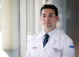 Fourman Mitchell S合作论文数Department of Orthopaedic Surgery, University of Pittsburgh Medical Center9
 Han Jo Kim合作论文数Hospital for Special Surgery;Orthopaedic Surgery Department, Weill Cornell Medical College9Izzet Akosman合作论文数8
 Overley Samuel C合作论文数Department of Orthopaedics, The Icahn School of Medicine at Mount Sinai8
 Bovonratwet Patawut合作论文数Department of Biomedical Engineering and the Translational Tissue Engineering Center, The Johns Hopkins University;Clarice Smith Building, 400 North Broadway, Baltimore, MD 21231, USA.;Department of Biomedical Engineering and the Translational Tissue Engineering Center, The Johns Hopkins University8
 Takashi Hirase合作论文数University of California, Los Angeles, University of California,UCLA8Kevin Phan合作论文数7Stephane Owusu-Sarpong合作论文数7
 John Di Capua合作论文数From the Department of Scholarly Engagement, Harvard Medical School;Department of Radiology, Massachusetts General Hospital;and Department of Radiology, Mayo Clinic Florida;Massachusetts General Hospital, Harvard Medical School7Gang Catherine H合作论文数7Myles R. J. Allen合作论文数6




