Diabetes mellitus is a metabolic disorder characterized by high blood sugar levels which give rise to complications in the eye, kidneys and the brain. Diabetes triggers the development of ocular diseases like diabetic retinopathy and cataracts which are the leading cause of blindness around the world. The most common method for the diagnosis of diabetes involves measuring the blood sugar levels in the body. One major disadvantage of this method is the fluctuating blood sugar levels which contribute to false negative results. This leads to delay in treatment, eventually causing permanent damage to the organs. Therefore, diagnosis of diabetes at an early stage is very crucial. One biomarker for diabetes related diseases is the formation of Advanced Glycation End-products (AGEs) that result from the Maillard reaction of proteins with glucose. α-crystallin in the ocular lens is a small heat shock protein with no protein turnover and hence acts as a record for post-translational modifications especially glycation which forms AGEs. We have used steady state and time resolved fluorescence measurements to study the spectroscopic changes in α-crystallin with increase in time of glycation and the intact lenses from diabetic and nondiabetic donors. Overall, this study was focused on developing a noninvasive diagnostic tool for early detection of diabetes mellitus.
Alpha crystallin, a small heat-shock protein, has been studied extensively for its chaperone function. Alpha crystallin subunits are expressed in stress conditions and have been found to prevent apoptosis by inhibiting the activation of caspase pathway. Non-enzymatic glycation of protein leads to the formation of advanced glycation end-products (AGEs). These AGEs bind to receptors and lead to blocking the signaling pathways or cause protein precipitation as observed in aggregation-related diseases. Methylglyoxal (MGO) is one of the major glycating agents expressed in pathological conditions due to defective glycolysis pathway. MGO reacts rapidly with proteins, forms AGEs and finally leads to aggregation. The goal of this study was to understand the non-enzymatic glycation-induced structural damage in alpha crystallin using biophysical and spectroscopic characterization. This will help to develop better disease models for understanding the biochemical pathways and also in drug discovery.
Purpose To obtain a better understanding of oxygen and nutrient transport within the lens, the viscosity of whole lenses was investigated using a fiber optic oxygen sensor (optode). The diffusion coefficient of oxygen was calculated using the Stokes-Einstein equation at the slip boundary condition. Methods The optode was used to measure the oxygen decay signal in samples consisting of different glycerol/water solutions with known viscosities. The oxygen decay signal was fitted to a double exponential decay rate equation, and the lifetimes (tau) were calculated. It was determined that the tau-viscosity relationship is linear, which served as the standard curve. The same procedure was applied to fresh bovine lenses, and the unknown viscosity of the bovine lens was calculated from the tau-viscosity relationship. Results The average viscosity in a whole bovine lens was determined to be 5.74±0.88 cP by our method. Using the Stokes-Einstein equation at the slip boundary condition, the diffusion coefficient for oxygen was calculated to be 8.2 × 10⁻⁶ cm2/s. Conclusions These data indicate a higher resistance to flow for oxygen and nutrients in the lens than what is currently assumed in the literature. Overall, this study allows a better understanding of oxygen transport within the lens.
Lipofuscin accumulates normally with age and is more pronounced in retinal dystrophies such as age-related macular degeneration. The major bis-retinoid component of lipofuscin is A2E. In addition to cell damage effects by A2E, we have previously demonstrated that blue-light-mediated A2E leads to modifications in the basement membrane protein laminin. Therefore, the purpose of this study was to advance the understanding of A2E photooxidation effects on fibronectin, the major glycoprotein of Bruch's membrane. In this study, A2E was irradiated with blue light in the presence of a fibronectin peptide consisting of amino acids from the integrin binding region. The modification sites were identified via LC/MS. Our research indicated that blue light irradiation caused cleavage throughout the A2E molecule closest to the pyridinium ring, and attached to the fibronectin peptide preferentially at lysine and arginine residues. All of these reactions are similar to the Maillard reaction. Altogether this study suggests that blue-light-irradiated A2E modifies peptides and forms advance glycation endproducts. Furthermore, these results can be used to identify modifications that occur in Bruch's membrane in vivo.
PURPOSETime and spectrally resolved measurements of autofluorescence have the potential to monitor metabolism at the cellular level. Fluorophores that emit with the same fluorescence intensity can be discriminated from each other by decay time of fluorescence intensity after pulsed excitation. We performed time-resolved autofluorescence measurements on fundus samples from a donor with significant extramacular drusen.METHODSTissue sections from two human donors were prepared and imaged with a laser scanning microscope. The sample was excited with a titanium-sapphire laser, which was tuned to 860 nm, and frequency doubled by a BBO crystal to 430 nm. The repetition rate was 76 MHz and the pulse width was 170 femtoseconds (fs). The time-resolved autofluorescence was recorded simultaneously in 16 spectral channels (445-605 nm) and bi-exponentially fitted.RESULTSRPE can be discriminated clearly from Bruch's membrane, drusen, and choroidal connective tissue by fluorescence lifetime. In RPE, bright fluorescence of lipofuscin could be detected with a maximum at 510 nm and extending beyond 600 nm. The lifetime was 385 ps. Different types of drusen were found. Most of them did not contain lipofuscin and exhibited a weak fluorescence, with a maximum at 470 nm. The lifetime was 1785 picoseconds (ps). Also, brightly emitting lesions, presumably representing basal laminar deposits, with fluorescence lifetimes longer than those recorded in RPE could be detected.CONCLUSIONSThe demonstrated differentiation of fluorescent structures by their fluorescence decay time is important for interpretation of in vivo measurements by the new fluorescence lifetime imaging (FLIM) ophthalmoscopy on healthy subjects as well as on patients.
The purpose of this study was to determine the transmission properties of the anterior segment of young primate eyes and potentially relate those changes to photochemical processes in the retina that lead to the early, rapid formation of lipofuscin. A simple method has been developed to determine the optical properties of the anterior segment of the intact eye. Using this technique, the transmission/absorption properties of primate cadaver eyes were determined. A young primate anterior segment has a maximum absorption at 365 nm due to the presence of the O‐β‐glucoside of 3‐hydroxykynurenine in the lens. This is synthesized in the last trimester of gestation. Although this compound filters out most of the UV light from reaching the retina, there is a small window of transmission centered on an absorption minimum at 320 nm. This closes by the second decade of life. The window of transmission of UV light to the primate retina may explain the initial accelerated formation of lipofuscin in the young human retina by a photochemical process. This would be exacerbated by any decrease in the ozone layer with concomitant increase in UV‐B reaching the earth’s surface.
The physical properties of membranes derived from the total lipids extracted from the lens cortex and nucleus of a 2-year-old cow were investigated using EPR spin-labeling methods. Conventional EPR spectra and saturation-recovery curves show that spin labels detect a single homogenous environment in membranes made from cortical lipids. Properties of these membranes are very similar to those reported by us for membranes made of the total lipid extract of 6-month-old calf lenses (J. Widomska, M. Raguz, J. Dillon, E. R. Gaillard, W. K. Subczynski, Biochim. Biophys. Acta 1768 (2007) 1454-1465). However, in membranes made from nuclear lipids, two domains were detected by the EPR discrimination by oxygen transport method using the cholesterol analogue spin label and were assigned to the bulk phospholipid-cholesterol domain (PCD) and the immiscible cholesterol crystalline domain (CCD), respectively. Profiles of the order parameter, hydrophobicity, and the oxygen transport parameter are practically identical in the bulk PCD when measured for either the cortical or nuclear lipid membranes. In both membranes, lipids in the bulk PCD are strongly immobilized at all depths. Hydrophobicity and oxygen transport parameter profiles have a rectangular shape with an abrupt change between the C9 and C10 positions, which is approximately where the steroid ring structure of cholesterol reaches into the membrane. The permeability coefficient for oxygen, estimated at 35 degrees C, across the bulk PCD in both membranes is slightly lower than across the water layer of the same thickness. However, the evaluated upper limit of the permeability coefficient for oxygen across the CCD (34.4 cm/s) is significantly lower than across the water layer of the same thickness (85.9 cm/s), indicating that the CCD can significantly reduce oxygen transport in the lens nucleus.
The physical properties of membranes derived from the total lipid extract of porcine lenses before and after the addition of cholesterol were investigated using EPR spin-labeling methods. Conventional EPR spectra and saturation-recovery curves indicate that the spin labels detect a single homogenous environment in membranes before the addition of cholesterol. After the addition of cholesterol (when cholesterol-to-phospholipid mole to mole ratio of 1.55-1.80 was achieved), two domains were detected by the discrimination by oxygen transport method using a cholesterol analogue spin label. The domains were assigned to a bulk phospholipid-cholesterol bilayer made of the total lipid mixture and to a cholesterol crystalline domain. Because the phospholipid analogue spin labels cannot partition into the pure cholesterol crystalline domain, they monitor properties of the phospholipid-cholesterol domain outside the pure cholesterol crystalline domain. Profiles of the order parameter, hydrophobicity, and oxygen transport parameter are identical within experimental error in this domain when measured in the absence and presence of a cholesterol crystalline domain. This indicates that both domains, the phospholipid-cholesterol bilayer and the pure cholesterol crystalline domain, can be treated as independent, weakly interacting membrane regions. The upper limit of the oxygen permeability coefficient across the cholesterol crystalline domain at 35 degrees C had a calculated value of 42.5 cm/s, indicating that the cholesterol crystalline domain can significantly reduce oxygen transport to the lens center. This work was undertaken to better elucidate the major factors that determine membrane resistance to oxygen transport across the lens lipid membrane, with special attention paid to the cholesterol crystalline domain. (C) 2008 Elsevier B.V. All rights reserved.
 Elizabeth R. Gaillard合作论文数27David C. Paik合作论文数7
 Mai Thao合作论文数5devi kalyan karumanchi合作论文数Department of Chemistry and Biochemistry, Northern Illinois University4
 Joan E. Roberts合作论文数Department of Natural Sciences, Fordham University4Lei Zheng合作论文数3john c merriam合作论文数2Jc Merriam合作论文数2
 Raguz Marija合作论文数Department of Biophysics, Medical College of Wisconsin;School of Medicine, University of Split;Medical College of Wisconsin, University of Split2
 Zilu Wang合作论文数Tianjin University, Tianjin Univ.2Perez Daniel合作论文数2
 Zhen Wang (王震)合作论文数School of Artifcial Intelligence, OPtics and ElectroNics, Northwestern Polytechnical University2
 M.David Tilson合作论文数School of Medicine, Yale University2Stephen J. Atherton合作论文数2L.S. Murdaugh合作论文数2Stephen R Russell合作论文数1Brian S. Fuchs合作论文数1P.J.G. Cornelissen合作论文数1Kyung-Shin Suh合作论文数1Stephen L. Trokel合作论文数1Brendan C. Dutmer合作论文数1jennifer tournear合作论文数1H de Vries合作论文数1s lofgren合作论文数1quan wen合作论文数1S. Schauder合作论文数1Marcelo Ayala合作论文数1Victor Ryzhov合作论文数1Widomska Justyna合作论文数1Beijersbergen van Henegouwen G M合作论文数1Robert F Mullins合作论文数Chorioretinal Degenerations Lab, Department of Ophthalmology and Visual Sciences, Carver College of Medicine, University of Iowa1Peters Sven合作论文数1Mandal S合作论文数1Renfus Daniel J合作论文数1
 Karunaratne Vidanage Nuwan合作论文数1Dubertret L合作论文数1Santos Rodrigo A V合作论文数1Hoffmann Birgit合作论文数1Gilbert Thomas M合作论文数1
 John Douglas Simon合作论文数Harvard's Belfer Center1Lucian V. Del Priore合作论文数School of Medicine, Washington University1
 Witold Subczynski合作论文数1
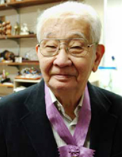 Koji Nakanishi (中西香爾)合作论文数Department of Chemistry, Columbia University1
 Christoph Biskup合作论文数1James C. Kennedy合作论文数1Lilia A. Rousseva合作论文数1Elisabeth R. Gaillard合作论文数1William G. Ramey合作论文数1




