In the pathophysiologic setting of cerebral ischemia, excitotoxic levels of glutamate contribute to neuronal cell death. Our previous work demonstrated the ability of glutamate oxaloacetate transaminase (GOT) to metabolize neurotoxic glutamate in the stroke-affected brain. Here, we seek to identify small-molecule inducers of GOT expression to mitigate ischemic stroke injury. From a panel of phytoestrogen isoflavones, biochanin A (BCA) was identified as the most potent inducer of GOT gene expression in neural cells. BCA significantly increased GOT mRNA and protein expression at 24 h and protected against glutamate-induced cell death. Of note, this protection was lost when GOT was knocked down. To validate outcomes in vivo, C57BL/6 mice were intraperitoneally injected with BCA (5 and 10 mg/kg) for 4 wk and subjected to ischemic stroke. BCA levels were significantly increased in plasma and brain of mice. Immunohistochemistry demonstrated increased GOT protein expression in the brain. BCA attenuated stroke lesion volume as measured by 9.4T MRI and improved sensorimotor function-this protection was lost with GOT knockdown. BCA increased luciferase activity in cells that were transfected with the pERRE(3)tk-LUC plasmid, which demonstrated transactivation of GOT. This increase was lost when estrogen-related receptor response element sites were mutated. Taken together, BCA represents a natural phytoestrogen that mitigates stroke-induced injury by inducing GOT expression.-Khanna, S., Stewart, R., Gnyawali, S., Harris, H., Balch, M., Spieldenner, J., Sen, C. K., Rink, C. Phytoestrogen isoflavone intervention to engage the neuroprotective effect of glutamate oxaloacetate transaminase against stroke.
Endothelial cell tumors are the most common soft tissue tumors in infants. Tumor-forming endothelial (EOMA) cells are able to escape cell death fate despite excessive nuclear oxidant burden. Our previous work recognized perinuclear Nox-4 as a key contributor to EOMA growth. The objective of this work was to characterize the mechanisms by which EOMA cells evade oxidant toxicity and thrive. In EOMA cells, compared with in the cytosol, the nuclear GSSG/GSH ratio was 5-fold higher. Compared to the ratio observed in healthy murine aortic endothelial (MAE) cells, GSSG/GSH was over twice as high in EOMA cells. Multidrug resistance-associated protein-1 (MRP-1), an active GSSG efflux mechanism, showed 2-fold increased activity in EOMA compared with MAE cells. Hyperactive YB-1 and Ape/Ref-1 were responsible for high MRP-1 expression in EOMA. Proximity ligand assay demonstrated MRP-1 and YB-1 binding. Such binding enabled the nuclear targeting of MRP-1 in EOMA in a leptomycin-B-sensitive manner. MRP-1 inhibition as well as knockdown trapped nuclear GSSG, causing cell death of EOMA. Disulfide loading of cells by inhibition of GSSG reductase (bischoloronitrosourea) or thioredoxin reductase (auranofin) was effective in causing EOMA death as well. In sum, EOMA cells survive a heavy oxidant burden by rapid efflux of GSSG, which is lethal if trapped within the cell. A hyperactive MRP-1 system for GSSG efflux acts as a critical survival factor for these cells, making it a potential target for EOMA therapeutics.
The objective of this study (clinicaltrials.gov NCT01858376) was to determine the effect of oral supplementation of a standardized extract of Phyllanthus emblica (CAPROS((R))) on cardiovascular disease (CVD) risk factors in overweight adult human subjects from the US population. Overweight/Class-1 obese (body-mass index: 25-35) adult subjects received 500 mg of CAPROS supplement b.i.d for 12 weeks. The study design included two baseline visits followed by 12 weeks of supplementation and then 2 weeks of washout. At all visits, peripheral venous blood was collected in sodium citrate tubes. Lipid profile measurements demonstrated a significant decrease in calculated low-density lipoprotein cholesterol and total cholesterol/high-density lipoprotein following 12 weeks of CAPROS supplementation when compared to averaged baseline visits. Circulatory high-sensitivity C reactive protein (hs-CRP) levels were significantly decreased after 12 weeks of supplementation. In addition, both ADP- and collagen-induced platelet aggregation was significantly downregulated following 12 weeks of supplementation. Overall, the study suggests that oral CAPROS supplementation may provide beneficial effects in overweight/Class-1 obese adults by lowering multiple global CVD risk factors.
Photoperiodism is a biological phenomenon in which environmental day length is monitored to ascertain time of year to engage in seasonally appropriate adaptations. This trait is common among organisms living outside of the tropics. White-footed mice (Peromyscus leucopus) are small photoperiodic rodents which display a suite of adaptive responses to short day lengths, including reduced hippocampal volume, impairments in hippocampal-mediated memory, and enhanced hypothalamic-pituitary-adrenal axis reactivity. Because these photoperiodic changes in brain and behavior mirror some of the etiology of post-traumatic stress disorder (PTSD), we hypothesized that photoperiod may also alter fear memory and neuronal morphology within the hippocampus–basolateral amygdala–prefrontal cortex fear circuit. Ten weeks of exposure to short days increased fear memory in an auditory-cued fear conditioning test. Short days also increased dendritic spine density of the neurons of the basolateral amygdala, without affecting morphology of pyramidal neurons within the infralimbic region of the medial prefrontal cortex. Taken together, photoperiodic phenotypic changes in brain morphology and physiology induced by a single environmental factor, exposure to short day lengths, affect responses to fearful stimuli in white-footed mice. These results have potential implications for understanding seasonal changes in fear responsiveness, as well as for expanding translational animal models for studying gene–environment interactions underlying psychiatric diseases, such as PTSD.
 Savita Khanna合作论文数Davis Heart and Lung Research Institute, The Ohio State University Medical Center4
 Chandan K. Sen合作论文数McGowan Institute for Regenerative Medicine, University of Pittsburgh School of Medicine;Mary Ann Liebert, Inc.;Bioengineering MDPI;JoVE;Genes and Nutrition;DNA & Cell Biology3Ayan Biswas合作论文数School of Medicine, University of Alabama at Birmingham2
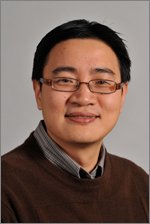 Pan Xueliang合作论文数Ohio State Univ2
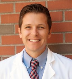 Cameron Rink合作论文数Davis Heart and Lung Research Institute, The Ohio State University Medical Center2
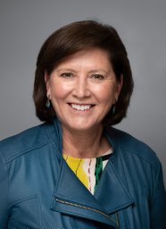 Gayle Gordillo合作论文数512 Davis Heart and Lung Research Institute, Ohio State University2Amitava Das合作论文数1
 Surya Gnyawali合作论文数1
 Hallie Harris合作论文数1
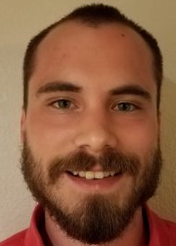 Richard Stewart合作论文数College of Literature, Science, and the Arts, University of Michigan1Maria Balch合作论文数1Haim Achikam合作论文数1Sashwati Roy合作论文数Division of General and Gastrointestinal Surgery, College of Medicine, The Ohio State University1
 Randy Nelson合作论文数Department of Neuroscience, School of Medicine, West Virginia University;Rockefeller Neuroscience Institute, School of Medicine, West Virginia University1James C. Walton合作论文数Department of Neuroscience, Ohio State University1




