Transcutaneous spinal stimulation aims to target dorsal spinal roots, which deliver sensory information to the spinal cord. However, the stimulation waveform that most effectively recruits sensory fibers at the lowest intensity has not been identified. The purpose of this study was to compare conventional and high-frequency burst-modulated stimulation waveforms in their ability to recruit sensory fibers, as assessed by H-reflex threshold, recruitment characteristics, and motor fiber activation. In participants with intact neurological function (n = 12), soleus H-reflex recruitment curves were recorded for 10 stimulation waveforms: a conventional waveform (400 μs), serving as the reference condition, high-frequency burst-modulated waveforms (2, 5, and 10 kHz) with the same total phase duration, and a longer conventional waveform (1,000 μs), each delivered as biphasic and monophasic pulses. H-reflex threshold was higher for high-frequency waveforms: 2 kHz (biphasic: +63%, monophasic: +35%); 5 kHz (biphasic: +179%, monophasic: +109%); 10 kHz (biphasic: +307%, monophasic: +249%); and lower for the conventional 1,000 μs waveform (biphasic: -39%, monophasic: -44%). Similarly, recruitment curve peak slope (mV/mA) was less steep for high-frequency waveforms: 2 kHz (biphasic: -43%, monophasic: -40%); 5 kHz (biphasic: -77%, monophasic: -40%); 10 kHz (biphasic: -82%, monophasic: -48%); and increased (+38%) for the monophasic conventional 1,000 μs waveform. At stimulus intensity near the H-reflex threshold, M-wave amplitude was larger for high-frequency waveforms. Overall, high-frequency waveforms were less effective than conventional waveforms of equal phase duration at eliciting H-reflexes and showed more motor activation at near-threshold intensities.NEW & NOTEWORTHY Compared with high-frequency burst-modulated waveforms (2, 5, and 10 kHz), conventional waveforms (400 μs) elicited H-reflexes more efficiently, as evidenced by lower thresholds and steeper recruitment curves, and less motor activation, as evidenced by smaller M-waves near threshold and larger Hmax/Mmax ratios. Monophasic 5 and 10 kHz waveforms also produced lower H-reflex thresholds and steeper slopes than their biphasic versions, suggesting that the anodal phase impairs sensory fiber recruitment with high-frequency stimulation.
[This corrects the article DOI: 10.1016/j.eclinm.2026.103802.].
Persistent inward currents (PICs) are known to prolong firing of limb-muscle motoneurons. Although there is immunohistochemical evidence of PIC channels in respiratory motoneurons, it is unknown whether PICs contribute to their firing prolongation. Intramuscular electromyographic signals were recorded from human inspiratory muscles to identify motor unit (MU) activity. Diaphragm MUs were identified during quiet breathing (n = 7; one female) and MUs from the 1st, 3rd, and 5th parasternal intercostal muscles during quiet (no lung volume feedback) and voluntary (triangular-shaped lung volume feedback) breathing (n = 5 males). PIC contribution to firing prolongation was estimated via quantification of firing hysteresis (paired MU analysis; ∆F) and firing symmetry in relation to peak volume (duration ratio). Diaphragm was the only muscle in which MUs exhibited ∆F scores significantly greater than 1 Hz, suggesting a possible PIC contribution. The proportion of MUs firing into expiration (duration ratio < 1) was higher in diaphragm than in the 1st and 3rd intercostal muscles, but not different to the 5th. Duration ratios were higher in voluntary compared to quiet breaths in the 3rd and 5th intercostals, suggesting less firing prolongation. However, ∆F was not different between quiet and voluntary breaths in parasternal intercostal MUs. These findings suggest that PICs contribute to firing prolongation in diaphragm MUs during quiet breathing. However, PIC-like behaviours were not evident in parasternal intercostal MUs firing during either quiet or voluntary breathing. These data underscore potentially different levels of neuromodulation across inspiratory muscles and further advance our understanding of the neural mechanisms regulating respiratory muscle control. KEY POINTS: Spinal motoneurons transmit signals to muscles to regulate their contraction, and the intrinsic excitability of motoneurons is enhanced by persistent inward currents (PICs). PICs consist of a persistent flow of sodium and calcium ions into the motoneuron, which can help initiate, accelerate and prolong its firing. We investigated PIC contributions to motoneuron firing in human inspiratory muscles by analysis of diaphragm and parasternal intercostal motor unit activity during involuntary (quiet) and voluntary breathing. Diaphragm motor units showed a magnitude of firing prolongation consistent with a significant role of PICs in this muscle, whereas no such effect was observed in parasternal intercostal muscles. These findings suggest a differential contribution of PICs to motoneuron firing across different inspiratory muscles. Our study enhances understanding of respiratory muscle control and motivates future efforts to validate, further estimate or modulate possible PIC contributions to respiratory motoneuron firing in humans and animal models.
STUDY DESIGN:Sub-analysis of a randomised controlled trial. OBJECTIVES:Respiratory muscle training (RMT) in people with tetraplegia yields marked improvements in inspiratory muscle strength. The present study investigated whether RMT also modifies sleep disordered breathing (SDB) and daytime sleepiness. SETTING:Independent research institute in Sydney, Australia. METHODS:Sixty-two adults with tetraplegia underwent six weeks of supervised RMT. The active-RMT group trained the respiratory muscles through progressive threshold loading to a mean intensity of 50% maximal inspiratory pressure whereas the sham-RMT group experienced the same training protocol, but their device had no progressive threshold load. Primary measures of SDB were obtained using level II ambulatory polysomnograms and daytime sleepiness was assessed using the Epworth Sleepiness Scale (ESS) at baseline and after 6-weeks RMT. RESULTS:Forty-eight participants completed two home-based polysomnograms. Maximal inspiratory pressure (primary outcome) increased more after active intervention than sham, between-group difference 11.8cmH2O (95%CI, 5.2-18.4, p = 0.001). There were no between group differences for any SDB parameter; mean apnoea-hypopnea index (± SD) (active 46 ± 21 vs. 45 ± 22 events/h; sham 44 ± 25 vs. 40 ± 25 events/h; p = 0.553) nor ESS (active 9.5 ± 5.7 vs. 9.2 ± 6.7; sham 10.8 ± 6.2 vs. 10.1 ± 5.8; p = 0.952). CONCLUSIONS:Despite significant increases in inspiratory muscle strength, 6 weeks of supervised respiratory muscle training at a mean intensity of 50% maximal inspiratory pressure does not reduce SDB or daytime sleepiness in people with tetraplegia and severe SDB.
This study examined the effects of nasal and oral breathing routes on genioglossus electromyographic activity (EMG), inspiratory (nadir) epiglottic pressures, and the pressure-dependent reflex regulation of inspiratory genioglossus EMG in awake supine participants. We hypothesized that oral breathing reduces negative pharyngeal pressure swings during inspiration and, via a pressure-dependent reflex mechanism, reduces phasic inspiratory genioglossus EMG. Twenty participants with obstructive sleep apnea (OSA) and 8 people without OSA were recruited into this study. Measurements included multiunit genioglossus EMG via percutaneous bipolar electrodes, airflow via nasal pneumotach, epiglottic pressure, and end-tidal CO2. Participants were studied while in supine position, breathing quietly via nose, then switched to oral breathing for ∼5 min/route, and then repeated the protocol. Oral breathing reduced inspiratory epiglottic negative pressures by ∼50% in all participants [OSA nasal: -4 ± 1.8 (SD) cmH2O; oral: -1.8 ± 0.8 cmH2O; non-OSA nasal: -2.4 ± 1.6 cmH2O; oral: -1.1 ± 0.8 cmH2O]. In people without OSA and a third of people with OSA, oral breathing was associated with reduced phasic inspiratory genioglossus EMG and correlated with reduced inspiratory negative epiglottic pressure swings. Unexpectedly, in the remaining two-thirds of people with OSA, phasic inspiratory EMG was maintained and/or increased with oral breathing, despite reduced inspiratory negative pharyngeal pressure swings. Remarkably, the increased inspiratory genioglossus EMG during oral breathing in these participants with OSA was inversely correlated with epiglottic pressure. The divergent responses in most people with OSA may represent a reflex adaptation to oral breathing, perhaps due, at least in part, to increased and persistent nocturnal oral breathing.NEW & NOTEWORTHY Breathing is supposed to occur through the nose. The switch to oral breathing occurs during exercise, with nasal congestion, or during sleep. This study reports that oral breathing is associated with different patterns of genioglossus muscle activity in individuals with and without obstructive sleep apnea (OSA). A subset of participants with OSA showed increased inspiratory genioglossus muscle activity with oral breathing, despite reduced airway resistance, which was unexpected, and may represent a neural adaptation to increased oral breathing in OSA.
Spinal root magnetic stimulation can be used to assess diaphragm voluntary activation (VA), but its validity during inspiratory efforts remains unclear. In addition, its use to evaluate abdominal muscle VA during expiratory efforts has not been explored. This study investigates spinal root magnetic stimulation as a method to assess VA of inspiratory and expiratory muscles. In 13 healthy participants (8 female, 18-40 yr), magnetic stimulation was delivered over the cervical spine (C6) and lower thoracic spine (T10) to activate the diaphragm (n = 11) during inspiratory efforts and the abdominal muscles (n = 11) during expiratory efforts, respectively. A nasogastric catheter recorded inspiratory transdiaphragmatic and expiratory gastric pressures. During voluntary 25/50/75/100% maximal inspiratory and expiratory efforts, stimuli were delivered at 100% stimulator output to produce a superimposed twitch and, ∼5 s later, a resting twitch, to calculate VA. Good utility of the technique was assessed per individual as r2 ≥ 0.7 and 95% prediction interval (95% PI) limits within 25% of the predicted VA. For inspiratory muscles, VA had poor correspondence and predictability (r2 = 0.26-0.85; 95% PI limits: 27.1%-74.5%) with contraction intensity in 8/11 participants and good correspondence and predictability (r2 = 0.83-0.90; 95% PI limits: 22.7%-23.8%) in 3/11 participants. For expiratory muscles, VA had good correspondence and predictability (r2 = 0.73-0.97; 95% PI limits: 10.4%-20.8%) with contraction intensity in 7/11 participants and poor correspondence and predictability (r2 = 0.13-0.72; 95% PI limits: 19.0%-40.0%) in 4/11 participants. Spinal root magnetic stimulation can be used with caveats to assess expiratory muscle VA only, requiring individualized testing across multiple contraction intensities and a comparison of estimated versus measured resting twitches.NEW & NOTEWORTHY Magnetic stimulation of the cervical spinal roots cannot be used to assess voluntary activation of the diaphragm reliably during inspiratory efforts. However, magnetic stimulation of the thoracic spinal roots could be used to assess abdominal muscle voluntary activation during expiratory efforts but, importantly, required estimation of the twitch response at rest. This novel, less invasive method may be used to assess expiratory muscle activation, but requires testing across a range of contraction intensities.
Inspiratory-related genioglossus EMG activity is crucial to maintain upper airway patency. However, whether this activity differs between the oblique (middle) and horizontal (base) compartments or if they vary in people with obstructive sleep apnoea (OSA) is unknown. Here, intramuscular electrodes were inserted into the anterior and posterior regions of the horizontal and oblique genioglossal compartments in nine controls [apnoea-hypopnoea index (AHI) ≤ 5 events/h] and 45 OSA participants (AHI range 5-94.3 events/h). Multiunit EMG patterns were categorised as phasic (respiratory modulation) or tonic (no respiratory modulation) during nasal breathing in awake, supine participants. The effects of OSA status and genioglossus compartments were assessed through linear mixed models, controlling for nadir epiglottic pressure (Pepi) and repeated measures within participants. Phasic patterns occurred in 57.6% (n = 106/184) of compartments. Within phasic compartments, log10-transformed peak, phasic and tonic EMG (% maximum) were higher in the oblique than in the horizontal compartments. Additionally, more pronounced negative Pepi correlated with increased log10-transformed phasic EMG in the anterior oblique (beta = -0.075, P = 0.002) and posterior oblique compartments (beta = -0.080, P = 0.027), but not in the horizontal compartments. Effects of OSA severity on activity patterns or EMG measurements were not significant. To conclude, the genioglossus exhibited regional (oblique-horizontal) variation in neural drive during awake inspiration. This compartmental activity appears to be driven by reflex activation in the oblique compartments, which increase phasic EMG. People with and without OSA have similar drive during wakefulness. Understanding the mechanisms driving efficient genioglossus dilatory activity is essential to develop targeted treatments for OSA that focus on pharyngeal muscle activity. KEY POINTS: Inspiratory genioglossus multiunit EMG activity is thought to vary across different neuromuscular compartments. However, it remains unclear whether obstructive sleep apnoea (OSA) affects this compartmental variability. During quiet nasal breathing in awake supine individuals, inspiratory genioglossus EMG normalised to maximum EMG was measured in four genioglossus neuromuscular compartments in individuals with and without OSA. Both tonic (no respiratory modulation, 42%) and phasic (respiratory modulation, 58%) activity patterns were observed during breathing. When a genioglossus compartment showed phasic activity, peak, phasic and tonic EMG activities were higher in the oblique than in the horizontal compartments. Furthermore, greater phasic activity was associated with a more negative nadir epiglottic pressure only in the oblique compartments. There was no additive effect of OSA severity on top of the more negative nadir epiglottic pressure, suggesting people with and without OSA received similar drive during inspiration during wakefulness.
Lumbar transcutaneous spinal cord stimulation (TSS) evokes synchronized muscle responses, termed spinally evoked motor response (sEMR). Whether the structures TSS activates to evoke sEMRs differ when TSS intensity and waveform are varied is unknown. In 15 participants (9 F, 6 M), sEMRs were evoked by TSS over L1-L3 (at sEMR threshold and suprathreshold intensities) with conventional (one 400-mu s biphasic pulse) or high-frequency burst (ten 40-mu s biphasic pulses at 10 kHz) stimulus waveforms in vastus medialis (VM), tibialis anterior (TA), and medial gastrocnemius (MG) muscles. TSS was paired with transcranial magnetic stimulation (TMS) over the contralateral motor cortex at relative interstimulus intervals (ISIs) (-10 ms to 11 ms), centered on the ISI when TSS and TMS inputs simultaneously activated VM motoneurons. Doublet TSS was delivered at 80-ms ISI. For VM, the area of the combined response evoked by paired TMS and TSS was not facilitated at any ISI. For TA and MG, combined responses were facilitated by similar to 40-100% when TMS activated the motoneurons before or at a similar time as TSS, particularly with suprathreshold TSS. Additionally, for TA, there was greater suppression of the second sEMR evoked by TSS doublets using suprathreshold conventional TSS compared to high-frequency burst TSS (P < 0.001). The results suggest that for VM TSS activated predominantly motor axons, but for TA and MG facilitation of the sEMR by TMS suggests that TSS activated sensory axons. Stimulation waveforms had similar outcomes in most conditions.
Survival relies on neural respiratory drive from the medullary respiratory centres to activate the respiratory muscles for breathing. Accurate assessment of respiratory muscle activity, as an estimate of neural respiratory drive in humans, will facilitate a greater understanding of respiratory physiology, the pathophysiology of diseases and injuries, clinical investigations and the neurophysiology of breathlessness perception. Here, we highlight the methodologies to measure respiratory muscle activity, their appropriate application, the potential limitations of the data and their safety and feasibility considerations. These recommendations can be applied to all skeletal muscles.
Acute intermittent hypoxia (AIH) can increase maximal strength of limb muscles in people with incomplete spinal cord injury (SCI), but it is mostly untested in people without SCI. Acute intermittent hypercapnia (AIC) may engage similar respiratory circuits to AIH, but the effects of AIC on human limb motor output are unknown. We examined whether single sessions of AIH or AIC improved motor output to a hand muscle in neurologically intact people. Twelve adults completed a single 30-min session of AIH (breathing alternate 1-min low oxygen air and 1-min normal air), AIC (alternate 1-min high carbon dioxide air and 1-min normal air), or SHAM (normal air). At baseline and for 80 min post-intervention, participants performed repeated isometric maximal voluntary thumb adductions. Transcranial magnetic stimulation elicited motor evoked potentials (MEPs) from first dorsal interosseous and adductor pollicis at each time point. Generalised linear mixed models were compared between conditions (AIH, AIC, SHAM). Normalised to baseline, voluntary activation was higher after AIC than SHAM (5.9%, P < 0.001) and AIH (5.5%, P < 0.001); MVC force was higher after AIC than SHAM (7.7%, P < 0.001), whereas maximal EMG was higher after AIH than SHAM (14.3%, P < 0.001). MEPs and maximal M-waves did not differ between conditions for either muscle (P > 0.25). Thus, single sessions of AIC induced small motor output improvements in people without SCI, but AIH did not. AIC increased maximal voluntary activation, but the mechanisms for this remain unclear because the MEPs provided no evidence for corticospinal facilitation.
This review covers the knowledge gains made about human respiratory neural drive resulting from ∼30 years of single motor unit recordings from human inspiratory muscles. Section 2 illustrates the non-uniformity of output across the various inspiratory motoneurone pools innervating diaphragm, scalene, intercostal, and genioglossus muscle activity during quiet breathing and during voluntary breathing. Section 3 describes the rostrocaudal graded timing and magnitude of respiratory neural drive across the parasternal intercostal and external intercostal muscles, which identified a principle of motoneurone recruitment termed the principle of neuromechanical matching that has been since identified as a principle of motor control more generally. Section 4 focuses on the changes in diaphragm motor unit discharge and morphology in ageing, chronic obstructive pulmonary disease and cervical spinal cord injury, linking increased drive to breathe to the changes in respiratory mechanics, with the intriguing findings that this is not always associated with a cortical contribution to breathing (i.e. change in the central control of breathing). Our studies have revealed an elaborate organisation of respiratory neural drive to the motoneurones to match the anatomical and functional complexity of the muscles themselves.
Transcutaneous spinal cord stimulation, as used for rehabilitation of impaired motor function after spinal cord injury, often involves a 10-kHz waveform modulated to produce repetitive bursts of stimulation. Kilohertz-frequency waveforms may facilitate the summation of subthreshold depolarisations, but the optimal burst duration for nerve stimulation has not been systematically investigated. In 11 adults, the ulnar nerve was stimulated transcutaneously with a 10-kHz waveform that contained 1, 2, 4, 6, 8 or 10 pulses, in random order. Compound muscle action potentials (CMAPs) and sensory nerve action potentials (SNAPs) were measured from motor threshold up to the maximal CMAP (M-max). The efficacy of each waveform was determined at M-max as CMAP amplitude divided by total phase charge. For CMAPs and SNAPs, increasing the number of pulses shifted the stimulus-response curves to the left for current and to the right for total charge. Accordingly, an increase in the number of pulses decreased the current but increased the total charge at sensory and motor thresholds and M-max. Efficacy decreased as the number of pulses increased. Onset latencies were delayed for waveforms with six or more pulses compared to a single pulse. These findings provide evidence of the summation of subthreshold depolarisations in sensory and motor axons in humans. However, the optimal number of pulses for summation remains unclear due to the opposing changes in current and total charge. It is clear, though, that more than six pulses is suboptimal, as there were no further decreases in threshold current while total charge continued to increase.
Single centre training study. To investigate, in a group of people with spinal cord injury (SCI), the effect of transcutaneous functional electrical stimulation of the abdominal muscles (abdominal FES) during cough training on blood pressure (BP), and how it is affected by injury characteristics and alters over time. Laboratory and community. Sixteen participants with SCI (C4-T5) underwent 25 of abdominal FES cough training (5 sets of 10 stimulated coughs) over 6 weeks as part of a previously published study on the effect of abdominal FES training on cough. Systolic BP (SBP), diastolic BP (DBP) and calculated mean arterial pressure (MAP) were measured at the completion of each set. Abdominal FES coughing resulted in an average 30
Intramuscular recordings of single motor unit activity from parasternal intercostal muscles show a rostrocaudal gradient in timing and amplitude of inspiratory activity. This study determined the feasibility of surface electromyographic activity (EMG) to measure graded parasternal intercostal activity in young females and males during quiet breathing and breathing with inspiratory resistive loads. Surface EMGs were recorded from the 1st-to-5th parasternal intercostal muscles during 10 min of breathing. EMGs were processed to remove 50 Hz and electrocardiogram artifacts and integrated. Amplitude and onset time of inspiratory activity were measured from waveform averages triggered at the onset of inspiratory flow. Onset times were measured independently by two assessors, blinded to interspace and EMG scale, with excellent agreement (ICC3,k = 0.86). The onset of inspiratory activity in the 1st-to-3rd interspaces was at or within ∼400 ms of the start of inspiratory airflow, but activity in the caudal (4th and 5th) spaces was delayed by up to ∼1,000 ms (P < 0.001). There was no main effect of sex on onset time (P = 0.07), but an interaction with interspace (P < 0.001) revealed that inspiratory activity in the caudal interspaces was delayed by 15% of inspiratory time in female participants compared with 30% of inspiratory time in male participants. Inspiratory loads did not affect EMG onset time (P = 0.31). Thus, surface EMG is feasible to assess the onset time of inspiratory activity as a marker of inspiratory neural drive and pattern of activation across spaces, in both females and males.NEW & NOTEWORTHY We demonstrated that surface EMG is a valid method to measure graded inspiratory EMG in the parasternal intercostal muscles in healthy young male and female participants during quiet breathing and loaded breathing. Across the 1st-to-5th interspaces, there was more homogenous activation in women and more graded activity in men across parasternal intercostal muscles during breathing. By recording surface EMG from both male and female participants, we have revealed sex differences in inspiratory activity across intercostal muscles.
Abstract Introduction Inspiratory-related genioglossus electromyography (EMG) activity is crucial to maintain upper airway patency during inspiration. Recent findings suggest variations in anterior/posterior genioglossus multiunit EMG activation during awake supine quiet breathing in healthy adults. However, whether such differences extend to EMG across oblique (middle) and horizontal (base) genioglossus compartments, and whether activation differ in obstructive sleep apnoea (OSA) is unclear. Methods Prior to in-lab polysomnography, intramuscular electrodes were placed into the horizontal and oblique compartments of the genioglossus in 9 controls (AHI≤5 events/hr) and 45 age- and BMI- matched OSA patients (AHI range 5-94.3 events/hr). Multiunit EMG patterns were classified as phasic or tonic during awake supine quiet nasal breathing. Phasic pattern recordings were quantitatively analysed (% maximum of swallowing or tongue protrusion). The effects of OSA status and genioglossus compartments were assessed through mixed linear analysis, controlling for nadir Pepi and repeated measures in participants. Results More than half (57.6%, n=106/184) of the recording sites had a phasic pattern. A more negative nadir Pepi correlated with reduced likelihood of tonic activity (OR: 0.819, P=0.042). Within phasic patterns, a more negative nadir Pepi correlated with increased peak EMG (beta=-0.847, P=0.011), phasic EMG (beta=-0.654, P=0.005), and tonic EMG (beta=-0.253, P=0.048). No significant effects of OSA status or genioglossus compartment on activity patterns or EMG measurements were observed. Conclusions No compartmental disparity in EMG or differences between OSA and non-OSA individuals were found. Understanding mechanisms driving efficient dilatory activity of the genioglossus compartments is pivotal to develop OSA treatments targeting pharyngeal muscle activity.
Transcutaneous electrical stimulation with repetitive bursts of a kilohertz carrier frequency is thought to be less painful than conventional pulsed currents by reducing the sensitivity of pain receptors. However, no purported benefit has been shown unequivocally. We compared the effects of carrier-frequency stimulation and conventional stimulation on pain tolerance and the thresholds for sensory and motor axons in twelve participants. The ulnar nerve was stimulated transcutaneously with a conventional single pulse and 5 and 10 kHz carrier-frequency waveforms that had 5 and 10 pulses, respectively, when delivered in bursts of ∼1 ms duration. Phase durations were adjusted across waveform types to match the total charge for a given current amplitude. Single bursts of stimulation were delivered from 1 mA up until no longer tolerable. This was repeated with repetitive bursts of stimulation at 20 Hz for 1 s. Participants tolerated higher current amplitudes with both carrier-frequency waveforms than conventional stimulation, with repetitive bursts more painful than single bursts. However, compared to conventional stimulation, carrier-frequency waveforms required more current to produce sensory and motor-threshold responses and to obtain a maximal motor response (Mmax). When the current at pain tolerance was normalised to the current at Mmax, participants tolerated lower stimulus intensities with carrier-frequency waveforms than conventional stimulation. These findings indicate that there is little to no benefit in using carrier-frequency waveforms to minimise the discomfort from electrical stimulation as the increase in stimulus intensity at pain tolerance is more than offset by reduced effectiveness in the activation of sensory and motor axons. KEY POINTS: Transcutaneous electrical stimulation with repetitive bursts of a kilohertz carrier-frequency waveform is thought to be less painful than conventional pulsed currents. For ulnar nerve stimulation, when stimulus waveforms were matched for total phase charge, participants tolerated higher current amplitudes with carrier-frequency stimulation than conventional stimulation. However, compared to conventional stimulation, carrier-frequency waveforms required more current to produce a threshold response in both sensory and motor axons and to produce a maximal motor response (Mmax). When current at pain tolerance was normalised to current at Mmax, participants tolerated lower stimulus intensities with carrier-frequency waveforms than conventional stimulation. Carrier-frequency waveforms provide little to no benefit in minimising the discomfort from transcutaneous electrical stimulation as the increase in stimulus intensity at pain tolerance is more than offset by reduced effectiveness in activating sensory and motor axons.
Acute intermittent hypoxia (AIH) can induce sustained facilitation of motor output in people with spinal cord injury (SCI). Most studies of corticospinal tract excitability in humans have used 9% fraction inspired oxygen ([Formula: see text]) AIH (AIH-9%), with inconsistent outcomes. We investigated the effect of single sessions of 9% [Formula: see text] and 12% [Formula: see text] AIH (AIH-12%) on corticospinal excitability of a hand and leg muscle in able-bodied adults. Ten naïve participants completed three sessions on separate days comprising 15 epochs of 1 min of AIH-9%, AIH-12%, or sham (SHAM-21%) followed by 1 min of room air (21% [Formula: see text]) in a randomized crossover design. Motor-evoked potentials (MEPs; n = 30, ∼1 mV) elicited at rest by transcranial magnetic stimulation and maximal M-waves (Mmax) evoked by peripheral nerve stimulation were measured from the first dorsal interosseous (FDI) and tibialis anterior (TA) muscles at baseline and at ∼0, 20, 40, and 60 min post intervention. AIH-9% induced the greatest reduction in peripheral oxygen saturation (to 85% vs. 93% and 100% in AIH-12% and SHAM-21%, respectively; P < 0.001) and the greatest increase in ventilation [by 22% vs. 12% and -3% in AIH-9%, AIH-12%, and SHAM-21%, respectively (P < 0.001)]. There was no difference in MEP amplitudes (%Mmax) after any of the three conditions (AIH-9%, AIH-12%, SHAM-21%) for both the FDI (P = 0.399) and TA (P = 0.582). Despite greater cardiorespiratory changes during AIH-9%, there was no evidence of corticospinal facilitation (tested with MEPs) in this study. Further studies could explore variability in response to AIH between individuals and other methods to measure motor facilitation in people with and without spinal cord injuries.NEW & NOTEWORTHY This is the first study that tests whether acute intermittent hypoxia (AIH) induces motor output facilitation in humans after two different doses of AIH (9% and 12% [Formula: see text]) and the reproducibility of participant responses after a repeat AIH intervention at 9% AIH. There was no motor output facilitation in response to either dose of AIH. The results question the effectiveness of a single 30-min session of AIH in inducing motor output facilitation, tested in this way.
Connections link a sequence of three related research papers. The central article which links the other two papers has been published in Experimental Physiology. In a Connections article, an author (or authors) of the central article outlines its principal novel findings, tracing how they were influenced by the first article and how the central article has contributed to the developments made in the third article. The author(s) may also speculate on the direction of future research in the field. Connections articles aim to set the research in a wide context. Motoneurone output is crucial for all reflex and voluntary movements. Furthermore, this output is compromised in conditions affecting motoneurones, their local intraspinal circuits and descending corticofugal drives. This justifies the need to understand the physiology of motoneurones and to search for therapeutic strategies to repair and rehabilitate their output when impaired. Of course, outputs from respiratory motoneurones are prioritised in any hierarchy of output because they drive pulmonary ventilation. Short alternating periods of hypoxia and normoxia (acute intermittent hypoxia, AIH) have been shown in animal studies, first done more than 30 years ago, to enhance the output of respiratory motoneurones, particularly those in the phrenic motor nucleus. The responsible post-synaptic pathways and neurophysiological mechanisms have been well explored (e.g., Navarrete–Opazo & Mitchell, 2014). This seemingly straightforward way to boost motoneurone output has been taken up and used to enhance (or not) motoneurone output in humans with disorders such as stroke and spinal cord injury. This Connections piece begins with a description of a test of a major corollary of up-regulation of the output from human motoneurones by AIH via post-synaptic mechanisms (in healthy humans without a spinal cord injury). In a simple, double-blinded, randomised, cross-over design, Finn et al. (2022) asked whether the Hoffman (H) reflex in the soleus muscle, which is largely monosynaptic, was altered by a period of intermittent hypoxic breathing. The duration and severity of the hypoxia (9% O2) was typical of prior studies. The central dogma would say this intervention would enhance the post-synaptic effect of the primary muscle spindle input on soleus motoneurones. They were studied under controlled conditions with the participants at rest. In this study there was no consistent change in the size of the reflex over the 60 min after the AIH intervention. This was based on an assessment of the H reflex recruitment curve and the maximal H-reflex. The maximal M-wave was unchanged. In addition, Finn et al. (2022) asked whether the motor potential evoked by transcranial magnetic stimulation (MEP) in an intrinsic hand muscle changed after the AIH intervention. Again, the evoked responses include a strong monosynaptic component (corticospinal) and they were measured during relaxation. This was motivated by the prior study of Christiansen et al. (2018) who had also studied this response and showed it increased in all participants. Across the group of participants, Finn et al. (2022) found AIH failed to produce a coherent change in corticospinal evoked responses. However, despite these generally negative findings, Finn et al. (2022) talked up some 'statistically significant' amplifications of evoked reflex and corticospinal responses which 'were inconsistent and only at distinct time intervals'. Of the 120 pre-planned post hoc tests of AIH efficacy, five were statistically significant at the conventional 5% level (Finn et al., 2022; supplementary information S1)—hardly a reason to head to the casino! So, what were the dramatic findings of Christiansen et al. (2018)? They focused on the corticospinal pathway to a hand muscle—from motor cortex to motoneurone. First, in the hour after the AIH intervention, the effect of motor cortical stimulation, but also importantly subcortical stimulation of corticospinal axons, was facilitated significantly above baseline (by ∼40%, SD ∼30%, and by ∼75–100%, SD ∼75%, respectively). (This was estimated crudely from their Figs 2 and 4 for the 15–75 min interval after the intervention.) Second, another expression of motoneurone plasticity (spike timing-dependent plasticity) based on the temporal interaction between a descending (corticospinal) input to the motoneurones and separate (antidromic) activation of the motoneurone showed upregulation (>50%, their Fig. 6) after the AIH intervention. Finally, an indirect, albeit controversial, measure of the 'intrinsic excitability' of the motoneurone (Ozyurt et al., 2024), its ability to be activated by an antidromic action potential (the F-wave) was not changed by the AIH intervention. The simplest unifying conclusion from Christiansen et al. (2018) is that AIH acts at the level of the corticospinal synapses onto motoneurones. Disappointingly, while this may have prompted the study by Finn et al. (2022) described earlier, any facilitation of corticospinal action on motoneurones seems not to occur uniformly within studies or between them. Radia et al. (2022) also found no influence of AIH on corticospinal output to a hand motoneurone pool. They used a slightly different intermittent protocol, but it still involved 15 min of hypoxia in total. More recently, Mathew et al. (under review) 1found no change in MEPs after single sessions of AIH on corticospinal output to the hand or to the lower limb. So far, the only published study on changes in a mostly monosynaptic spinal reflex by AIH is that by Finn et al. (2022) with minimal to no effect. However, the failure of AIH to enhance reflex output has recently been confirmed for the stretch reflex of soleus (Tan et al., 2024). Where does this leave us? Perplexed. But, a study by Welch et al. (2022) adds a further dimension to the physiological processes being explored with AIH. It is the level of CO2. In a typical AIH protocol, the mild over-ventilation, due to an increased tidal volume, reduces end-tidal CO2 in some cases (e.g., Finn et al. 2022) but not others (Welch et al., 2022). How does the CO2 level interact with the putative neuroplasticity at the motoneurone pool? Using transcranial magnetic stimulation to elicit MEPs in the diaphragm with indirect chest-wall recordings, Welch et al. (2022) found that the typical AIH intervention did not alter the size of the MEPs, but if they added 4% CO2 to the hypoxic gas (9% O2), facilitation of the corticospinal output to phrenic motoneurones appeared. It is not known whether this would occur for limb motoneurones. It is disappointing that a proposal based on a bevy of animal studies has not translated more convincingly into studies of neurophysiology and pathophysiology in humans. While it may yet achieve some therapeutic place, for now, we are left in an uncertain and problematic place. It is uncertain because there is always the possibility that subtle differences in experimental protocols and methods of measurement may obscure a genuine effect, and that to achieve a clinically useful effect (if it exists), the number of participants in studies may need to be increased many fold. It is problematic because early published clinical and other studies in humans pointed to a beneficial effect of even a single session of AIH on motor outcomes, but it is doubtful that CO2 levels were high enough to help promote the potential effect of AIH at motoneuronal level. As a result, we are unable to decide for or against the corollaries of the central dogma surrounding AIH. Mindful that early studies in a field usually amplify the size of any real effect and that any P-value of 0.05 has only a 50% chance of replication (Gandevia et al., 2021), we have therefore subjected the three papers (Christiansen et al. 2018; Finn et al. 2022; Welch et al., 2022) in this Connections piece to a formal Quality Output Checklist and Content Assessment (QuOCCA) (Héroux et al., 2022). The results are given in a Supplementary file S1. The findings of this independent assessment are likely typical of those in neuroscience and physiology, and they point to key areas for improvement which should enhance the reproducibility of the sorts of human studies discussed here. Both authors have read and approved the final version of this manuscript and agree to be accountable for all aspects of the work in ensuring that questions related to the accuracy or integrity of any part of the work are appropriately investigated and resolved. All persons designated as authors qualify for authorship, and all those who qualify for authorship are listed. We are grateful to Annie Butler and Marel Parono for independent compilation of the supplementary file and to Harrision Finn for comments on the manuscript. Simon C. Gandevia is supported by the National Health and Medical Research Council. None. Please note: The publisher is not responsible for the content or functionality of any supporting information supplied by the authors. Any queries (other than missing content) should be directed to the corresponding author for the article.
Study design/setting Secondary analysis. Objectives To compare the change in maximal inspiratory pressure (PI max ) over the first 4 weeks of two different inspiratory muscle training (IMT) protocols and explore if either method is more effective for people with spinal cord injury. Methods Data originated from two published studies. Participants completed flow-resistive IMT (F-IMT) at 80% daily PI max, 7 days/week (supervised weekly), or threshold IMT (T-IMT) at 30–80% weekly PI max, twice-daily, 5 days/week (supervised every session). Seven participants from each trial were matched by training adherence, level of spinal cord injury, impairment grade (A–C), and height. Differences between F-IMT and T-IMT groups in training intensity, breaths taken, inspiratory work, and the change in the PI max from baseline at the end of week four were analysed. Results Over 4 weeks, there was no difference in the change in PI max between groups (Absolute change in PI max (cmH 2 O): p = 0.456, Percent change in PI max relative to baseline: p = 0.128). F-IMT participants trained at a higher intensity (median: 77 vs 22 cmH 2 O, p = 0.001 and 80% baseline vs 61% baseline, p = 0.038) but took fewer breaths (840 vs 1404 breaths, p = 0.017) than T-IMT participants. Inspiratory work was similar between groups (64,789 vs 65,910 (% PI max × number of breaths), p = 0.535). Conclusions Our findings support both methods of IMT as the change in PI max and inspiratory work were similar between groups. However, daily high-intensity F-IMT with intermittent supervision, required fewer breaths and less participant and therapist time. Future studies should examine optimal dosage and supervision required to achieve increased PI max .
Background The demand for physiotherapy clinical placements is rising which requires innovative approaches and an understanding of clinical placement models. Objective To determine physiotherapy student contribution to direct patient care activity during a collaborative clinical placement model. Secondary aims determined the impact of clinical area and clinical educator to student (CE:student) ratio and if a group of students could reach equivalent direct patient care activity of a junior or senior physiotherapist. Method Physiotherapy student, and junior and senior physiotherapist occasions of service (OOS) were collected from five Queensland Public Health Sector hospital information management systems from four physiotherapy clinical areas (i.e. cardiorespiratory, musculoskeletal, neurorehabilitation, and orthopedics). Number of days of clinical activity was recorded to provide average OOS/day. Results Across a 5-week clinical placement a group of physiotherapy students in a collaborative clinical placement model provided on average 10.6 OOS/day (95%CI 10.1-11.2). In three (75%) clinical areas, a group of students participating in higher CE:student ratios produced more OOS/day. Clinical area and CE:student ratio predicted 39% of the variance in student average OOS/day. On average a group of students reached the equivalent direct patient care activity of a junior and senior physiotherapist by week two of a 5-week clinical placement. Conclusion Physiotherapy students in a collaborative clinical placement model met or exceeded the direct patient care activity of a physiotherapist, irrespective of clinical area and CE:student ratio.
 Simon Gandevia合作论文数Neuroscience Research Australia158
 Janet Taylor合作论文数School of Medical and Health Sciences, Edith Cowan University;Neuroscience Research Australia;University of New South Wales52
 Danny Eckert合作论文数School of Biomedical Sciences, University of New South Wales47
 Anna L Hudson合作论文数Neuroscience Research Australia, University of New South Wales44
 Lynne Bilston合作论文数Neuroscience Research Australia, UNSW42
 Claire Boswell-Ruys合作论文数Spinal Cord Injuries Research Centre, Neuroscience Research Australia;University of New South Wales;Prince of Wales Hospital28P.G.R. Burke合作论文数28Fiona L Knapman合作论文数24Luu Billy L合作论文数19Rachel A. McBain合作论文数18Brown Elizabeth C合作论文数17
 Julian P Saboisky合作论文数Div Sleep Med, Brigham & Womens Hosp16David K McKenzie合作论文数15
 Martin Heroux合作论文数15
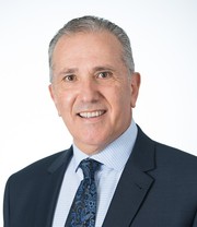 Peter Cistulli合作论文数School of Medicine, Faculty of Medicine and Health, The University of Sydney13
 Joachim Ngiam合作论文数12
 Euan McCaughey合作论文数School of Biomedical Sciences, Faculty of Medicine and Health Sciences, University of New South Wales;Liberate Medical11L. Juge合作论文数10E. Brown合作论文数10
 Robert C. Gorman合作论文数Department of Surgery, Perelman School of Medicine, University of Pennsylvania10
 Lauriane Juge合作论文数10K Sutherland合作论文数9Jayne C Carberry合作论文数9N Petersen合作论文数Dept Neurosci, Univ Copenhagen8
 Lee Bonsan B合作论文数Australia Prince of Wales Hospital, Australia University of New South Wales8
 David K Mckenzie合作论文数Department of Respiratory Medicine, Prince of Wales Hospital8
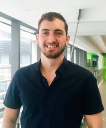 Harrison T Finn合作论文数Neurosci Res Australia7
 Paul Hodges合作论文数School of Health and Rehabilitation Sciences, Faculty of Health and Behavioural Sciences, The University of Queensland7
 Adam Jaffe合作论文数School of Women's and Children's Health, Faculty of Medicine, The University of New South Wales7Ethel Cosmos合作论文数7Jade Yeung合作论文数6
 Sandra Ya chu Chuang合作论文数School of Women's and Children's Health, Faculty of Medicine, The University of New South Wales6David A T Nguyen合作论文数5J. Yeung合作论文数5
 Gabrielle Todd合作论文数UniSA Clin & Hlth Sci, Univ South Australia5
 Joanna Diong合作论文数School of Medicine Sciences, Faculty of Medicine and Health, The University of Sydney5
 Amy Jordan合作论文数Melbourne School of Psychological Sciences, Faculty of Medicine, Dentistry and Health Sciences, The University of Melbourne5
 David P. White合作论文数Division of Sleep Medicine, Harvard Medical School;Philips Home Healthcare Solutions5John Trinder合作论文数5
 Peter Humburg合作论文数Department of Statistics, Macquarie University;CSIRO Mathematical and Information Sciences;The Wellcome Trust Centre for Human Genetics;The Wellcome Trust Centre for Human Genetics, Macquarie University5
 Shaokoon Cheng合作论文数Neuroscience Research Australia, University of New South Wales5Chaminda Lewis合作论文数4
 Elizabeth Ann Bye合作论文数Sydney Med Sch Northern, Univ Sydney4peter g martin合作论文数4
 Sophie Carter合作论文数4
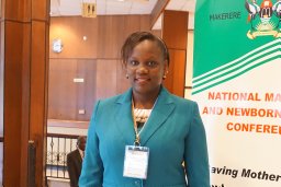 Isabella Epiu合作论文数Prince Wales Hosp4Kate Sutherland合作论文数School of Medicine, Faculty of Medicine and Health, The University of Sydney4
 Kylie Tucker合作论文数School of Biomedical Sciences, Faculty of Medicine, The University of Queensland4Monica A Perez合作论文数Department of Physical Medicine & Rehabilitation, Feinberg School of Medicine, Northwestern University4




