Objective Patients living with pituitary neuroendocrine tumours (PitNETs) present with a spectrum of clinical manifestations and often follow a circuitous route to diagnosis, resulting in diagnostic delays. The objective of this study is to identify and report the various sources of referrals for patients who underwent pituitary resection for PitNETs in a tertiary referral centre for pituitary disease. Design Retrospective population-based cohort study. Patients Patients undergoing a first surgery for management of PitNET in Northern Ireland between 01/01/2000-19/07/2019. Measurements Demographics, referral sources, incidentaloma rates, apoplexy rates, symptoms, age at diagnosis and diagnostic delay according to referral source. Results Data were retrospectively analysed for 520 patients in whom the referral source leading to diagnosis was known. Fifty-seven percent of patients were male. Median age at diagnosis was 54 years (range 18-85). Patients were referred from 29 different sources. The majority of referrals came from general practice (23%), ophthalmology (14%), emergency medicine (9%), optician/optometry (9%) and internists (8%). Twenty-nine patients were referred following radiological discovery of an incidentaloma. Twenty-seven patients were referred with an initial presentation of pituitary apoplexy, with emergency medicine accounting for 70% of these referrals. Visual disturbance and headache were the most frequently documented symptoms. Median diagnostic delay was 2 years (range 0-25 years). Conclusions Patients with PitNETs encounter a range of clinical services in their journey to pituitary surgery. Raising awareness of these tumours and educating healthcare professionals across all disciplines could reduce diagnostic delays, thereby minimising the adverse sequelae of delayed diagnosis.
Cushing’s disease is a rare condition that occurs due to an adrenocorticotrophin-producing corticotrophinoma arising from the pituitary gland. The consequent hypercortisolaemia results in multisystem morbidity and mortality. This study aims to report incidence, clinicopathological characteristics, remission outcomes and mortality in a regional pituitary neurosurgical cohort of patients diagnosed with Cushing’s disease in Northern Ireland (NI) from 2000 to 2019. Clinical, biochemical and radiological data from a cohort of patients operated for Cushing’s disease were retrospectively collected and analysed. Fifty-three patients were identified, resulting in an estimated annual incidence of Cushing’s disease of 1.39–1.57 per million population per year. Females accounted for 72% (38/53) of the cohort. The majority (74%, 39/53) of corticotrophinomas were microadenomas and in 44% (17/39) of these no tumour was identified on preoperative magnetic resonance imaging. Histopathological characterisation was similarly difficult, with no tumour being identified in the histopathological specimen in 40% (21/53) of cases. Immediate postoperative remission rates were 53% and 66% when considering serum morning cortisol cut-offs of ≤ 50 nmol/L (1.8 µg/dL) and ≤ 138 nmol/L (5 µg/dL), respectively, in the week following pituitary surgery. Approximately 70% (37/53) of patients achieved longer-term remission with a single pituitary surgery. Three patients had recurrent disease. Patients with Cushing’s disease had a significantly higher mortality rate compared to the NI general population (standardised mortality ratio 8.10, 95% CI 3.3–16.7, P < 0.001). Annual incidence of Cushing’s disease in NI is consistent with other Northern European cohorts. Functioning corticotrophinomas are a clinically, radiologically and histopathologically elusive disease with increased mortality compared to the general population.
Tumours of the anterior part of the pituitary gland represent just 1% of all childhood (aged <15 years) intracranial neoplasms, yet they can confer high morbidity and little evidence and guidance is in place for their management. Between 2014 and 2022, a multidisciplinary expert group systematically developed the first comprehensive clinical practice consensus guideline for children and young people under the age 19 years (hereafter referred to as CYP) presenting with a suspected pituitary adenoma to inform specialist care and improve health outcomes. Through robust literature searches and a Delphi consensus exercise with an international Delphi consensus panel of experts, the available scientific evidence and expert opinions were consolidated into 74 recommendations. Part 1 of this consensus guideline includes 17 pragmatic management recommendations related to clinical care, neuroimaging, visual assessment, histopathology, genetics, pituitary surgery and radiotherapy. While in many aspects the care for CYP is similar to that of adults, key differences exist, particularly in aetiology and presentation. CYP with suspected pituitary adenomas require careful clinical examination, appropriate hormonal work-up, dedicated pituitary imaging and visual assessment. Consideration should be given to the potential for syndromic disease and genetic assessment. Multidisciplinary discussion at both the local and national levels can be key for management. Surgery should be performed in specialist centres. The collection of outcome data on novel modalities of medical treatment, surgical intervention and radiotherapy is essential for optimal future treatment.
Pituitary adenomas are rare in children and young people under the age of 19 (hereafter referred to as CYP) but they pose some different diagnostic and management challenges in this age group than in adults. These rare neoplasms can disrupt maturational, visual, intellectual and developmental processes and, in CYP, they tend to have more occult presentation, aggressive behaviour and are more likely to have a genetic basis than in adults. Through standardized AGREE II methodology, literature review and Delphi consensus, a multidisciplinary expert group developed 74 pragmatic management recommendations aimed at optimizing care for CYP in the first-ever comprehensive consensus guideline to cover the care of CYP with pituitary adenoma. Part 2 of this consensus guideline details 57 recommendations for paediatric patients with prolactinomas, Cushing disease, growth hormone excess causing gigantism and acromegaly, clinically non-functioning adenomas, and the rare TSHomas. Compared with adult patients with pituitary adenomas, we highlight that, in the CYP group, there is a greater proportion of functioning tumours, including macroprolactinomas, greater likelihood of underlying genetic disease, more corticotrophinomas in boys aged under 10 years than in girls and difficulty of peri-pubertal diagnosis of growth hormone excess. Collaboration with pituitary specialists caring for adult patients, as part of commissioned and centralized multidisciplinary teams, is key for optimizing management, transition and lifelong care and facilitates the collection of health-related quality of survival outcomes of novel medical, surgical and radiotherapeutic treatments, which are currently largely missing. This article is the second part of the first-ever comprehensive, two-part, consensus guideline to cover the care of children and young people under 19 years of age with pituitary adenoma. Part 2 details 57 recommendations for CYP with prolactinomas, Cushing disease, growth hormone excess, clinically non-functioning adenomas and TSHomas.
Introduction Cushing’s disease presents major diagnostic and management challenges. Although numerous preoperative and intraoperative imaging modalities have been deployed, it is unclear whether these investigations have improved surgical outcomes. Our objective was to investigate whether advances in imaging improved outcomes for Cushing’s disease. Methods Searches of PubMed and EMBASE were conducted. Studies reporting on imaging modalities and clinical outcomes after surgical management of Cushing’s disease were included. Multilevel multivariable meta-regressions identified predictors of outcomes, adjusting for confounders and heterogeneity prior to investigating the effects of imaging. Results 166 non-controlled single-arm studies were included, comprising 13181 patients over 44 years. The overall remission rate was 77.0% [CI: 74.9%-79.0%]. Cavernous sinus invasion (OR: 0.21 [CI: 0.07-0.66]; p=0.010), radiologically undetectable lesions (OR: 0.50 [CI: 0.37–0.69]; p<0.0001), previous surgery (OR=0.48 [CI: 0.28–0.81]; p=0.008), and lesions ≥10mm (OR: 0.63 [CI: 0.35–1.14]; p=0.12) were associated with lower remission. Less stringent thresholds for remission was associated with higher reported remission (OR: 1.37 [CI: 1.1–1.72]; p=0.007). After adjusting for this heterogeneity, no imaging modality showed significant differences in remission compared to standard preoperative MRI. The overall recurrence rate was 14.5% [CI: 12.1%-17.1%]. Lesion ≥10mm was associated with greater recurrence (OR: 1.83 [CI: 1.13–2.96]; p=0.015), as was greater duration of follow-up (OR: 1.53 (CI: 1.17–2.01); p=0.002). No imaging modality was associated with significant differences in recurrence. Despite significant improvements in detection rates over four decades, there were no significant changes in the reported remission or recurrence rates. Conclusion A lack of controlled comparative studies makes it difficult to draw definitive conclusions. Within this limitation, the results suggest that despite improvements in radiological detection rates of Cushing’s disease over the last four decades, there were no changes in clinical outcomes. Advances in imaging alone may be insufficient to improve surgical outcomes. Systematic Review Registration https://www.crd.york.ac.uk/PROSPERO/ , identifier CRD42020187751.
Background Commonly used methods of CTG classification do not reliably predict neonatal hypoxic-ischemic encephalopathy (HIE). Objective To examine whether a relationship exists between the types of hypoxia as identified on the cardiotocograph using novel physiology-based CTG classification and patterns of injury on neonatal cerebral MRI and later neurodevelopmental outcomes. Study design A retrospective study of term-born infants admitted to four neonatal units with HIE as part of a brain injury biomarkers study between January 2014 and December 2015. Intrapartum CTG traces were analyzed by two obstetricians trained in physiological CTG classification, blind to neonatal outcomes. Neonatal cerebral MR images were assessed independently by a neuroradiologist and an expert neonatologist. CTG traces were classified into types of hypoxia and allocated to groups; (1) chronic hypoxia or antepartum injury; (2) gradually evolving or subacute hypoxia; and (3) acute hypoxia. Results Of 106 infants recruited to the study, records were available for 58 cases. Of these, CTGs were available for 37. All 37 had abnormal CTGs. Twenty-four infants, all of whom had received therapeutic hypothermia had cerebral MRI. Fourteen of the 24 (58%) infants had abnormal MRI. In group 1 (chronic hypoxia/antenatal injury), total brain injury was most predominant (4/6 infants). Group 2 (gradually evolving/subacute hypoxia) was associated with peripheral brain injury (5/5 infants). Group 3 (acute hypoxia) was associated with basal-ganglia thalamic injury pattern (3/3 infants). Later neurodevelopmental outcomes were available for 35 cases. Infants suspected to have a pre-labor injury on CTG (group 1) had a higher proportion of adverse neurodevelopmental outcomes (4/10, 40%) compared to groups 2 and 3 (4/25, 16%). Conclusion Using this novel physiology-based CTG classification, we demonstrate an association between types of hypoxia observed on the CTG and MRI patterns of hypoxic brain injury. Infants with CTG trace suggestive of chronic hypoxia or other antenatal injuries were overrepresented in this cohort and were also more likely to have a poor neurodevelopmental outcome.
Summary A male patient with a germline mutation in MEN1 presented at the age of 18 with classical features of gigantism. Previously, he had undergone resection of an insulin-secreting pancreatic neuroendocrine tumour (pNET) at the age of 10 years and had subtotal parathyroidectomy due to primary hyperparathyroidism at the age of 15 years. He was found to have significantly elevated serum IGF-1, GH, GHRH and calcitonin levels. Pituitary MRI showed an overall bulky gland with a 3 mm hypoechoic area. Abdominal MRI showed a 27 mm mass in the head of the pancreas and a 6 mm lesion in the tail. Lanreotide-Autogel 120 mg/month reduced GHRH by 45% and IGF-1 by 20%. Following pancreaticoduodenectomy, four NETs were identified with positive GHRH and calcitonin staining and Ki-67 index of 2% in the largest lesion. The pancreas tail lesion was not removed. Post-operatively, GHRH and calcitonin levels were undetectable, IGF-1 levels normalised and GH suppressed normally on glucose challenge. Post-operative fasting glucose and HbA1c levels have remained normal at the last check-up. While adolescent-onset cases of GHRH-secreting pNETs have been described, to the best of our knowledge, this is the first reported case of ectopic GHRH in a paediatric setting leading to gigantism in a patient with MEN1. Our case highlights the importance of distinguishing between pituitary and ectopic causes of gigantism, especially in the setting of MEN1, where paediatric somatotroph adenomas causing gigantism are extremely rare. Learning points It is important to diagnose gigantism and its underlying cause (pituitary vs ectopic) early in order to prevent further growth and avoid unnecessary pituitary surgery. The most common primary tumour sites in ectopic acromegaly include the lung (53%) and the pancreas (34%) (1): 76% of patients with a pNET secreting GHRH showed a MEN1 mutation (1). Plasma GHRH testing is readily available in international laboratories and can be a useful diagnostic tool in distinguishing between pituitary acromegaly mediated by GH and ectopic acromegaly mediated by GHRH. Positive GHRH immunostaining in the NET tissue confirms the diagnosis. Distinguishing between pituitary (somatotroph) hyperplasia secondary to ectopic GHRH and pituitary adenoma is difficult and requires specialist neuroradiology input and consideration, especially in the MEN1 setting. It is important to note that the vast majority of GHRH-secreting tumours (lung, pancreas, phaeochromocytoma) are expected to be visible on cross-sectional imaging (median diameter 55 mm) (1). Therefore, we suggest that a chest X-ray and an abdominal ultrasound checking the adrenal glands and the pancreas should be included in the routine work-up of newly diagnosed acromegaly patients.
ABSTRACTINTRODUCTIONCushing’s disease is a condition of hypercortisolaemia caused by a functional adrenocorti-cotropic hormone secreting pituitary adenoma. Surgical management is the first line of treatment for Cushing’s disease, but remains challenging in some cases. Various imaging modalities have been tried in an attempt to improve surgical remission rates. However, the sizes and certainties of any effect, in comparison to standard preoperative MRI imaging, remain unclear.METHODSThis protocol has been developed according to the PRISMA-P 2015 standards. Searches of PubMed and Embase were conducted on 21 May 2020, and abstract screening commenced. The eligibility criteria, items for data extraction, and the analysis plan are laid out in this protocol, and uploaded prior to commencement of formal data extraction.DISCUSSIONThe results of this review will inform decision-making and priority-setting at the clinical, organisational and research levels. The results will be disseminated throughout the international neurosurgical community through presentations and publications.
Searchable abstracts of presentations at key conferences in endocrinology ISSN 1470-3947 (print) | ISSN 1479-6848 (online)
Meningiomas are the most common dural tumour. They are regularly being seen as an incidental finding on brain imaging and treated conservatively. However, there are many other dural masses which mimic their appearances, including primary neoplastic processes, metastases, granulomatous diseases and infection. While some of these are rare, others such as metastases and tuberculosis arise relatively frequently in practice. Although not pathognomonic, key features which increase the probability of a lesion being a meningioma include intralesional calcifications, skull hyperostosis, local dural enhancement and increased perfusion. It is important to have an awareness of these entities as well as their main imaging findings, as they have a wide range of prognoses and differing management strategies. This review outlines several of the most important mimics along with their imaging findings on both standard and advanced techniques with key features which may be used to help differentiate them from meningiomas.
AUTHORS: Peter E. Hall, Rachel Lewis, Nelofer Syed, Richard Shaffer, Jane Evanson, Stephen Ellis, Matthew Williams, Xiaoxing Feng, Amanda Johnston, Jim Thomson, Fiona Harris, Raj Jena, Tomasz Matys, Sarah Jefferies, Kate Smith, Bor-Wen Wu, John Bomalaski, Timothy Crook, Kevin O’Neill, Dimitris Paraskevopoulos, Ramsay Khadeir, Michael T. Sheaff, Simon Pacey, Piers N. Plowman, and Peter W. Szlosarek
Searchable abstracts of presentations at key conferences in endocrinology ISSN 1470-3947 (print) | ISSN 1479-6848 (online)
Searchable abstracts of presentations at key conferences in endocrinology ISSN 1470-3947 (print) | ISSN 1479-6848 (online)
MRI is the primary imaging modality for the pituitary gland. This chapter reviews and illustrates the normal anatomy and MRI appearances of the pituitary gland and hypothalamic region. The optimal MRI technique relies on thin section T1-weighted sequences in the sagittal and coronal planes before and after gadolinium contrast enhancement. T2-weighted sequences can add useful additional information in some cases but are not a substitute for T1-weighted sequences. Congenital abnormalities with characteristic imaging appearances including the ectopic posterior pituitary and hypothalamic hamartoma are illustrated, along with images of all the common primary pituitary pathologies: adenomas, Rathke’s cysts and hypophysitis (infundibular, lymphocytic, and immunotherapy drug-induced). Non-pituitary origin pathologies which may involve the pituitary or parasellar region are also illustrated, including meningioma, arachnoid cysts, germinoma, craniopharyngioma and chondrosarcoma; they all have distinct imaging appearances. Inflammatory processes such as sarcoid or Langerhans cell histiocytosis can also involve the pituitary stalk or hypothalamus and in these cases diabetes insipidus may be a clinical feature, correlating with stalk involvement on imaging. More recently, IgG4- and immune checkpoint inhibitor-associated hypophysitis have emerged as new diseases entities. Metastases or lymphoma can occasionally involve the pituitary gland and stalk. The granular cell tumor is a rare tumor arising in the pituitary stalk and has a typical appearance, distinct from other inflammatory or infiltrative stalk processes. For complete coverage of all related areas of Endocrinology, please visit our on-line FREE web-text, WWW.ENDOTEXT.ORG .
AbstractPurpose: Patients with recurrent high-grade gliomas (HGG) are usually managed with alkylating chemotherapy ± bevacizumab. However, prognosis remains very poor. Preclinically, we showed that HGGs are a target for arginine depletion with pegargiminase (ADI-PEG20) due to epimutations of argininosuccinate synthetase (ASS1) and/or argininosuccinate lyase (ASL). Moreover, ADI-PEG20 disrupts pyrimidine pools in ASS1-deficient HGGs, thereby impacting sensitivity to the antifolate, pemetrexed. Patients and Methods: We expanded a phase I trial of ADI-PEG20 with pemetrexed and cisplatin (ADIPEMCIS) to patients with ASS1-deficient recurrent HGGs (NCT02029690). Patients were enrolled (01/16–06/17) to receive weekly ADI-PEG20 36 mg/m2 intramuscularly plus pemetrexed 500 mg/m2 and cisplatin 75 mg/m2 intravenously once every 3 weeks for up to 6 cycles. Patients with disease control were allowed ADI-PEG20 maintenance. The primary endpoints were safety, tolerability, and preliminary estimates of efficacy. Results: Ten ASS1-deficient heavily pretreated patients were treated with ADIPEMCIS therapy. Treatment was well tolerated with the majority of adverse events being Common Terminology Criteria for Adverse Events v4.03 grade 1-2. The best overall response was stable disease in 8 patients (80%). Plasma arginine was suppressed significantly below baseline with a reciprocal increase in citrulline during the sampling period. The anti–ADI-PEG20 antibody titer rose during the first 4 weeks of treatment before reaching a plateau. Median progression-free survival (PFS) was 5.2 months (95% confidence interval (CI), 2.5–20.8) and overall survival was 6.3 months (95% CI, 1.8–9.7). Conclusions: In this recurrent HGG study, ADIPEMCIS was well tolerated and compares favorably to historical controls. Additional trials of ADI-PEG20 in HGG are planned.
Searchable abstracts of presentations at key conferences in endocrinology ISSN 1470-3947 (print) | ISSN 1479-6848 (online)
 Marta Korbonits合作论文数17
 Ashley Grossman合作论文数Green Templeton College, University of Oxford;Royal Free Hospital;Oxford Centre for Diabetes, Endocrinology and Metabolism, University of Oxford9W M Drake合作论文数8
 Helen L Storr合作论文数8Federico Roncaroli合作论文数Division of Neuroscience and Experimental Psychology, School of Biological Sciences, Faculty of Biology, Medicine and Health, The University of Manchester6Jacqueline Trouillas合作论文数6Ian Sabin合作论文数5Rachel Lewis合作论文数5Joan Grieve合作论文数5
 Sian Ellard合作论文数College of Medicine and Health, University of Exeter;University of Exeter Medical School5Dimitrios Paraskevopoulos合作论文数Neurosurgical Department Im Neuenheimer Feld, University of Heidelberg5Scott A. Akker合作论文数5Ramsay Khadeir合作论文数4Amanda Johnston合作论文数4Syed Nelofer合作论文数4
 Matthew Williams合作论文数4Sheaff Michael合作论文数4Feng Xiaoxing合作论文数4
 Raj Jena合作论文数Department of Ocology, University of Cambridge4
 Simon Pacey合作论文数Department of Ocology, University of Cambridge4
 Donato Iacovazzo合作论文数4Fiona Harris合作论文数4Wu Bor-Wen合作论文数4
 Martin O Savage合作论文数4Tim Crook合作论文数4Richard Shaffer合作论文数4
 John Monson合作论文数Centre for Endocrinology, Queen Mary University of London;Centre for Endocrinology, William Harvey Research Institute4Matthew B. Matson合作论文数4Maralyn Druce合作论文数Imperial College Healthcare NHS Trust, Hammersmith Hospital4
 Anant Krishnan合作论文数4John S. Bomalaski合作论文数4
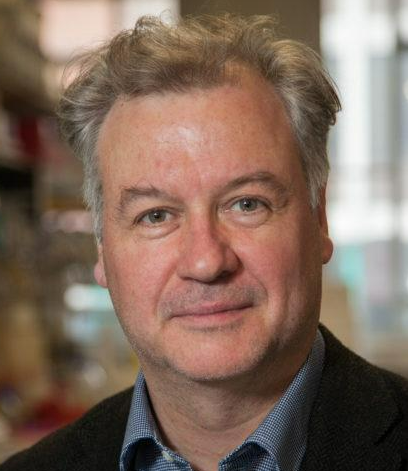 Peter Szlosarek合作论文数Barts Cancer Institute, Queen Mary University of London4Farhad Afshar合作论文数4Paul Benjamin Loughrey合作论文数3Peter E Hall合作论文数3Brian Herron合作论文数3Kate Smith合作论文数3Philip Weir合作论文数3Ajith Kumar合作论文数3Kevin O'Neill合作论文数3
 Stephanie Craig合作论文数3Lee Martin合作论文数3Plowman Piers N合作论文数3
 Harvinder Chahal合作论文数3
 Pinaki Dutta合作论文数Department of Radiation Oncology, University of Pennsylvania3
 Stephen Ellis合作论文数Department of Cardiovascular Medicine, Cleveland Clinic Foundation3N Glynn合作论文数3
 Sarah Jefferies合作论文数Addenbrooke's Hospital;Genesis Cancer Care UK Limited;CNS3Jacqueline A. James合作论文数3




