In the last thirty years, tissue engineering (TI) has emerged as an alternative method to regenerate tissues and organs and restore their function by implanting specific lineage cells, growth factors, or biomolecules functionalizing a matrix scaffold. Recently, several pathologies have led to bone loss or damage, such as malformations, bone resorption associated with benign or malignant tumors, periodontal disease, traumas, and others in which a discontinuity in tissue integrity is observed. Bone tissue is characterized by different stiffness, mechanical traction, and compression resistance as a function of the different compartments, which can influence susceptibility to injury or destruction. For this reason, research into repairing bone defects began several years ago to find a scaffold to improve bone regeneration. Different techniques can be used to manufacture 3D scaffolds for bone tissue regeneration based on optimizing reproducible scaffolds with a controlled hierarchical porous structure like the extracellular matrix of bone. Additionally, the scaffolds synthesized can facilitate the inclusion of bone or mesenchymal stem cells with growth factors that improve bone osteogenesis, recruiting new cells for the neighborhood to generate an optimal environment for tissue regeneration. In this review, current state-of-the-art scaffold manufacturing based on the use of polycaprolactone (PCL) as a biomaterial for bone tissue regeneration will be described by reporting relevant studies focusing on processing techniques, from traditional—i.e., freeze casting, thermally induced phase separation, gas foaming, solvent casting, and particle leaching—to more recent approaches, such as 3D additive manufacturing (i.e., 3D printing/bioprinting, electrofluid dynamics/electrospinning), as well as integrated techniques. As a function of the used technique, this work aims to offer a comprehensive overview of the benefits/limitations of PCL-based scaffolds in order to establish a relationship between scaffold composition, namely integration of other biomaterial phases’ structural properties (i.e., pore morphology and mechanical properties) and in vivo response.
The use of electrospun fibers as anti-inflammatory drug carriers is currently one of the most interesting approaches for the design of drug delivery systems. In recent years, biodegradable polymers blended with naturally derived ones have been extensively studied to fabricate bioinspired platforms capable of driving biological responses by releasing selected molecular/pharmaceutical signals. Here, sodium diclofenac (DicNa)-loaded electrospun fibers, consisting of polycaprolactone (PCL) or gelatin-functionalized PCL, were studied to evaluate fibroblasts’ in vitro and in vivo response. In vitro studies demonstrated that cell adhesion of L929 cells (≈70%) was not affected by the presence of DicNa after 4 h. Moreover, the initial burst release of the drug from PD and PGD fibers, e.g., 80 and 48%, respectively, after 5 h—combined with its sustained release—did not produce any cytotoxic effect and did not negatively influence the biological activity of the cells. In particular, it was demonstrated that the addition of gelatin concurred to slow down the release mechanism, thus limiting the antiproliferative effect of DicNa, as confirmed by the significant increase in cell viability and collagen deposition after 7 days, with respect to PCL alone. In vivo studies in a rat subcutaneous model also confirmed the ability of DicNa-loaded fibers to moderate the inflammatory/foreign body response independently through the presence of gelatin that played a significant role in supporting the formation of small-caliber vessels after 10 days of implantation. All of these results suggest using bicomponent fibers loaded with DicNa as a valid therapeutic tool capable of supporting the wound healing process and limiting in vivo inflammation and rejection phenomena.
La impresión 3D es una herramienta innovadora que ha experimentado un crecimiento significativo en diversas disciplinas, tanto industriales como médicas. En particular, el campo odontológico ha aprovechado las características únicas que esta tecnología ofrece, entre las cuales destacan: (i) La personalización de diferentes dispositivos o modelos dentales; mediante el uso de imágenes médicas obtenidas de tomografías, resonancias y escaneos intraorales y extraorales, es posible diseñar y fabricar modelos dentales adaptados a las necesidades específicas de cada paciente. (ii) La precisión en la fabricación de los dispositivos dentales; las diferentes técnicas de manufacturación aditiva, como la estereolitografía (SLA), el modelado por deposición fundida (FDM), la sinterización por láser (SLS), el procesado digital de luz (DLP) y la pantalla de cristal líquido (LCD), ofrecen una alta resolución que garantiza la precisión en la fabricación de dispositivos dentales. (iii) Mejora en la eficiencia clínica; debido a la disminución en los tiempos de fabricación, al estandarizar y controlar el flujo de trabajo digital en comparación con los métodos tradicionales. (iv) Los avances en materiales para la impresión 3D, en particular el uso de resinas biocompatibles en las técnicas de fotopolimerización (SLA, DLP y LCD), que son ampliamente empleadas en el campo dental. Además, se utilizan otros materiales con menor frecuencia, como polímeros termoplásticos, aleaciones metálicas, cerámicas e hidrogeles. (v) Variedad de aplicaciones según la especialidad odontológica. Por consiguiente, se exploran las contribuciones de la impresión 3D en cada especialidad odontológica con el propósito de analizar los avances tecnológicos y su impacto en la atención de los pacientes.
The E6 and E7 oncoproteins of high-risk types of human papillomavirus (HR-HPV) are crucial for the development of cervical cancer (CC). Small interfering RNAs (siRNAs) are explored as novel therapies that silence these oncogenes, but their clinical use is hampered by inefficient delivery systems. Modification (pegylation) with polyethylene glycol (PEG) of liposomal siRNA complexes (siRNA lipoplexes) may improve systemic stability. We studied the effect of siRNA targeting HPV16 E6, delivered via cationic liposomes (lipoplexes), on cellular processes in a cervical carcinoma cell line (CaSki) and its potential therapeutic use. Lipoplexes-PEG-HPV16 E6, composed of DOTAP, Chol, DOPE, and DSPE-PEG2000 were prepared. The results showed that pegylation (5% DSPE-PEG2000) provided stable siRNA protection, with a particle size of 86.42 ± 3.19 nm and a complexation efficiency of over 80%; the siRNA remained stable for 30 days. These lipoplexes significantly reduced HPV16 E6 protein levels and restored p53 protein expression, inhibiting carcinogenic processes such as proliferation by 25.74%, migration (95.7%), and cell invasion (97.8%) at concentrations of 20 nM, 200 nM, and 80 nM, respectively. In conclusion, cationic lipoplexes-PEG-HPV16 E6 show promise as siRNA carriers for silencing HPV16 E6 in CC.
During the preparation of fixed prosthesis (including individual bridges and crowns) it is important to select the materials that have the best features and properties to predict a successful clinical treatment. The objective of this study was to determine if the chemical and structural characteristics could cause to increase the fracture resistance, we used four bis-acryl resins Luxatemp, Protemp, Structur and Telio. Three-points bending by Flexural test were performed in ten bars and they were carried out to compare with Anova test. In addition, the bis-acryl resins were analyzed by scanning electron microscopy, to analyze microstructure and morphology and the molecular structure were performed by Infrared Spectroscopy through Attenuated Total Reflectance. A higher flexural strength was found in Luxatemp and Structur with, no significant differences between this study groups. Regarding Protemp and Telio, these study groups showed a lower flexural strength when were compared with Luxatemp and Structur. These results corroborate SEM and ATR analysis because Luxatemp sample showed a regular size particle on the surface and chemically presents a long cross-linkage polymer chain. The presence of CO3, SiO2 and N-H groups as a fillers particle interacting with OH groups cause a higher flexural strength compared with another groups.
Este trabajo muestra la obtención de osteoesferoides con la capacidad de formar nódulos mineralizados, puediendo utilizarse como modelos de estudio para probar nanofármacos o nanomateriales. Se realizaron cultivos celulares en suspensión para obtener esferoides de osteoblastos fetales, probando diferentes concentraciones celulares/mL durante 5 días. Se seleccionó la condición donde el diámetro va de 80 a 150 μM para realizar ensayos a 3, 7, 14 y 21 días. La viabilidad celular de los osteoesferoides se cuantificó mediante exclusión con azul tripano y se realizaron ensayos clonogénicos para determinar el efecto del medio mineralizante en la formación de estas estructuras. La integridad de los osteoesferoides se observó por H&E y la formación de nódulos mineralizados fue detectada mediante tinción con alizarina roja. Los resultados muestran osteoesferoides regulares sin coagregados a una concentración de 5 x 10² células /mL y viables por arriba del 70% a los 7 días de formación. Los ensayos clonogénicos no muestran diferencias significativas en morfología ni el número de colonias entre el control y el medio para inducir mineralización. Las tinciones con H&E dejan ver núcleos y citoplasma definido a los 3, 7 y 14 días, y a los 7 y 14 días la tinción con alizarina roja sugiere que están formando deposiciones de calcio.
Andamios impresos en 3D con poros heterogéneos, surge como estrategia para la regeneración de tejidos. En este estudio, se evaluó la regeneración ósea en defectos críticos de ratas Wistar, debido a la osteoconducción de andamios de poliácido-láctico (PAL), impresos en 3D con diferentes tamaños de poros; 250-300 µm en la periferia, seguido de 350-400 µm y en el centro 400-740 µm, debido a que los pequeños promueven adhesión celular, mientras que los grandes la angiogénesis. Los andamios se imprimieron en 3D con PLA, un material termoplástico, biocompatible, biorreabsorbible, aprobado por la Administración de Alimentos y Medicamentos de los Estados Unidos (FDA, por sus siglas en inglés), evaluando tamaño de poro y porosidad, in vivo, en defectos de 9 mm de diámetro en calvarias de ratas, calculando el tejido mineralizado por la radiodensidad de las unidades Hounsfield (UH) en imágenes microtomográficas a 8, 30, 60 y 90 días. Los resultados demostraron rango de poros de 200-800 µm (como el diseño), la porosidad fue del 98%, favoreciendo el flujo de nutrientes, oxígeno y eliminación de desechos. Se observó in vivo tejido radiodenso al día 30, evidentemente al 90, concordando con las UH 93.66 y 118.31, respectivamente. Los andamios 3D con poros heterogéneos, demostraron su capacidad osteoconductora en la regeneración ósea, abriendo alternativas en la bioingeniería tisular.
The challenge of three-dimensional (3D) printing by polymeric extrusion in tissue bioengineering is to control with precision the microarchitecture and porous interconnectivity of scaffolds, as well as search for models that allow and facilitate the development of personalized constructs that meet optimal characteristics for the regeneration of significant bone defects. In this study, anatomically accurate scaffolds were designed and printed to a critical size defect from a microtomography image of the rat calvaria. Different software is used to design a scaffold with exact anatomy. With Ultimaker Cura software, distinct printing parameters were standardized, permitting the printing of different types of pores and graded porosity scaffolds, with exact adaptation to the bone defect, utilizing a commercial 3D printer with a fused deposition modeling technique and compensating for the limitations of the method. The scaffolds were characterized by evaluating their mechanical properties and surface characteristics (pore size and porosity), employing scanning electron microscopy images, verifying that the size and shape of the pores were controlled, and evaluating cell viability and cell distribution on the 3D printed scaffold. Therefore, this work proves that by standardizing the printing parameters, it was possible to print a unique customized scaffold, controlling the shape and size of pores.
Maintaining pulp vitality and function is a priority of the medicaments employed in pulp therapy to preserve tooth integrity. Aim: This study evaluated inflammatory response and reparative dentin bridge formation after direct pulp capping with two different bioceramics. Materials and Method: This was an in vivo controlled experimental study on 12 male Wistar rats. Pulpotomies were performed and the exposed pulps were capped with Biodentine or Neo MTA. After 15, 45 and 90 days, maxillary segments were obtained and prepared for histologic analysis and Micro-CT. Hounsfield Units (HU) were quantified. Results: Micro-CT analysis showed greater mineralization at 90 days with Neo MTA than with Biodentine. HU did not differ significantly (p >0.05) between molars treated with Biodentine and Neo MTA at 15 and 45 days, but at 90 days, there was statistically significant difference (p <0.05) between them. Reparative dentin was observed near the pulp exposure and canal orifice with both bioceramics. At 45 and 90 days, molars treated with Neo MTA showed mineralized tissue filling the canal orifice. Molars treated with Biodentine showed mineralized tissue and dentin bridge at the site of exposure at 45 days, and total pulp exposure coverage and mineralized entin matrix at 90 days. Conclusions: Biodentine and Neo MTA induce the formation of reparative dentin bridge after 45 days with inflammatory cell infiltrate.
The advent of 3D printing technologies has led to a new era in tissue regeneration, offering unparalleled precision and versatility in fabricating scaffolds for various biomedical applications. Robocasting and stereolithography stand out among these technologies for their distinct advantages in engineering complex tissue structures. Robocasting, an extrusion-based 3D printing technique, has shown significant promise in cartilage tissue engineering. Its ability to precisely deposit biomaterials layer-by-layer allows the creation of porous scaffolds with encapsulated cells and bioactive molecules that improve mechanical properties and biological functionality. These scaffolds support the proliferation and differentiation of chondrocytes or mesenchymal stem cells, facilitating the regeneration of damaged cartilage. Stereolithography, a light-based 3D printing technique, offers remarkable resolution and accuracy with highly detailed structures that closely resemble the complex architecture of tissues. This makes it particularly suitable for new treatments for injuries and diseases of cartilage and ocular tissue regeneration. Integrating robocasting and stereolithography presents a synergistic approach to replicating cartilage and ocular tissues' complex physical and biological environments, offering innovative regenerative therapies. This review highlights the recent advancements and potential of robocasting and stereolithography in regenerative medicine, focusing on their applications in cartilage and ocular tissue regeneration.
Abstract Objective To determine if the chemical and structural characteristics could cause to increase the fracture resistance using four bis-acryl resins Luxatemp, Protemp, Structur and Telio. Materials and Methods Three-points bending by Flexural test, SEM, and FTIR were carried out to compare the Kruskal-Wallis test. Results The Kruskal-Wallis test indicated a significant difference in the materials (< 0.0001) that showed higher flexural strength found in Luxatemp, while comparing all bis-acryl resins by Dunn's multiple comparison test indicate that there was no significant difference between Luxatemp VS Structur, and Protemp VS Telio. SEM analysis showed that different sizes of filler particles were observed in all samples, which causes them to present irregular surfaces. By FTIR, typical C = O and CH2 groups were observed corresponding to methyl methacrylate and methylene groups respectively, and different bands located at 1460 − 1350 cm− 1 assigned to pigment materials, stabilizers, and filler particles. In conclusion, a long cross-linkage polymer chain, and the presence of CO3, SiO2 and N-H groups as a fillers particle interacting with OH groups could be the cause of our results. Conclusion It is necessary a longer storage under a different condition that would emulate the oral environment and allow us to determine changes in the chemical compounds as well as changes in ultrastructural characteristics related to mechanical properties to identify their correct clinical use. Clinical Relevance During the preparation of fixed prosthesis (including individual bridges and crowns) it is important to select the materials that have the best features and properties to predict a successful clinical treatment.
In the few decades, tissue engineering has been a compelling emerging technology that is expected to impact the fields of diagnostics, therapeutic sciences, and the prevention of diseases in a fundamental way. Most of the current research has been focused on optimizing the biomechanical and physicochemical properties of nanomaterials to induce regeneration. However, the clear benefits of tissue engineering are often confronted by concerns about the lack of adequate information regarding their interactions with the immune system. The present manuscript explores such properties and interactions in materials used in craniofacial tissue regeneration.
The design of scaffolds that could adjust and adapt greatly to bone defects is a significant challenge to bone tissue engineering. Recently, 3D printing technology emerged as a process that could precisely control the architecture and design an exact geometry of the scaffold to the site or defect where it will be implanted. Thus, this research aimed to design and synthesize individualized constructs of polylactic acid (PLA) by 3D printing using microtomographic images to fit the edge of Wistar rat calvaria's critical size defects. The 3D-printed construct using the DICOM data of microtomographic images process to STL manipulated and designed by various software showed an excellent geometry. The in vitro biocompatibility assay of the 3D printing scaffold was evaluated by WST-1, showing an excellent biological response. Moreover, the in vivo evaluation of the bone regeneration process onto the rat calvaria defect model measured by the bone mineral density (BMD) at 8, 30, 60, and 90 days via micro-CT showed that at the end of the evaluation period, the 3D construct was integrated into the edges of the bone tissue, and new tissue deposited was in the process of mineralization. These findings suggest that the 3D construct matches the calvaria defect, allowing the novo mineral tissue to form. These individualized printed scaffolds may be a promising candidate in bone tissue engineering for future regeneration strategies.
Bone is one of the most affected tissue during radiation therapy treatments. In postmenopausal women, the risk of a hip and femoral head fracture due to osteoporosis induced by ionizing radiation increases from 65% to 216% after receiving radiation treatment for cervical, anal, or rectal cancer. To date, there is no preventive or curative treatment for this damage; this situation promotes to explore preclinical models of radio-osteoporosis. In this work, the pelvic region of adult (male and female) Wistar rats were irradiated with a 6 MV photon beam. Four schemes of two fractions (2 fx) and one scheme of four fractions (4 fx), all with different fractional doses, were evaluated. microCT images were acquired two and four months after irradiations to evaluate changes in the hydroxyapatite concentration [HA] in the hip and femoral head. After imaging, animals were sacrificed, and the femurs were removed to perform histology and scanning electron microscopy studies to evaluate changes in trabecular bone microarchitecture. Low [HA] in the femoral head was observed in male rats (30-weeks-old) after two months post-irradiations. In the hip, a reduction of [HA] was observed after four months. No significant changes were detected in the bone microarchitecture. In female rats, no significant differences (experimental vs. control) in [HA] were observed. This result was associated with the age of rats (12-week-old) at the time of irradiations; it is known that the repair of radiation damage in bone tissue is more efficient in younger rats than in older rats. Visual inspection in the shape of the microarchitecture shows structural differences in the irradiated group; however, quantitative differences in the parameters of the evaluation were not detected. The fractionation schemes of irradiation implemented in this work result in bone damage that could promote radio-osteoporosis. Work is in progress with different image analysis techniques to quantify the damage in the microarchitecture.
The FDA's approval of peptide drugs such as Ziconotide or Exendin for pain relief and diabetes treatment, respectively, enhanced the interest to explore novel conotoxins from Conus species venom. In general, conotoxins can be used in pathologies where voltage-gated channels, membrane receptors, or ligands alter normal physiological functions, as in metabolic diseases such as Type 2 diabetes. In this study, the synthetic cal14.2b (s-cal14.2b) from the unusual Californiconus californicus demonstrated bioactivity on NIT-1 insulinoma cell lines stimulating insulin secretion detecting by high performance liquid chromatography (HPLC). Accordingly, s-cal14.2b increased the CaV1.2/1.3 channel-current by 35 ± 4% with a recovery τ of 10.3 ± 4 s in primary cell culture of rat pancreatic β-cells. The in vivo results indicated a similar effect of insulin secretion on mice in the glucose tolerance curve model by reducing the glucose from 500 mg/dL to 106 mg/dL in 60 min, compared to the negative control of 325 mg/dL at the same time. The PET-SCAN with radiolabeling 99mTc-s-cal14.2b demonstrated biodistribution and accumulation in rat pancreas with complete depuration in 24 h. These findings show the potential therapeutic use of s-cal14.2b in endocrinal pathologies such as early stages of Type 2 Diabetes where the pancreas's capability to produce insulin is still effective.
Bone tissue engineering is a multidisciplinary field that includes biology, medicine and engineering areas, of which the main objective is bone regeneration. The craniofacial bone defects are challenges for tissues engineering because of the presence of complex physiological structures, including cartilage, facial skeletal features, muscles, skin, ligaments, blood vessels and nerves. This chapter describes the most important etiologies of craniofacial defects. The most common birth congenital anomalies, include orofacial clefts, craniosynostoses, the mandibulofacial dysostoses and craniofacial macrosomia. One common cause of craniofacial defects is trauma including acute trauma, falls, assaults, sport injuries and vehicle accidents. Bone grafting is one of the most commonly used surgical methods to augment bone regeneration, and the second most frequent tissue transplantation just after blood transfusion. The main objective is to have promising and efficient treatments that achieve regeneration anatomically and physiologically of the craniofacial area, thus promoting personalized therapies, which can be custom designed using tomographic images.
In the last two decades, alginate scaffolds have been variously studied as extracellular matrix analogs for tissue engineering. However, relevant evidence is still lacking concerning their ability to mimic the microenvironment of hierarchical tissues such as bone. Hence, an increasing amount of attention has recently been devoted to the fabrication of macro/microporous sponges with pore anisotropy able to more accurately replicate the cell niche structure as a trigger for bioactive functionalities. This paper presents anin vivostudy of alginate sponges with anisotropic microporous domains (MAS) formed by ionic crosslinking in the presence of different fractions (30 or 50% v) of hydroxyapatite (HA). In comparison with unloaded sponges (MAS0), we demonstrated that HA confers peculiar physical and biological properties to the sponge, depending upon the inorganic fraction used, enabling the sponge to bio-mimetically support the regeneration of newly formed bone. Scanning electron microscopy analysis showed a preferential orientation of pores, ascribable to the physical constraints exerted by HA particles during the pore network formation. Energy dispersive spectroscopy (EDS) and X-Ray diffraction (XRD) confirmed a chemical affinity of HA with the native mineral phase of the bone.In vitrostudies via WST-1 assay showed good adhesion and proliferation of human Dental Pulp-Mesenchymal Stem Cells (hDP-MSC) that increased in the presence of the bioactive HA signals. Moreover,in vivostudies via micro-CT and histological analyses of a bone model (e.g., a rat calvaria defect) confirmed that the maximum osteogenic response after 90 days was achieved with MAS30, which supported good regeneration of the calvaria defect without any evidence of inflammatory reaction. Hence, all of the results suggested that MAS is a promising scaffold for supporting the regeneration of hard tissues in different body compartments.
Fibers have received increased attention in biomedical applications for their ability to mimic the morphological and/or chemical features or the native extracellular matrix of native tissues. In recent years, different technologies have been optimized to manipulate biomaterials in the form of fibers. In this chapter, an overview of the main technologies used to produce fiber at micro- and submicrometric scale are discussed, underlining key points, benefits, and limitations of electric and nonelectric-assisted processing techniques.
3D printing with controlled microarchitectures has gained traction in a wide variety of fields, including bone tissue engineering, because it represents an exciting alternative for the synthesis of new scaffolds due to its rapid manufacturing process, high precision, cost-effectiveness, and ease of use. Thus, this study is aimed at evaluating the biocompatibility response of a 3D-printed tubular scaffold coated by a layer of 7% PLA nanofibers. The morphology, structure, and chemical composition of the 3D-printed tubular scaffold were characterized by scanning electron microscopy (SEM), X-ray diffraction (XRD), Fourier Transform Infrared (FTIR), thermogravimetric analysis (TGA), differential scanning calorimetry (DSC), and surface property analysis by profilometry. The biocompatibility response of the scaffold was assessed by cell adhesion, proliferation, and cell-material interactions of human fetal osteoblasts. Our results showed that 3D printing allowed obtaining similar and reproducible structures and the biocompatibility assays showed that nanofiber coating of the surface of the 3D tubular scaffold promoted an improvement on cell attachment, proliferation, and the morphology of osteoblast cells when compared with a noncoated scaffold. In conclusion, the surface of the 3D-printed tubular scaffold could be improved by the deposition of a nanofiber layer to render a more mimetic and active topography with excellent cellular biocompatibility for bone tissue applications.
 Marco Antonio Alvarez-Perez合作论文数11
 Luis A Medina合作论文数8Osmar Alejandro Chanes-Cuevas合作论文数5Marco A. Alvarez-Perez合作论文数5
 Vincenzo Guarino合作论文数Federico II Interdisciplinary Research Centre in Biomaterials (CRIB), University of Naples5
 Patricia Gonzalez-Alva合作论文数5Febe Carolina Vázquez-Vázquez合作论文数4Monserrat Llaguno合作论文数4
 Rodrigo Correa-Prado合作论文数3Enrique Romo合作论文数3Iriczalli Cruz-Maya合作论文数Institute of Polymers, Composites and Biomaterials, National Research Council of Italy3
 Margarita Zeichner-David合作论文数School of Dentistry, USC3Higinio Arzate合作论文数Laboratorio de Biología Celular y Molecular, Universidad Nacional Autónoma de México3christian guerra合作论文数2Norma O Uribe-Uribe合作论文数2Elias Nahum Salmeron-Valdes合作论文数Ctr Res & Adv Studies Dent, Autonomous Univ Mexico State2Patricia García-López合作论文数Lab Farmaco Oncol, Inst Nacl Cancerol2
 Gabriela E Mercado合作论文数Subdirection of Basic Research, National Institute of Genomic Medicine;Subdirection of Basic Research, National Institute of Genomic Medicine2Romero-Piña Mario合作论文数2Beatriz Marcela Millan Malo合作论文数Universidad Nacional Autónoma de México2
 Eric Rivera-Muñoz合作论文数, FATA, UNAM2O. Alvarez-Fregoso合作论文数Instituto de Investigaciones en Materiales, UNAM2
 Jose Luis Suarez Franco合作论文数2Silvia Maldonado-Frías合作论文数2
 David Masuoka合作论文数Rheumatic Disease Section, University of Southern California2daniel chavarria bolanos合作论文数1carlos alvarez gayosso合作论文数1Noemí Sánchez-Campos合作论文数1Fernando Suaste-Olmos合作论文数1Julia Buján-Varela合作论文数1Iñigo Gaitán-Salvatella合作论文数1
 Alexei Licea合作论文数1Linda Nuñez-Garcia合作论文数1Manuel Aguilar-Franco合作论文数Univ Nacl Autonoma Mexico, Ciudad Univ1
 Mercedes Bermudez合作论文数Fac Odontol, Autonomous Univ Chihuahua1Pavel Lugo合作论文数1
 Carmen L Peza-Ledesma合作论文数Centro de Física Aplicada y Tecnología Avanzada, Universidad Nacional Autónoma de México1
 Ciresthel Bello合作论文数Lab Biomed Mol, Univ Autonoma Guerrero1
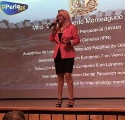 Cynthia Trejo合作论文数Dental Research Laboratory Almaraz/UBIMED., DDS1
 Saul Ernesto Cifuentes-Mendiola合作论文数Lab Dent Res, Univ Nacl Autonoma Mexico1Isabel Arenas合作论文数1
 Berenice Illades-Aguiar合作论文数Laboratorio de Biomedicina Molecular, Universidad Autónoma de Guerrero1As Narayanan合作论文数1Antonio Barajas-Martínez合作论文数1Marine Ortiz-Magdaleno合作论文数1
 Lizbeth de la Cruz合作论文数1Johanna Bernáldez-Sarabia合作论文数1
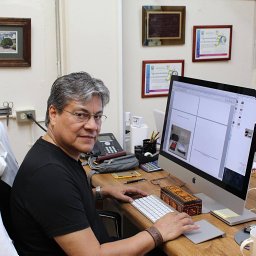 Marco Antonio Leyva-Vazquez合作论文数Laboratorio de Biomedicina Molecular, Universidad Autónoma de Guerrero1
 Gómez-Gómez Yazmín合作论文数Laboratories of Molecular Biomedicine, and.1




