IntroductionNeuropathic pain is one of the most common and debilitating complications following spinal cord injury (SCI), frequently surpassing motor and sensory deficits as the symptom patients most want treated. Despite advances in understanding the molecular and physiological mechanisms underlying central neuropathic pain, effective treatments remain lacking and show wide variability in efficacy. Previous reports have indicated that early intervention represents the most effective pain management strategy, underscoring the clinical importance of identifying patients at risk during the acute care phase.MethodsWe utilized the TRACK-SCI prospective clinical research database to assess neuropathic pain outcomes in all enrolled SCI patients and identify acute care variables predictive of chronic neuropathic pain development. Pain status was evaluated at 6 and 12 months post-injury. Candidate predictors were analyzed using multidimensional analytics, and a logistic regression model was constructed and validated using repeated 5-fold cross-validation.ResultsOf 61 patients in the study cohort, 36 (59%) reported neuropathic pain in the chronic stages after SCI. Four acute care variables were identified as significant predictors of chronic neuropathic pain development: (1) the total number of systemic injuries sustained, (2) the injury severity score (ISS), (3) the lower limb total motor score, and (4) the sensory pinprick total score. The logistic regression model achieved a balanced accuracy of 74.3%, and repeated 5-fold cross-validation yielded an AUC of 0.708.DiscussionThese findings highlight a crucial role of polytrauma in the development of chronic pain after SCI. The four identified predictors are parameters routinely measured in every trauma center, making the proposed model readily translatable to clinical practice. This predictive tool may enable earlier, targeted intervention for at-risk patients, addressing the clinical need for proactive pain management strategies in the acute post-SCI setting. Future work should validate this model in larger, independent cohorts and explore its utility in guiding early treatment decisions.
We assessed the extent to which initial injury severity, functional independence, and health-related quality of life (QoL) predict overall QoL in adults during their first year of recovery following a traumatic spinal cord injury (SCI). Data were collected from two level-I trauma centers and outpatient follow-up care as part of the longitudinal, prospective, and multicenter Transforming Research and Clinical Knowledge in SCI study. Participants included adults with traumatic SCI presenting acutely within 24-h of injury (N = 115). Functional independence was measured using the Spinal Cord Independence Measure Version III, health-related QoL was measured using 11 short-form questionnaires from the QoL in Neurological Disorders (Neuro-QoL) measurement system, and overall QoL was measured using the International SCI QoL Basic Data Set. Injury severity and functional independence measures were not significant predictors of overall QoL. Ten of the 11 Neuro-QoL questionnaires were strongly associated with overall QoL (p ≤ 0.001-0.015). In a multivariable regression model, depression (p = 0.002) and satisfaction with social roles/activities (p < 0.001) maintained significance with overall QoL at 6-12 months post-SCI. Findings indicate that patient-reported mental and social well-being may be more important to overall QoL than injury severity, functional independence, or physical health-related QoL during the first year of recovery following traumatic SCI. This reveals the importance of incorporating mental and social health care plans during early SCI rehabilitation.
The RSNA Lumbar Degenerative Imaging Spine Classification dataset is the largest publicly available adult MRI lumbar spine dataset for degenerative disease. The dataset includes multisequence, multiplanar MRIs from 2,697 patients through contributions from 8 institutions across 6 countries and 5 continents. ©RSNA, 2026.
Traumatic brain injury can lead to venous sinus injury and thrombosis, which may be associated with elevated intracranial pressure and poor outcomes. We sought to examine the risk factors, management, and clinical outcomes of traumatic venous sinus thrombosis (tVST). We conducted a comprehensive search of our institutional radiology database for final radiology reports from 2013 to 2022 that contained the terms “venous sinus thrombosis,” “sinus thrombosis,” or “venous occlusion.” tVST was detected on computed tomography and confirmed by a board-certified neuroradiologist. We identified 135 patients on initial screening and entered 112 into our final analysis. Patients were predominantly male (76.8
Traumatic spinal injuries are common and carry a high risk for severe morbidity and mortality. While computed tomography remains the primary screening imaging method for acute spinal trauma, MR imaging plays an important complementary role in emergency management and triage for cervical spine trauma. This review examines the benefits and limitations of MR imaging, focusing on its indications, structured assessment of spinal stability, and evolving role for evaluation of spinal cord injuries.
Deep learning models have achieved remarkable success in segmenting brain white matter lesions in multiple sclerosis (MS), becoming integral to both research and clinical workflows. While brain lesions have gained significant attention in MS research, the involvement of spinal cord lesions in MS is relatively understudied. This is largely owing to the variability in spinal cord magnetic resonance imaging (MRI) acquisition protocols, high individual anatomical differences, the complex morphology and size of spinal cord lesions, and lastly, the scarcity of labeled datasets required to develop robust segmentation tools. As a result, automatic segmentation of spinal cord MS lesions remains a significant challenge. Although some segmentation tools exist for spinal cord lesions, most have been developed using sagittal T2-weighted (T2w) sequences primarily focusing on cervical spines. With the growing importance of spinal cord imaging in MS, axial T2w scans are becoming increasingly relevant due to their superior sensitivity in detecting lesions compared to sagittal acquisition protocols. However, most existing segmentation methods struggle to effectively generalize to axial sequences due to differences in image characteristics caused by the highly anisotropic spinal cord scans. To address these challenges, we developed a robust, open-source lesion segmentation tool tailored specifically for axial T2w scans covering the whole spinal cord. We investigated key factors influencing lesion segmentation, including the impact of stitching together individually acquired spinal regions, straightening the spinal cord, and comparing the effectiveness of 2D and 3D convolutional neural networks (CNNs). Drawing on these insights, we trained a multi-center model using an extensive dataset of 582 MS patients, resulting in a dataset comprising an entirety of 2,167 scans. We empirically evaluated the model's segmentation performance across various spinal segments for lesions with varying sizes. Our model significantly outperforms the current state-of-the-art methods, providing consistent segmentation across cervical, thoracic, and lumbar regions. To support the broader research community, we integrate our model into the widely-used Spinal Cord Toolbox (v7.0 and above), making it accessible via the command sct_deepseg lesion_ms_axial_t2 -i .
OBJECTIVES:Fentanyl has become the primary drug responsible for fatal overdoses in most urban US regions. Information about the impact of fentanyl-related overdose in neurological outcomes after cardiac arrest (CA) compared with other etiologies of CA is limited. METHODS:Retrospective review of medical records from adult patients with out-of-hospital CA who had admission drug testing for fentanyl and opioids from August 2019 to June 2021. Good outcome was defined as a Cerebral Performance Category score of 1-2 at discharge. χ 2 was used for group comparison. RESULTS:Neurological prognosis evaluation was pursued for 233 patients, and 61 (26.2%) met criteria for good outcome. Thirty-six (15.45%) patients tested positive for fentanyl and 13 for other opioids (5.58%). The proportion of good outcomes was similar between groups (fentanyl 22.2%, other opioids 38.5%, nonopioid 26.1%, P = 0.52). Fewer fentanyl-related CA had bystander cardiopulmonary resuscitation (19.4% vs other opioids 38.5% vs nonopioid 43.8%, P = 0.02) shockable rhythms (2.9%, 16.7%, 25%, P = 0.01) or corneal reflexes 72 hours after CA (25.8%, 66.7%, 39.8%, P = 0.046), but no difference was seen for pupillary response at 72 hours ( P = 0.17). More fentanyl-related CA cases had signs of severe brain dysfunction on EEG with burst suppression (54.8%, 0%, 39.4%, P = 0.01). CONCLUSIONS:Cardiac arrest associated with fentanyl use was linked to decreased rates of bystander cardiopulmonary resuscitation, increased incidence of nonshockable Rhythms, and greater neurological injury as indicated by electroencephalography (EEG) suppression measures. However, the proportion of good neurological outcomes (CPC: 1-2) was similar across groups.
Spinal cord pathologic condition often presents as a neurologic emergency where timely and accurate diagnosis is critical to expedite appropriate treatment and minimize severe morbidity and even mortality. MR imaging is the gold standard imaging technique for diagnosing patients with suspected spinal cord pathologic condition. This review will focus on the basic principles of diffusion imaging and how spinal anatomy presents technical challenges to its application. Both the promises and shortcomings of spinal diffusion imaging will then be explored in the context of several clinical spinal cord pathologies for which diffusion has been evaluated.
BACKGROUND AND PURPOSE:Cerebral venous sinus thrombosis (CVST) is an underrecognized cause of morbidity in acute traumatic brain injury (TBI). Radiologic diagnosis is challenging in the setting of concurrent extra-axial injury and a lack of standardized diagnostic criteria. The prevalence of traumatic thrombosis versus compression is unknown. Treatment with anticoagulation is often determined by the appropriate classification of the type of traumatic venous injury.METHODS:We developed a two-part radiologic grading method for standardized assessment of traumatic CVST based on (1) the degree of flow limitation through the affected sinus and (2) the location of venous pathology (ie, external compression vs. intrinsic thrombosis) based on computed tomography venography. We applied this grading method to a retrospective cohort of TBI patients presenting to a Level 1 Trauma center. Chart review was performed to identify potential clinical correlates. A senior neuroradiologist graded the entire cohort and a random subsample was selected for blinded rating by two independent neuroradiologists.RESULTS:Seventy-six of 221 patients were identified for inclusion after excluding nontraumatic mechanisms. Seven unique grades were employed to characterize the full extent of venous injuries. The plurality of patients from the cohort (43/76 = 43.4%) suffered compressive injuries. Inter-rater reliability was moderate for the combined grade, kappa = 0.48, p<.05, and substantial for the flow limitation component, kappa = 0.69, p<.05.CONCLUSIONS:We introduce a standardized two-part classification system for traumatic venous sinus injury with moderate-substantial inter-rater reliability. Compressive injuries were more common than thrombotic injuries. Further prospective work is needed to validate the clinical significance of this classification system.
AbstractBackgroundAlthough many molecules have been investigated as biomarkers for spinal cord injury (SCI) or ischemic stroke, none of them are specifically induced in central nervous system (CNS) neurons following injuries with low baseline expression. However, neuronal injury constitutes a major pathology associated with SCI or stroke and strongly correlates with neurological outcomes. Biomarkers characterized by low baseline expression and specific induction in neurons post‐injury are likely to better correlate with injury severity and recovery, demonstrating higher sensitivity and specificity for CNS injuries compared to non‐neuronal markers or pan‐neuronal markers with constitutive expressions.MethodsIn animal studies, young adult wildtype and global Atf3 knockout mice underwent unilateral cervical 5 (C5) SCI or permanent distal middle cerebral artery occlusion (pMCAO). Gene expression was assessed using RNA‐sequencing and qRT‐PCR, while protein expression was detected through immunostaining. Serum ATF3 levels in animal models and clinical human samples were measured using commercially available enzyme‐linked immune‐sorbent assay (ELISA) kits.ResultsActivating transcription factor 3 (ATF3), a molecular marker for injured dorsal root ganglion sensory neurons in the peripheral nervous system, was not expressed in spinal cord or cortex of naïve mice but was induced specifically in neurons of the spinal cord or cortex within 1 day after SCI or ischemic stroke, respectively. Additionally, ATF3 protein levels in mouse blood significantly increased 1 day after SCI or ischemic stroke. Importantly, ATF3 protein levels in human serum were elevated in clinical patients within 24 hours after SCI or ischemic stroke. Moreover, Atf3 knockout mice, compared to the wildtype mice, exhibited worse neurological outcomes and larger damage regions after SCI or ischemic stroke, indicating that ATF3 has a neuroprotective function.ConclusionsATF3 is an easily measurable, neuron‐specific biomarker for clinical SCI and ischemic stroke, with neuroprotective properties.Highlights ATF3 was induced specifically in neurons of the spinal cord or cortex within 1 day after SCI or ischemic stroke, respectively. Serum ATF3 protein levels are elevated in clinical patients within 24 hours after SCI or ischemic stroke. ATF3 exhibits neuroprotective properties, as evidenced by the worse neurological outcomes and larger damage regions observed in Atf3 knockout mice compared to wildtype mice following SCI or ischemic stroke.
INTRODUCTION: Venous thromboembolism (VTE) following traumatic spinal cord injury (SCI) is a major clinical concern. Current guidelines recommend initiation of chemical prophylaxis within 72 hours of injury or surgery. Given the high reported incidence of VTE in this population, the safety and efficacy of earlier initiation is an important clinical question. METHODS: We analyzed prospectively collected data in a cohort of 162 SCI patients from a single quaternary center. Demographic and clinical data were recorded. Univariate and multivariate logistic regression analyses were performed to identify predictors of VTE in SCI patients treated with LMWH within 24 hours of injury or surgery. RESULTS: Mean age was 56.9 years (28% females). One hundred and thirty (87.8%) patients underwent SCI surgery. There was an extremity fracture in 18.2% and lumbar drain was placed in 22.3%. DVT occurred in 7.4%, PE in 6.1%, and any VTE in 12.2%. A multivariable regression model including age, sex, race, injury severity score, level of SCI, SCI surgery status, admission lower extremity motor score, and extremity fracture showed that only lower extremity motor score (OR 0.94, p=0.03) was significantly associated with VTE. There were 1.5% patients with post-SCI surgery-related bleeds requiring surgery takeback. There were 5.6% non-surgery related bleeds, and 1.2% wound dehiscence. CONCLUSIONS: Initiation of LMWH within 24 hours of injury or surgery maintained the incidence of any VTE event within the lower end of previously reported ranges with 1.5% of patients who underwent SCI surgery requiring surgery takeback due to bleeding complications. Admission lower extremity motor score was the sole predictor of VTE in this patient cohort.
PURPOSE:Timely identification of intracranial blood products is clinically impactful, however the detection of subdural hematoma (SDH) on non-contrast CT scans of the head (NCCTH) is challenging given interference from the adjacent calvarium. This work explores the utility of a NCCTH bone removal algorithm for improving SDH detection. METHODS:A deep learning segmentation algorithm was designed/trained for bone removal using 100 NCCTH. Segmentation accuracy was evaluated on 15 NCCTH from the same institution and 22 NCCTH from an independent external dataset using quantitative overlap analysis between automated and expert manual segmentations. The impact of bone removal on detecting SDH by junior radiology trainees was evaluated with a reader study comparing detection performance between matched cases with and without bone removal applied. RESULTS:Average Dice overlap between automated and manual segmentations from the internal and external test datasets were 0.9999 and 0.9957, which was superior to other publicly available methods. Among trainee readers, SDH detection was statistically improved using NCCTH with and without bone removal applied compared to standard NCCTH alone (P value <0.001). Additionally, 12/14 (86 %) of participating trainees self-reported improved detection of extra axial blood products with bone removal, and 13/14 (93 %) indicated that they would like to have access to NCCTH bone removal in the on-call setting. CONCLUSION:Deep learning segmentation-based NCCTH bone removal is rapid, accurate, and improves detection of SDH among trainee radiologists when used in combination with standard NCCTH. This study highlights the potential of bone removal for improving confidence and accuracy of SDH detection.
OBJECTIVES/GOALS: Burst suppression is a neurophysiological marker associated with severe hypoxic-ischemic injury following cardiac arrest. The goal of this study is to identify the anatomical regions of the brain associated with burst suppression post-cardiac arrest. METHODS/STUDY POPULATION: 86 comatose patients post-cardiac arrest admitted to the neurological-ICU from Massachusetts General Hospital and Brigham and Women’s Hospital were included in this study. EEG data after return of spontaneous circulation were preprocessed and artifact was rejected. Burst segments were extracted for source localization analysis from epochs with burst suppression. Four bursts for each patients were manually selected. The source of the bursts were obtained using the Champagne algorithm and mapped on the Desikan-Killiany atlas. The source for each burst was defined as any region of interest (ROI) with power > = 75th percentile relative to all ROIs. The power of the bursts at each source was correlated with the burden of brain injury measured using apparent diffusion coefficient (ADC) per ROI. RESULTS/ANTICIPATED RESULTS: 48 (56%) patients had burst suppression. 5 (10.4%) of patients with burst suppression were independent at the time of hospital discharge. Preliminary analyses was performed on 6 patients (24 bursts in total). ROI’s determined to be sources in a majority of the burst (>=13) were bilateral superior frontal, rostral middle frontal, parstriangularis precentral, superior parietal, inferior parietal, right post central, superior temporal, lateral occipital, and left middle temporal ROI. A lower mean ADC intensity was associated with a higher EEG power in the bilateral superior frontal (r = -0.80, p < 0.0001; r = -0.677, p < 0.001, respectively), left superior parietal (r = -0.53, p = 0.009), left middle temporal (r = -0.43, p = 0.042) ROI. DISCUSSION/SIGNIFICANCE: The source of bursts in patients post-cardiac arrest experiencing burst suppression is not well defined. This study will improve our understanding of how burst suppression is a measure of cortical injury, how it may relate to the burden of injury found on ADC imaging, and patient outcomes.
OBJECTIVE:The International Standards for Neurological Classification of Spinal Cord Injury (ISNCSCI) assessment is the gold standard for evaluation of neurological function after spinal cord injury (SCI). Although it is an invaluable tool for diagnostic and research purposes, it is time consuming and can be impractical in acute injury settings. Clinical neurosurgery motor examinations (NMEs) could serve as an expeditious surrogate for SCI research when ISNCSCI motor examinations are not feasible. The aim of this study was to evaluate the agreement between motor examinations performed by the neurosurgery clinical team and ISNCSCI examiners. METHODS:The multicenter prospective Transforming Research and Clinical Knowledge in Spinal Cord Injury (TRACK-SCI) registry was queried to identify patients with recorded neurosurgery and research motor examinations within 24 hours of each other. Pearson correlations and modified Bland-Altman analyses were performed using data from matching upper-extremity, lower-extremity, and combined examinations. Kappa analysis was used to test interrater reliability with respect to determination of American Spinal Injury Association Impairment Scale (AIS) grade. RESULTS:There were 72 pairs of matching clinical and research examinations in 63 patients. NME scores were strongly correlated with ISNCSCI motor scores (R = 0.962, p < 0.001). Both upper- and lower-extremity NME scores were strongly correlated with upper- and lower-extremity ISNCSCI motor scores, respectively (R = 0.939, p < 0.001; and R = 0.959, p < 0.001, respectively). In modified Bland-Altman analyses, total, upper-extremity, and lower-extremity NME scores and ISNCSCI motor scores showed low systematic bias and high agreeability (total: bias = 0.3, limit of agreement [LoA] = 36.6; upper extremity: bias = -0.5, LoA = 17.6; lower extremity: bias = 0.8, LoA = 24.0). There were 66 pairs of examinations that had thorough sensory and rectal examinations for AIS grade calculation. Using kappa analysis to test the interrater reliability of AIS grade calculation using NME versus ISNCSCI motor scores, the authors found a weighted kappa of 0.883 (SE 0.061, 95% CI 0.736-0.976), indicating strong agreement. CONCLUSIONS:Overall, this study suggests that ISNCSCI motor scores and NME scores are strongly correlated and highly agreeable. When conducting SCI research, a thorough clinical motor examination may be a useful surrogate when ISNCSCI examinations are missing.
Purpose To evaluate and report the performance of the winning algorithms of the Radiological Society of North America Cervical Spine Fracture AI Challenge. Materials and Methods The competition was open to the public on Kaggle from July 28 to October 27, 2022. A sample of 3112 CT scans with and without cervical spine fractures (CSFx) were assembled from multiple sites (12 institutions across six continents) and prepared for the competition. The test set had 1093 scans (private test set: n = 789; mean age, 53.40 years ± 22.86 [SD]; 509 males; public test set: n = 304; mean age, 52.51 years ± 20.73; 189 males) and 847 fractures. The eight top-performing artificial intelligence (AI) algorithms were retrospectively evaluated, and the area under the receiver operating characteristic curve (AUC) value, F1 score, sensitivity, and specificity were calculated. Results A total of 1108 contestants composing 883 teams worldwide participated in the competition. The top eight AI models showed high performance, with a mean AUC value of 0.96 (95% CI: 0.95, 0.96), mean F1 score of 90% (95% CI: 90%, 91%), mean sensitivity of 88% (95% Cl: 86%, 90%), and mean specificity of 94% (95% CI: 93%, 96%). The highest values reported for previous models were an AUC of 0.85, F1 score of 81%, sensitivity of 76%, and specificity of 97%. Conclusion The competition successfully facilitated the development of AI models that could detect and localize CSFx on CT scans with high performance outcomes, which appear to exceed known values of previously reported models. Further study is needed to evaluate the generalizability of these models in a clinical environment. Keywords: Cervical Spine, Fracture Detection, Machine Learning, Artificial Intelligence Algorithms, CT, Head/Neck Supplemental material is available for this article. © RSNA, 2024
The Radiological Society of North America (RSNA) has held artificial intelligence competitions to tackle real-world medical imaging problems at least annually since 2017. This article examines the challenges and processes involved in organizing these competitions, with a specific emphasis on the creation and curation of high-quality datasets. The collection of diverse and representative medical imaging data involves dealing with issues of patient privacy and data security. Furthermore, ensuring quality and consistency in data, which includes expert labeling and accounting for various patient and imaging characteristics, necessitates substantial planning and resources. Overcoming these obstacles requires meticulous project management and adherence to strict timelines. The article also highlights the potential of crowdsourced annotation to progress medical imaging research. Through the RSNA competitions, an effective global engagement has been realized, resulting in innovative solutions to complex medical imaging problems, thus potentially transforming health care by enhancing diagnostic accuracy and patient outcomes. Keywords: Use of AI in Education, Artificial Intelligence © RSNA, 2024.
Purpose: To evaluate the performance of the top models from the RSNA 2022 Cervical Spine Fracture Detection challenge on a clinical test dataset of both noncontrast and contrast-enhanced CT scans acquired at a level I trauma center. Materials and Methods: Seven top-performing models in the RSNA 2022 Cervical Spine Fracture Detection challenge were retrospectively evaluated on a clinical test set of 1828 CT scans (from 1829 series: 130 positive for fracture, 1699 negative for fracture; 1308 noncontrast, 521 contrast enhanced) from 1779 patients (mean age, 55.8 years +/- 22.1 [SD]; 1154 [64.9%] male patients). Scans were acquired without exclusion criteria over 1 year (January-December 2022) from the emergency department of a neurosurgical and level I trauma center. Model performance was assessed using area under the receiver operating characteristic curve (AUC), sensitivity, and specificity. False-positive and false-negative cases were further analyzed by a neuroradiologist. Results: Although all seven models showed decreased performance on the clinical test set compared with the challenge dataset, the models maintained high performances. On noncontrast CT scans, the models achieved a mean AUC of 0.89 (range: 0.79-0.92), sensitivity of 67.0% (range: 30.9%-80.0%), and specificity of 92.9% (range: 82.1%-99.0%). On contrast-enhanced CT scans, the models had a mean AUC of 0.88 (range: 0.76-0.94), sensitivity of 81.9% (range: 42.7%-100.0%), and specificity of 72.1% (range: 16.4%-92.8%). The models identified 10 fractures missed by radiologists. False-positive cases were more common in contrast-enhanced scans and observed in patients with degenerative changes on noncontrast scans, while false-negative cases were often associated with degenerative changes and osteopenia. Conclusion: The winning models from the 2022 RSNA AI Challenge demonstrated a high performance for cervical spine fracture detection on a clinical test dataset, warranting further evaluation for their use as clinical support tools.
 Michael S. Beattie合作论文数Brain and Spinal Injury Center, University of California, San Francisco50
 Adam Ferguson合作论文数Department of Neurological Surgery, School of Medicine, University of California, San Francisco;Brain and Spinal Injury Center, Weill Institute for Neurosciences, University of California, San Francisco;San Francisco VA Medical Center;San Francisco General Hospital;UCSF Medical Center47
 Sanjay S Dhall合作论文数Dept Neurol Surg, Univ Calif San Francisco46
 Jacqueline C. Bresnahan合作论文数46William D. Whetstone合作论文数43Jonathan Z. Pan合作论文数Center for Collaborative Neuroscience32
 Vineeta Singh合作论文数Dept Neurol Surg, Univ Calif San Francisco29
 Geoffrey Manley合作论文数Department of Neurological Surgery, School of Medicine, University of California, San Francisco;Brain And Spinal Injury Center;Zuckerberg San Francisco General Hospital27
 Huie J Russell合作论文数Brain and Spinal Injury Center, University of California San Francisco San Francisco25
 Anthony M DiGiorgio合作论文数Institute for Health Policy Studies, University of California20Lisa U Pascual合作论文数19Xuan Duong-Fernandez合作论文数18
 Abel Torres Espin合作论文数Institute of Neurosciences, Universitat Autònoma de Barcelona18Debra D Hemmerle合作论文数16
 Philip R. Weinstein合作论文数Department of Neurological Surgery, University of California, San Francisco;Institute for Neurodegenerative Diseases, University of California, San Francisco;School of Medicine, University of California, San Francisco;Medical Center, University of California San Francisco14
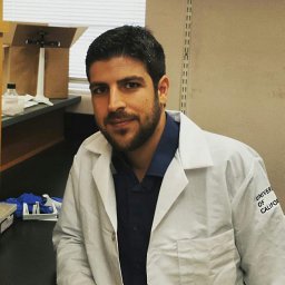 Kyritsis Nikos合作论文数DFG Center for Regenerative Therapies Dresden - Cluster of Excellence (CRTD),, Technische Universität Dresden13
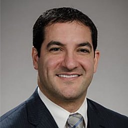 Rajiv Saigal合作论文数Electromyography Section;National Institute of Neurological;National Institutes of Health;National Institute of Neurological, National Institutes of Health13Praveen V. Mummaneni合作论文数Dept. of Neurosurgery, Emory University12Jared Narvid合作论文数12John F. Burke合作论文数11Leigh H Thomas合作论文数9Chou Austin合作论文数Brain & Spinal Injury Ctr, Univ Calif San Francisco9
 Jenny Haefeli合作论文数9
 Marc Mabray合作论文数Department of Radiology and Biomedical Imaging, University of California San Francisco9Vinil Shah合作论文数9Scott R. Whittemore合作论文数8
 Alina Uzelac合作论文数8
 Julien Cohen-Adad合作论文数Département de génie électrique, Polytechnique;Montréal8Nikolaos Kyritsis合作论文数7
 John Yue合作论文数Weill Institute for Neurosciences, University of California, San Francisco;The Philip R. Lee Institute for Health Policy Studies, University of California, San Francisco7
 William Dillon合作论文数Department of Radiology and Biomedical Imaging, University of California, San Francisco7
 Ethan Winkler合作论文数Weill Institute for Neurosciences, University of California, San Francisco;School of Medicine, University of California, San Francisco7Phiroz Tarapore合作论文数6Rachel E Tsolinas合作论文数6Sara M. Dupont合作论文数NeuroPoly Lab, Polytechnique Montreal, Montreal, Canada6Huang Michael C合作论文数6Callen Andrew L合作论文数6William J Readdy合作论文数5
 Felipe Campos Kitamura合作论文数Diagnosticos da America SA (Dasa)5
 Errol Colak合作论文数Department of Medical Imaging, Temerty Faculty of Medicine, University of Toronto5
 Prevedello Luciano M合作论文数Wexner Med Ctr, Ohio State Univ5
 Adam Flanders合作论文数Division of Neuroradiology, Thomas Jefferson University Hospital;ENT, Thomas Jefferson University Hospital;Sidney Kimmel Medical College, Thomas Jefferson University5
 Rohit Bakshi合作论文数Department of Neurology, Brigham and Women's Hospital, Harvard Medical School5
 Robyn L. Ball合作论文数Texas A and M University, Texas A&M Univ5Bhavya Rehani合作论文数5
 Cynthia Chin合作论文数Department of Radiology and Biomedical Imaging, University of California, San Francisco5
 Gregory Hawryluk合作论文数Lerner College of Medicine, Cleveland Clinic;Center for Spine Health, Neurological Institute, Cleveland Clinic5
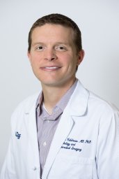 Evan Calabrese合作论文数5Debra P. Hemmerle合作论文数4




