The hindlimb representation in the somatosensory cortex of two-week old Wistar rats has been a valuable model system for dissecting the microcircuitry of neurons and their synaptic connections. In this study, we present a comprehensive experimental dataset quantifying the fiber length per cortical volume and the density of varicosities for cholinergic, catecholaminergic, and serotonergic neuromodulatory systems within the cortical neuropil using immunocytochemical staining and stereological techniques, along with a methodological framework for generating biophysically detailed computational models from these data. Acquired data were integrated into a biophysically detailed computational model of the somatosensory cortex to explore the anatomical organization and functional implications of neuromodulatory innervation. We found that neuromodulatory innervation, although sparse, substantially impacts network activity. Network simulations support the hypothesis that acetylcholine suppresses slow oscillations and promotes the desynchronization of cortical networks, consistent with the extensive findings in existing literature. Additionally, the temporal properties of acetylcholine modulation are consistent with synaptic rather than volume release. Furthermore, we found that the release of dopamine and serotonin in sensory cortices induces network desynchronization by inhibiting delta oscillations and that serotonin also initiates the emergence of theta oscillations, pointing to previously unexplored aspects of their function in governing cortical network activity. The experimental data and the biophysical computational model are available as an open-access community resource.
Accurate quantification of cellular composition is fundamental to understanding the structural and functional organization of the cerebral cortex. In the present study, we quantified the proportions of neurons, glia, and vascular cells (primarily endothelial cells) across the full cortical thickness (layers I-VI) of the mouse primary somatosensory cortex (S1HL) and the human temporal cortex (BA21) using immunocytochemical techniques and a direct 3D counting method. Over 25,000 cells in the mouse and 13,000 cells in the human cortex were individually identified and classified. For this purpose, we utilized EspINA software, which enables precise cell identification and volumetric analysis while preserving laminar and spatial context. Our results reveal marked species-specific differences in cellular proportions: neurons represent approximately 60% of all cells in the mouse S1HL but only 30% in the human BA21. These differences are reflected in the glia-to-neuron ratio (GNR) and non-neuron-to-neuron ratio (nNNR), which were consistently below 1.0 in the mouse (GNR: 0.4; nNNR: 0.6) but significantly higher in the human samples (GNR: 1.5; nNNR: 2.3). By overcoming the limitations of traditional stereological and tissue-homogenization techniques, this study provides a detailed laminar characterization of the cellular composition in these particular cortical regions (mouse S1HL and human BA21).
The neocortex underlies cognitive abilities that set humans apart from other species. Although Ramon y Cajal initiated its study in the 19th century, much about its fundamental properties remain poorly understood. Biologically detail modeling, has been shown to serve as a tool to understand the modeled system better. By comparing computational models for different species we can highlight functional differences between them, find their anatomical or physiological basis and thus improve our understanding of cortical function. In this study we built a detailed model of a human cortical microcircuit following an established workflow. We compared the human data and results against a previously published reconstruction of rat cortical circuitry. To parametrize the human model, we gathered new original data on human morphological reconstructions, axonal bouton densities, single cell and synaptic recordings. We combined them with data available in the literature and open-sourced databases. We also developed various strategies to overcome the missing data, such as generalizing or adapt- ing data from rodents. The resulting model consists of seven columnar units with similar characteristics. Each column has a radius of 476 um, a height of 2622 um, a volume of 1.86 mm3, a total cell density of 24,186 cells/mm3, on the order of 35,000 cells, around 12 million connections and approximately 47 million synapses. Comparing the rat and the human model showed that the human cortex is less dense in terms of cell bodies than the rodent cortex. Human cells have more complex branching, but lower bouton densities than rodent cells. However, the number of connections between cell types is similar. ### Competing Interest Statement The authors have declared no competing interest. This study was supported by funding to the Blue Brain Project, a research center of the École polytechnique fédérale de Lausanne (EPFL), from the Swiss governments ETH Board of the Swiss Federal Institutes of Technology.
Volume electron microscopy (vEM) has become a powerful tool for 3D ultrastructural analysis of neural circuits, yet its application to human brain organoids remains limited, particularly for connectomic studies. Here, we established a comprehensive and scalable workflow for applying vEM to human cortical organoids, integrating correlative light and electron microscopy, large-area SEM (scanning electron microscopy) mosaic imaging, focused ion beam–SEM (FIB-SEM), and transmission electron microscopy (TEM) validation. By systematically comparing two embedding protocols in use, we demonstrated that the DeFelipe and Fairén (1993); Cano-Astorga et al. (2024a) method provides optimal compatibility with toluidine blue–stained semithin sectioning and enables reliable synapse segmentation and neurite tracing. In contrast, the Deerinck’s protocol (2010) offers enhanced membrane contrast but limits postsynaptic density visualization. Using FIB-SEM imaging of peripheral, neuropil-like regions of cortical organoids, we achieved accurate 3D reconstruction of synapses, neurites and intracellular organelles, enabling quantitative assessment of synaptic apposition surfaces, neurite trajectories, and organelle distribution across defined cellular compartments. Together, our results demonstrate for the first time the feasibility of micro-connectomic reconstruction in human cortical organoids at nanometer resolution. This methodological framework expands the applicability of vEM to organoid systems and provides a robust foundation for future studies of human brain development, disease modeling, and therapeutic evaluation at the synaptic and subcellular level. Workflow for vEM–based micro-connectomic analysis in human cortical organoids. Cortical organoids underwent sample preparation, resin embedding, and semithin/ultrathin sectioning. Large-area SEM mosaic imaging was used to identify regions of interest for targeted FIB-SEM acquisition. Ultrastructural observations were independently validated by TEM, establishing a scalable pipeline for connectome reconstruction and subcellular analysis in human brain organoids. This image was created with BioRender
This article explores the historical progression of studying neuronal connections, beginning with nineteenth-century advancements in light microscopy and histological techniques. Early methods were limited in terms of their capacity to trace neuronal connections, but a breakthrough came with Camillo Golgi's "black reaction" staining method, later refined and extensively used by Santiago Ramón y Cajal. Cajal's observations supported the Neuron Theory, which proposed that neurons communicate via specialized points of contact, contradicting the prevailing Reticular Theory of a continuous neural network, which was supported by Golgi. This contrast is particularly intriguing because, although Golgi and Cajal used the same black reaction technique and similar microscopes, their interpretations of the microscopic world diverged significantly. An important consequence of the Neuron Theory was Cajal's Law of Dynamic Polarization, which proposed that neurons generally consist of three functionally distinct regions: a receptor apparatus (dendrites and soma), an emission apparatus (axon), and a distribution apparatus (terminal axonal arborization). He applied this principle across various parts of the nervous system and to different neuron types, enabling the generation of the first detailed circuit diagrams of the brain. Cajal's observations, concepts, and theories had a profound impact-not only on researchers of his time, but also on modern neuroscience. This article reflects on the early studies of neuronal connections, highlighting the scientific climate in which Golgi and Cajal initiated their groundbreaking research.
Tau has a wide variety of essential functions in the brain, but this protein also plays a determining role in the development of Alzheimer's disease (AD) and other neurodegenerative diseases called tauopathies. This is due to its abnormal aggregation and the subsequent formation of neurofibrillary tangles. Tau hyperphosphorylation appears to be a critical step in its transformation into an aggregated protein. However, the exact process, including the cellular events that trigger it, remains unclear. In this study, we employed immunocytochemistry assays on hippocampal sections from AD cases and from tauopathy cases (Braak stage III) with no evidence of cognitive decline, and the P301S mouse model to investigate the colocalization patterns of Tau phosphorylated (p) at specific residues (S202-T205, S214, and T231) within the proline-rich region. Our results show pyramidal neurons in the hippocampus of P301S mice in which Tau is intensely phosphorylated at residues S202 and T205 (recognized by the AT8 antibody), but with no detectable phosphorylation at S214 or T231. These non-colocalizing neurons displayed intensely labeled aggregated pTau deposits distributed through the soma and dendritic processes. However, most of the hippocampal pyramidal neurons are labeled with pTauS214 or pTauT231 antibodies and typically showed a homogeneous and diffuse pTau distribution (not aggregated). This different labeling likely reflects a Tau conformational step, potentially related to the transition from a diffuse tau phosphorylation phenotype (Type 2) into an NFT-like or Type 1 phenotype. We further observed that dendrites of CA3 pyramidal cells are intensely labeled with pTau214 in the stratum lucidum, but not with AT8 or pTauT231. By contrast, analysis of tissue from AD patients or other human tauopathy cases (Braak stage III) with no evidence of cognitive decline revealed extensive colocalization with both antibody combinations in CA1. The complete or mature tangle development may follow a different mechanism in the P301S mouse model or may require more time to achieve the maturity state found in AD cases. Further studies would be necessary to address this question.
Dysfunction of the basal ganglia is implicated in a wide range of neurological and psychiatric disorders. Our understanding of the operation of the basal ganglia is largely derived on data from studies conducted on mice, which are frequently used as model organisms for various clinical conditions. The striatum, the largest compartment of the basal ganglia, consists of 90-95% striatal projection neurons (SPNs). It is therefore crucial to establish if human and mouse SPNs have distinct or similar properties, as this has implications for the relevance of mouse models for understanding the human striatum. To address this, we compared the general organization of the somato-dendritic tree of SPNs, the dimensions of the dendrites, the density and size of spines (spine surface area), and ion channel subtypes in human and mouse SPNs. Our findings reveal that human SPNs are significantly larger, but otherwise the organisation of the dendritic tree (dendrogram) with an average of approximately 5 primary dendrites, is similar in both species. Additionally in both humans and mice, over 90% of the spines are located on the terminal branches of each dendrite. Human spines are somewhat larger (4.3 versus 3.1 μm2) and the terminal dendrites have a uniform diameter in both humans and mice, although somewhat broader in the latter (1.0 versus 0.6 μm). The composition of ion channels is also largely conserved. These data have been used to simulate human SPNs building on our previous detailed simulation of mouse SPNs. We conclude that the human SPNs essentially appear as enlarged versions of the mouse SPNs. This similarity suggests that both species process information in a comparable manner, supporting the relevance of mouse models for studying the human striatum.
Dendritic spines of pyramidal neurons are the targets of most excitatory synapses in the cerebral cortex, and dendritic spine morphology directly reflects their function. However, there are scarce data available regarding both the detailed morphology of these structures for the human cerebral cortex and the extent to which they differ in comparison with other species. Thus, in the present study, we used intracellular injections of Lucifer yellow to reconstruct-in three dimensions-the morphology of basal dendritic spines from pyramidal cells in the human and mouse CA1 hippocampal region and compared these spines with those of the human temporal and cingular cortex. We found that human hippocampal dendrites show lower spine density, larger volume, and longer length of dendritic spines than mouse CA1 spines. Furthermore, human hippocampal dendrites show higher spine density, smaller spine volume, and shorter length compared to dendritic spines from the human temporal and cingular cortex. This morphological diversity suggests an equally large variability of synaptic strength and learning rules across these brain regions in humans and between humans and mice. Nevertheless, a balance between size and density was found in all cases, which may be a cortical rule maintained across cortical areas and species.
The entorhinal cortex (EC) plays a pivotal role in memory function and spatial navigation, connecting the hippocampus with the neocortex. The EC integrates a wide range of cortical and subcortical inputs, but its synaptic organization in the human brain is largely unknown. We used volume electron microscopy to perform a 3D analysis of the microanatomical features of synapses in all layers of the medial EC (MEC) from the human brain. Using this technology, 12,974 synapses were fully 3D reconstructed at the ultrastructural level. The MEC presented a distinct set of synaptic features, differentiating this region from other human cortical areas. Furthermore, ultrastructural synaptic characteristics within the MEC was predominantly similar, although layers I and VI exhibited several synaptic characteristics that were distinct from other layers. The present study constitutes an extensive description of the synaptic characteristics of the neuropil of all layers of the EC, a crucial step to better understand the connectivity of this cortical region, in both health and disease.
Intracranial potentials are used as functional biomarkers of neural networks. As potentials spread away from the source populations, they become mixed in the recordings. In humans, interindividual differences in the gyral architecture of the cortex pose an additional challenge, as functional areas vary in location and extent. We used source separation techniques to disentangle mixing potentials obtained by exploratory deep arrays implanted in epileptic patients of either sex to gain access to the number, location, relative contribution, and dynamics of coactive sources. The unique spatial profiles of separated generators made it possible to discern dozens of independent cortical areas for each patient, whose stability maintained even during seizure, enabling the follow up of activity for days and across states. Through matching these profiles to MRI, we associated each with limited portions of sulci and gyri and determined the local or remote origin of the corresponding sources. We also plotted source-specific 3D coverage across arrays. In average, individual recording sites are contributed to by 3-5 local and distant generators from areas up to several centimeters apart. During seizure, 13-85% of generators were involved, and a few appeared anew. Significant bias in location assignment using raw potentials is revealed, including numerous false positives when determining the site of origin of a seizure. This is not amended by bipolar montage, which introduce additional errors of its own. In this way, source disentangling reveals the multisource nature and far intracranial spread of potentials in humans, while efficiently addressing patient-specific anatomofunctional cortical divergence.
Santiago Ramón y Cajal’s pioneering work laid the foundations for modern neuroscience and continues to impact the development of artificial intelligence, particularly deep learning. His neuron theory, the principle of dynamic polarization, and his insights into brain plasticity and network organization have significantly influenced both our understanding of the nervous system and the design of artificial neural networks. This article reviews Cajal’s key contributions, explores their role in the evolution of AI, and emphasizes the enduring links between neuroscience and machine learning in the digital era.
Elucidating human cerebral cortex function is essential for understanding the physiological basis of both healthy and pathological brain states. We obtained extracellular local field potential recordings from slices of neocortical tissue from refractory epilepsy patients. Multi-electrode recordings were combined with histological information, providing a two-dimensional spatiotemporal characterization of human cortical dynamics in control conditions and following modulation of the excitation/inhibition balance. Slices expressed spontaneous rhythmic activity consistent with slow wave activity, comprising alternating active (Up) and silent (Down) states (Up-duration: 0.08 ± 0.03 s, Down-duration: 2.62 ± 2.12 s, frequency: 0.75 ± 0.39 Hz). Up states propagated from deep to superficial layers, with faster propagation speeds than in other species (vertical: 64.6 mm/s; horizontal: 65.9 mm/s). GABAA blockade progressively transformed the emergent activity into epileptiform discharges, marked by higher firing rates, faster network recruitment and propagation, and infraslow rhythmicity (0.01 Hz). This dynamical characterization broadens our understanding of the mechanistic organization of the human cortical network at the micro- and mesoscale.
Alzheimer’s disease (AD) is the most common neurodegenerative disorder in the elderly. Recent efforts have centered on understanding early events that trigger AD, aiming to facilitate early diagnosis and intervention for improved patient outcomes. The traditional histopathological features observed in AD encompass the extracellular accumulation of amyloid-beta protein and the intracellular abnormal phosphorylation of Tau protein (pTau). However, elucidating how these pathological hallmarks ultimately contribute to cognitive deficits remains a complex challenge. While AD is commonly conceptualized as a disorder characterized by synaptic failure, substantial knowledge gaps persist regarding the mechanisms underlying the onset and progression of the disease, underscoring the need for novel and more effective therapeutic approaches. In this context, the impairment of GABAergic paravalbumin (PV+) neurons has been proposed as a crucial factor contributing to neuronal network dysfunction and cognitive decline in AD. The presence of pTau in pyramidal neurons is directly linked to their impairment in AD; however, the effect of pTau in PV+ neurons remains unclear. In this present study, we analyzed the existence of PV+ neurons containing pTau using immunocytochemistry in the hippocampal formation and entorhinal cortex of human samples from diagnosed AD cases and individuals without neurological or psychiatric disorders. Two pTau isoforms, pTauAT8 and pTaupS396, corresponding to early and late stages of AD respectively, were examined. Our findings indicate that most PV+ neurons across the hippocampal formation and entorhinal cortex did not contain pTau in either group cases. Interestingly, while AD cases diagnosed with dementia exhibited a higher number of pTau+ neurons, the majority of PV+/pTau+ neurons were found in individuals with no neurological alterations. This suggests that the presence of pTau in PV+ neurons does not directly correlate with the overall abundance of pTau+ neurons. Given that PV+ neuron impairment is a key pathogenic mechanism in AD and is associated with cognitive decline, understanding the changes in PV+ neurons during AD progression could provide critical insights into the alterations of neuronal circuits underlying the disease.
The organizational principles that distinguish the human brain from other species have been a long-standing enigma in neuroscience. Focusing on the uniquely evolved human cortical layers 2 and 3, we computationally reconstruct the cortical architecture for mice and humans. Human neurons form highly complex networks demonstrated by their increased number and simplex dimension compared to mice. This is surprising because human pyramidal cells are much sparser. The number and size of neurons cannot account for this increased network complexity, suggesting that another morphological property is a key determinant of network connectivity. The topological comparison of the dendritic structure reveals higher perisomatic density in human pyramidal cells. We quantitatively show that this neuronal structural property directly impacts network complexity, including the formation of a rich subnetwork structure. Therefore, greater dendritic complexity, a defining attribute of human pyramidal cells, may provide the human cortex with enhanced computational capacity and cognitive flexibility.
Functional and structural studies investigating macroscopic connectivity in the human cerebral cortex suggest that high-order associative regions exhibit greater connectivity compared to primary ones. However, the synaptic organization of these brain regions remains unexplored. In the present work, we conducted volume electron microscopy to investigate the synaptic organization of the human brain obtained at autopsy. Specifically, we examined layer III of Brodmann areas 17, 3b, and 4, as representative areas of primary visual, somatosensorial, and motor cortex. Additionally, we conducted comparative analyses with our previous datasets of layer III from temporopolar and anterior cingulate associative cortical regions (Brodmann areas 24, 38, and 21). 9,690 synaptic junctions were 3D reconstructed, showing that certain synaptic characteristics are specific to particular regions. The number of synapses per volume, the proportion of the postsynaptic targets, and the synaptic size may distinguish one region from another, regardless of whether they are associative or primary cortex. By contrast, other synaptic characteristics were common to all analyzed regions, such as the proportion of excitatory and inhibitory synapses, their shapes, their spatial distribution, and a higher proportion of synapses located on dendritic spines. The present results provide further insights into the synaptic organization of the human cerebral cortex.
Investigating and modelling the functionality of human neurons remains challenging due to the technical limitations, resulting in scarce and incomplete 3D anatomical reconstructions. Here we used a morphological modelling approach based on optimal wiring to repair the parts of a dendritic morphology that were lost due to incomplete tissue samples. In Drosophila, where dendritic regrowth has been studied experimentally using laser ablation, we found that modelling the regrowth reproduced a bimodal distribution between regeneration of cut branches and invasion by neighbouring branches. Interestingly, our repair model followed growth rules similar to those for the generation of a new dendritic tree. To generalise the repair algorithm from Drosophila to mammalian neurons, we artificially sectioned reconstructed dendrites from mouse and human hippocampal pyramidal cell morphologies, and showed that the regrown dendrites were morphologically similar to the original ones. Furthermore, we were able to restore their electrophysiological functionality, as evidenced by the recovery of their firing behaviour. Importantly, we show that such repairs also apply to other neuron types including hippocampal granule cells and cerebellar Purkinje cells. We then extrapolated the repair to incomplete human CA1 pyramidal neurons, where the anatomical boundaries of the particular brain areas innervated by the neurons in question were known. Interestingly, the repair of incomplete human dendrites helped to simulate the recently observed increased synaptic thresholds for dendritic NMDA spikes in human versus mouse dendrites. To make the repair tool available to the neuroscience community, we have developed an intuitive and simple graphical user interface (GUI), which is available in the TREES toolbox (www.treestoolbox.org).
The brain contains thousands of millions of synapses, exhibiting diverse structural, molecular, and functional characteristics. However, synapses can be classified into two primary morphological types: Gray’s type I and type II, corresponding to Colonnier’s asymmetric (AS) and symmetric (SS) synapses, respectively. AS and SS have a thick and thin postsynaptic density, respectively. In the cerebral cortex, since most AS are excitatory (glutamatergic), and SS are inhibitory (GABAergic), determining the distribution, size, density, and proportion of the two major cortical types of synapses is critical, not only to better understand synaptic organization in terms of connectivity, but also from a functional perspective. However, several technical challenges complicate the study of synapses. Potassium ferrocyanide has been utilized in recent volume electron microscope studies to enhance electron density in cellular membranes. However, identifying synaptic junctions, especially SS, becomes more challenging as the postsynaptic densities become thinner with increasing concentrations of potassium ferrocyanide. Here we describe a protocol employing Focused Ion Beam Milling and Scanning Electron Microscopy for studying brain tissue. The focus is on the unequivocal identification of AS and SS types. To validate SS observed using this protocol as GABAergic, experiments with immunocytochemistry for the vesicular GABA transporter were conducted on fixed mouse brain tissue sections. This material was processed with different concentrations of potassium ferrocyanide, aiming to determine its optimal concentration. We demonstrate that using a low concentration of potassium ferrocyanide (0.1%) improves membrane visualization while allowing unequivocal identification of synapses as AS or SS.
The highly complex structure of the brain requires an approach that can unravel its connectivity. Using volume electron microscopy and a dedicated software we can trace and measure all nerve fibers present within different samples of brain tissue. With this software tool, individual dendrites and axons are traced, obtaining a simplified "skeleton" of each fiber, which is linked to its corresponding synaptic contacts. The result is an intricate meshwork of axons and dendrites interconnected by a cloud of synaptic junctions. To test this methodology, we apply it to the stratum radiatum of the hippocampus and layers 1 and 3 of the somatosensory cortex of the mouse. We find that nerve fibers are densely packed in the neuropil, reaching up to 9 kilometers per cubic mm. We obtain the number of synapses, the number and lengths of dendrites and axons, the linear densities of synapses established by dendrites and axons, and their location on dendritic spines and shafts. The quantitative data obtained through this method enable us to identify subtle traits and differences in the synaptic organization of the samples, which might have been overlooked in a qualitative analysis.
Purkinje cells in the cerebellum are among the largest neurons in the brain and have been extensively investigated in rodents. However, their morphological and physiological properties remain poorly understood in humans. In this study, we utilized high-resolution morphological reconstructions and unique electrophysiological recordings of human Purkinje cells ex vivo to generate computational models and estimate computational capacity. An inter-species comparison showed that human Purkinje cell had similar fractal structures but were larger than those of mouse Purkinje cells. Consequently, given a similar spine density (2/μm), human Purkinje cell hosted approximately 7.5 times more dendritic spines than those of mice. Moreover, human Purkinje cells had a higher dendritic complexity than mouse Purkinje cells and usually emitted 2–3 main dendritic trunks instead of one. Intrinsic electro-responsiveness was similar between the two species, but model simulations revealed that the dendrites could process ~6.5 times (n = 51 vs. n = 8) more input patterns in human Purkinje cells than in mouse Purkinje cells. Thus, while human Purkinje cells maintained spike discharge properties similar to those of rodents during evolution, they developed more complex dendrites, enhancing computational capacity.
 Ruth Benavides-Piccione合作论文数Universidad Politecnica Madrid;Cajal Institute74
 Ángel Merchán Pérez合作论文数Universidad Politécnica de Madrid56
 Lidia Alonso-Nanclares合作论文数Instituto Cajal (CSIC), Avenida Dr Arce 37, 28002 Madrid, Spain38
 Isabel Fernaud合作论文数Departamento de Inmunologı́a. Centro de Investigaciones Biológicas, Consejo Superior de Investigaciones Cientı́ficas32Josérodrigo Rodríguez合作论文数30Blazquez-Llorca Lidia合作论文数Lab Cajal Circuitos Cort, Univ Politecn Madrid29Guy N. Elston合作论文数27
 Asta Kastanauskaite合作论文数24
 Pedro Larrañaga合作论文数Departamento de Inteligencia Artificial, Universidad Politécnica de Madrid;Computational Intelligence Group, Universidad Politécnica de Madrid21
 Concha Bielza合作论文数Department of Artificial Intelligence, Universidad Polit´ecnica de Madrid;ELLIS Unit Madrid21
 Paula Merino-Serrais合作论文数20
 Catia Teixeira合作论文数Department of Chemistry, University of Porto18Fausto Ulloa合作论文数18
 Eduardo Soriano合作论文数Univ Barcelona, Dept Cell Biol, E-08028 Barcelona, Spain17
 Albert Martinez合作论文数16
 Carles Bosch合作论文数Sensory Circuits and Neurotechnology Lab., The Francis Crick Institute16Alberto Muñoz合作论文数Univ Autonoma Madrid, CSIC, Inst Invest Biomed Alberto Sols, Madrid, Spain16Masachs Nuria合作论文数16
 Edward G. Jones合作论文数Center for Neuroscience, University of California, Davis16
 Joan Comella Carnicé合作论文数Barcelona Children's Hospital Sant Joan de Déu;Universitat Autònoma de Barcelona16
 Rafael Yuste合作论文数Department of Biological Sciences, Faculty of Arts and Sciences, Columbia University;Data Science Institute, Columbia University15
 Carlos Lois合作论文数Division of Biology and Biological Engineering, California Institute of Technology15
 Inmaculada Ballesteros-Yáñez合作论文数Faculty of Medicine, University of Castilla-La Mancha14
 Silvia Tapia-González合作论文数CSIC, Inst Cajal13
 Henry Markram合作论文数Laboratory of Neural Microcircuitry, Brain Mind Institute, School of Life Sciences, Ecole Polytechnique Fédérale De Lausanne;Life Sciences Engineering Section, School of Life Sciences, Ecole Polytechnique Fédérale De Lausanne13
 Marta Turegano-Lopez合作论文数Ph.D. Program in Neuroscience, Universidad Autónoma de Madrid - Instituto Cajal12
 Jesús Ávila de Grado合作论文数Centro de Biología Molecular "Severo Ochoa", CSIC, Universidad Autónoma de Madrid12J. Gonzalez-Soriano合作论文数12
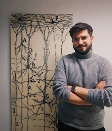 Sergio Plaza-Alonso合作论文数CSIC, Inst Cajal11esther perezmartinez合作论文数11
 Andrea Santuy合作论文数11
 Nicolas Cano-Astorga合作论文数CSIC, Inst Cajal10
 Luis Pastor合作论文数9León-Espinosa Gonzalo合作论文数9
 Juan Morales合作论文数9Kathleen S. Rockland合作论文数9
 Marta Domínguez-Álvaro合作论文数Lab Cajal Circuitos Cort, Univ Politecn Madrid8
 Angel Rodriguez合作论文数8Alejandra Elston合作论文数8
 Idan Segev合作论文数Institute of Life Sciences, Dept. of Neurobiology;The Hebrew University of Jerusalem8
 Alfonso Fairén合作论文数Institute of Neurosciences, Spanish National Research Council8SHC HENDRY合作论文数7Pilar Marco合作论文数7Mara Dierssen合作论文数7
 Miguel Miguens合作论文数Facultad de Psicología, UNED7R. G. Sola合作论文数6
 Oscar Herreras合作论文数CSIC, Cajal Inst6
 Patrick R Hof合作论文数Nash Family Department of Neuroscience, Icahn School of Medicine at Mount Sinai6




