BACKGROUND: Levetiracetam is commonly used as a prophylactic antiseizure medication in patients undergoing surgical resection of brain tumors. OBJECTIVE: To quantitate side effects experienced in patients treated with 1 week vs 6 weeks of prophylactic levetiracetam using validated measures for neurotoxicity and depression. METHODS: Patients undergoing surgical resection of a supratentorial tumor with no seizure history were randomized within 48 hours of surgery to receive prophylactic levetiracetam for the duration of either 1 or 6 weeks. Patients were given oral levetiracetam extended release 1000 mg during the first part of this study. Owing to drug backorder, patients enrolled later in this study received levetiracetam 500 mg BID. The primary outcome was the change in the neurotoxicity score 6 weeks after drug initiation. The secondary outcome was seizure incidence. RESULTS: A total of 81 patients were enrolled and randomized to 1 week (40 patients) or 6 weeks (41 patients) of prophylactic levetiracetam treatment. The neurotoxicity score slightly improved in the overall cohort between baseline and reassessment. There was no significant difference between groups in neurotoxicity or depression scores. Seizure incidence was low in the entire cohort of patients with 1 patient in each arm experiencing a seizure during the follow-up period. CONCLUSION: The use of prophylactic levetiracetam did not result in significant neurotoxicity or depression when given for either 1 week or 6 weeks. The incidence of seizure after craniotomy for tumor resection is low regardless of duration of therapy.
Meningiomas are the most common primary intracranial tumor. They are slow growing and often incidentally found tumors that arise from the arachnoid villi. As they grow, they have a greater likelihood of becoming symptomatic with seizures being one of the most clinically significant symptoms. Seizures are more likely to present as a symptom of larger meningiomas and meningiomas that compress cortical areas particularly those in non-skull base locations. These seizures are often managed medically, utilizing the same anti-seizure medications that are used to treat other causes of epilepsy. We discuss common anti-seizure medications used including valproate, phenobarbital, carbamazepine, phenytoin, lacosamide, lamotrigine, levetiracetam and topiramate and their common adverse effects. The goal of pharmacotherapy for seizure control is to maximize seizure control while minimizing the adverse effects of the medication. The decision to provide medical management is dependent on individual seizure history and plans for surgical treatment. Patients who did not require seizure prophylaxis before surgery are commonly prescribed seizure prophylaxis postoperatively. Symptomatic meningiomas not controlled by medical management alone are commonly evaluated for surgical resection. The efficacy of surgical resection in providing seizure freedom is dependent on several features of the tumor including tumor size, the extent of the peritumoral edema, the number of tumors, sinus infiltration and the degree of resection.
Introduction Continuous electroencephalography (EEG) is an important monitoring modality in the intensive care unit and a key skill for critical care fellows (CCFs) to learn. Our objective was to evaluate with CCFs an EEG educational curriculum on a web-based simulator. Methods This prospective cohort study was conducted at a major academic medical center in Florida. After Institutional Review Board approval, 13 CCFs from anesthesiology, surgery, and pulmonary medicine consented to take an EEG curriculum. A 25-item EEG assessment was completed at baseline, after 10 EEG interpretations with a neurophysiologist, and after 10 clinically relevant EEG-based simulations providing clinical EEG interpretation hints. A 50-minute tutorial podcast was viewed after the baseline assessment. Main assessment outcomes included multiple outcomes related to web-based simulator performance: percent of hints used, percent of first words on EEG interpretation correct, and percent hint-based EEG interpretation score correct, with higher scores indicating more correct answers. Participants completed a 25-item EEG assessment before (baseline) and after the web-based simulator. Results All 13 CCFs completed the curriculum. Between scenarios, there were differences in percent of hints used (F9,108 = 11.7, p < 0.001), percent of first words correct (F9,108 = 13.6, p < 0.001), and overall percent hint-based score (F9,108 = 14.0, p < 0.001). Nonconvulsive status epilepticus had the lowest percent of hints used (15%) and the highest hint-based score (87%). Overall percent hint-based score (mean across all scenarios) was positively correlated with change in performance as the number of correct answers on the 25-item EEG assessment from before to after the web-based simulator activity (Spearman's rho = 0.67, p = 0.023). Conclusions A self-paced EEG interpretation curriculum involving a flipped classroom and screen-based simulation each requiring less than an hour to complete significantly improved CCF scores on the EEG assessment compared to baseline.
We read with interest Dr. Philip Liu’s (1) review of the book titled Simulators in Anesthesiology Education (2), which contains the proceedings of a conference by the same name held in 1996. At the outset, we must disclose that one of us (DMG) contributed to this volume a chapter concerning research methodologies involving patient simulators. We were surprised by the rather abrupt transition at the end of Dr. Liu’s review from analyzing the strengths and weaknesses of the book to expressing his personal opinions about the value of realistic patient simulators to anesthesiology education. First, by mixing such opinions with his book review, Dr. Liu does a disservice to the editors and authors of this book and to the readers of Anesthesia & Analgesia. More importantly, Dr. Liu’s opinion that patient simulators are a luxury is unsubstantiated. To our knowledge, no claim has been made by anyone with first-hand experience using patient simulators in anesthesia education, or anywhere in the book under review, that every residency program must have a simulator. Each institution must, and does, judge the value of this (or any other) technology for its own purposes. Many institutions have decided that the value exceeds the costs. In fact, since Dr. Liu wrote his review stating that there are approximately 50 simulation centers around the world, there are now well over 100, and this number is increasing every month. Second, Dr. Liu argues that there is an enormous cost associated with simulators and their operation. However, the hardware and software costs are not excessive when compared with other clinical anesthesia devices, such as transesophageal echocardiography or evoked potential monitoring equipment, whose costs are similarly amortized over the useful life of the device. Importantly, in many simulation centers, these costs are commonly shouldered by a variety of users, not just by anesthesiologists. Dr. Liu ignores both the information provided in the book he reviewed and the now large body of literature on the wide spectrum of uses for patient simulators. To date, applications span education (medical students, classroom school teachers, college students) and clinical training (see below) to performance assessment, research on human factors (such as equipment design and the effects of fatigue), and even preprocurement testing of equipment. As for training, beyond anesthesiology, simulators have now been used for training residents and experienced clinicians in many domains including: combined delivery room teams (e.g., obstetricians, anesthesiologists, neonatologists, and neonatal intensive care unit nurses); emergency room teams; prehospital emergency responders, intensive care physicians, and intensive care unit teams; in-hospital resuscitation teams; and radiologists. As evidence of the widespread interest in simulator use outside anesthesiology, at a recent patient simulator workshop at the University of California San Diego, nonanesthesiologist participants out-numbered anesthesiologists by a ratio of 3 to 2, and virtually every clinical specialty was represented, including psychiatry, family medicine, internal medicine, surgery, radiology, orthopedics, pharmacology, physiology, bioengineering, emergency medicine, nursing, neurology, and reproductive medicine. Therefore, we believe that it is no longer an issue of whether every anesthesia residency can use a simulator—the question now is whether every health care institution can reap benefits from simulation technology. In fact, anesthesiologists are the acknowledged leaders in the use of simulation for clinical training, and we are sought by many other disciplines for advice and counsel in this regard. Dr. Liu points out that simulation usage would be bolstered by the adoption by the American Board of Anesthesiology of simulation-based examinations. No doubt this is true. The American Board of Anesthesiology has been studying the use of simulators for this purpose for over 10 yr. The road to simulation-based examinations will be long and slow, but this application, too, will probably be with us in the future. Interestingly, a member of the American Board of Internal Medicine who attended the University of California San Diego simulator workshop expressed the opinion that realistic simulators may similarly have a role in the certification process for internists. Finally, Dr. Liu suggests that we have “…successfully developed a ‘learn and earn’ system of resident education.” Few of us would argue that this system is perfect, or that its graduates are 100% optimal in all respects. Nearly every other high-hazard industry uses a similar “learn and earn” approach where more junior personnel operate under close supervision of those with more experience. Yet, many of these domains also conduct simulation training for these personnel, especially, as Dr. Liu himself recognizes, in “…situations that could not be offered safely in any other environment.” Thus, whether having access to a simulator is a luxury or a necessity has by no means been established. In our opinion, Dr. Liu’s judgements, grafted unnecessarily onto his book review, are biased, unsubstantiated, and, if nothing else, markedly premature. A more appropriate conclusion to his review would have been to congratulate the book’s editors and authors for their compilation of the state-of-the-art at the time of the conference. Another conference on simulation in anesthesiology education was held June 1999. Although it’s unlikely that the “quintessential education application for simulators” was delineated at this conference, the spectrum of simulation-based activities around the world was probably impressive. David M. Gaba MD Steve Howard MD Brian Smith MD Matthew B. Weinger MD
To report a case of a patient with intractable epilepsy from adult onset Rasmussen's encephalitis (RE) who responded to tocilizumab therapy.
Introduction The objective of the pilot study was to determine the association between learning preferences and improvement in the American Academy of Neurology Residency In-Service Training Examination (RITE) scores from postgraduate year 2 (PGY-2) to postgraduate year 3 (PGY-3) in neurology residents. Methods Neurology residents at the University of Florida were approached to participate, and their consent was obtained. VARK inventory, representing four modalities (visual, aural, read/write, kinesthetic) of learning preferences, was completed by participants. Participants could pick more than one modality. The number of responses in each sensory domain was recorded, with higher numbers indicating stronger preference. Residents’ performance on the RITE was recorded for PGY-2 and PGY-3. Results Seventeen residents completed the VARK inventory and 16 had data for RITE. Residents demonstrated overall positive change in RITE from PGY-2 to PGY-3 (mean change = 6%; 95%CI: 4%, 9%). The median number of responses was highest for the kinesthetic domain (median = 7, range = 1-12), followed by visual (median = 6, range = 2-12), aural (median = 4, range = 1-10), and read/write (median = 4, range = 1-10). Among VARK domains, the number of responses in read/write had the strongest correlation with mean change in RITE performance from PGY-2 to PGY-3 (r = 0.45; 95%CI: -0.08, 0.78); residents in the high read/write group (number of response above median) had greater mean change in RITE performance (9%; 95%CI: 6%, 12%) while those in the low read/write group showed little to no increase in RITE from PGY-2 to PGY-3 (2%; 95%CI: -1%, 6%). Conclusions Higher VARK survey responses in the read/write domain were related to greater change in RITE scores from PGY-2 to PGY-3. These findings seem intuitively obvious considering the format of the RITE. These pilot data permit further investigation of individual resident learning preference and how it relates to test performance. By understanding a resident’s learning style, both educators and the resident will have an awareness of areas that need to be improved to be successful, which may be via remedial curricula and self-study activities.
Develop and pilot test a simulator that presents ten commonly encountered representative clinical contexts for trainees to learn basic electroencephalogram (EEG) interpretation skills. We created an interactive web-based training simulator that allows self-paced, asynchronous learning and assessment of basic EEG interpretation skills. The simulator uses the information retrieval process via a free-response text box to enhance learning. Ten scenarios were created that present dynamic (scrolling) EEG tracings resembling the clinical setting, followed by questions with free-text answers. The answer was checked against an accepted word/phrase list. The simulator has been used by 76 trainees in total. We report pilot study results from the University of Florida’s neurology residents (N = 24). Total percent correct for each scenario and average percent correct for all scenarios were calculated and correlated with most recent In-training Examination (ITE) and United States Medical License Examination (USMLE) scores. Neurology residents’ mean percent correct scenario scores ranged from 27.1–86.0% with an average scenario score of 61.2% ± 7.7. We showed a moderately strong correlation r = 0.49 between the ITE and the average scenario score. We developed an online interactive EEG interpretation simulator to review basic EEG content and assess interpretation skills using an active retrieval approach. The pilot study showed a moderately strong correlation r = 0.49 between the ITE and the average scenario score. Since the ITE is a measure of clinical practice, this is evidence that the simulator can provide self-directed instruction and shows promise as a tool for assessment of EEG knowledge.
Objective: To describe a case of low-dose baclofen-related encephalopathy in the setting of renal disease, emphasizing the importance of medication reconciliation to avoid adverse events. Background: Baclofen is a gamma-aminobutyric acid (GABA) type-B receptor agonist used for muscle spasticity. Symptoms of baclofen toxicity include confusion, somnolence, nausea/vomiting, myoclonus, hypotonia, coma, seizures, and autonomic dysfunction. Baclofen is primarily renally excreted; renal impairment increases toxicity risk even with modest doses. EEG findings include generalized slowing, burst suppression, triphasic waves, rhythmic high-amplitude delta waves, and non-convulsive status epilepticus. Design/Methods: NA Results: A 27-year-old hemodialysis-dependent African-American female with spina bifida sustained second-degree lower extremity burns. Outpatient medications listed baclofen 10 milligrams three times daily and multiple other medications. On hospital day 2, cefepime, metronidazole, and vancomycin were begun for sepsis. Later that evening, she became progressively nonverbal and encephalopathic with no new focal deficits. Acyclovir was added for possible viral encephalitis and brain MRI was unremarkable. Continuous renal replacement therapy was instituted. On hospital day 4, she was intubated, and EEG showed burst suppression. On day 5, baclofen was discontinued and sedation changed from propofol to dexmedetomidine. By day 6, she followed commands and answered questions; EEG transformed to nearly continuous sharply contoured theta. By day 7, EEG background was 7–8 Hz. She was at baseline mental status by day 8, was extubated, and antimicrobials were discontinued. It was determined then she took baclofen only rarely as needed. Conclusions: Baclofen toxicity can cause adverse events in metabolically vulnerable patients, even at modest doses, and this may not be well known among clinicians. Electronic health record software can query outside pharmacy medication dispense reports which would have identified that this patient rarely took baclofen. This is especially useful for non-verbal patients often seen in neurology consultations. Disclosure: Dr. Smith has nothing to disclose. Dr. Fahy has nothing to disclose. Dr. Cibula has nothing to disclose.
Left ventricular assist devices are increasingly used as therapy for patients with severe congestive heart failure. These patients typically receive care in the intensive care unit when EEG monitoring is necessary. Identification of artifacts created by these devices is important for accurate EEG diagnosis, thus avoiding unnecessary therapies that may result in complications or require intubation of the patient. This report describes a case of a patient with altered mental status and history of congestive heart failure with a left ventricular assist device who developed seizures following left subdural hematoma and emergent evacuation. While being treated for seizures, the EEG showed a diffuse continuous and regular delta, predominant on the left that mirrored the electrocardiogram (EKG) rhythm strip and appeared to indicate an external artifact. Moving the amplifier farther away from the heart and repositioning the patient resolved the artifact. It is important to recognize potential sources of interference and artifacts when these devices are involved. This case illustrates how clinicians can prevent administration of unneeded therapies to these patients.
Learning Objectives: Introduction: The purpose of this study is to evaluate the effectiveness of an interdisciplinary electroencephalogram (EEG) educational module including a web-based module for critical care fellows with a variety of backgrounds during a NeuroICU rotation. EEG is increasingly utilized for diagnosis, monitoring, and treatment decisions with continuous EEG monitoring an additional evaluation tool in the ICU. Fellows were surveyed about experience with technology and web-based training modules. Methods: Methods: During a neuroICU rotation, critical care fellows of various backgrounds (pulmonary, surgery, and anesthesiology) participated in an EEG curriculum utilizing a web-based training module to cover EEG basics, clinical exposure, and EEG interpretations. Using 25-question evaluation tools that included EEG interpretations, participants were assessed before EEG instruction and after curriculum completion. Change in EEG scores from baseline to after 10 EEG readings was assessed using paired t-tests. Additionally, we assessed heterogeneity in this change associated with previous experience with web-based training modules. P < 0.05 was considered statistically significant. Results: Results: N=15 fellows completed the EEG instrument at baseline and after 10 EEG readings. The critical care fellow backgrounds included 27% (n=4) were surgery fellows, 33% (n=5) were pulmonary fellows, and 40% (n=6) were anesthesiology fellows. Overall, fellows’ performance on the EEG instrument improved from baseline to after 10 EEG readings (mean change= 5.3, 95%CI: 2.8 to 7.7). Previous educational experience with webbased training modules was associated with this improvement (p = 0.039). Fellows who had four or more previous web-based training education experiences showed more improvement (n=11, mean change= 6.6, 95%CI: 3.7 to 9.5) than those with fewer experiences (n=4, mean change= 1.5, 95%CI: -0.6 to 3.6). Conclusions: Conclusions: In summary, this pilot study implementing an interdisciplinary EEG curriculum for critical care fellows of varied backgrounds provided preliminary evidence that this educational curriculum may be an effective educational method for obtaining important EEG knowledge to optimize care for critically ill patients.
Not everything that shakes is an epileptic seizure. We present a patient who repeatedly exhibited severe shaking at emergence from general anesthesia. Her nonepileptic myoclonus was mistaken for a refractory seizure and treated with benzodiazepines and intravenous anesthetics. The resulting depressed level of consciousness rendered myoclonus clinically indistinguishable from refractory seizures. Over the course of 6 procedures, we found that levetiracetam, a first-line antiepileptic drug, effectively suppressed her myoclonus. The episodic nature of peri-operative anesthesia care presents a challenge for differentiating myoclonus from seizure while balancing the concerns raised by different surgical procedures, rare comorbidities, and the subjective patient experience.
INTRODUCTION:Topiramate (TPM), a frequently prescribed antiseizure medication, can cause severe cognitive side-effects. Though these side-effects have been studied behaviorally, the underlying neural mechanisms are unknown. In a double-blind, randomized, placebo-controlled, crossover study of TPM's impact on cognition, nine healthy volunteers completed three study sessions: a no-drug baseline session and two sessions during which they received either TPM or placebo. Electroencephalogram was recorded during each session while subjects performed a working-memory task with three memory-loads. RESULTS:Comparing TPM with baseline we found the following results. (a) TPM administration led to declines in behavioral performance. (b) Fronto-central event-related potentials (ERP) elicited by probe stimuli, representing the primary task network activity, showed strong memory-load modulations at baseline, but the magnitude of these load-dependent modulations was significantly reduced during TPM session, suggesting drug-induced impairments of the primary task network. (c) ERP responses over bilateral fronto-temporal electrodes, which were not load sensitive at baseline, showed significant memory-load modulations after TPM administration, suggesting the drug-related recruitment of additional neural resources. (d) At fronto-central scalp sites, there was significant increase in response amplitude for low memory-load during TPM session compared to baseline, and the amplitude increase was dependent on TPM plasma concentration, suggesting that the primary task network became less efficient under TPM impact. (e) At bilateral fronto-temporal electrodes, there were no ERP differences when comparing low memory-load trials, but TPM administration led to an increase in ERP responses to high load, the magnitude of which was positively correlated with task performance, suggesting that the recruited neural resources were beneficial for task performance. Placebo-TPM comparison yielded similar effects albeit with generally reduced significance and effect sizes. CONCLUSION:Our findings support the hypothesis that TPM impairs the primary task network by reducing its efficiency, which triggers compensatory recruitment of additional resources to maintain task performance.
A challenge in ICU EEG interpretation is identifying subclinical status epilepticus versus patterns on the ictal–interictal continuum versus other repetitive patterns. In the electrically noisy intensive care unit, identifying and eliminating interference and artifact allow accurate diagnoses from the EEG, avoiding unnecessary treatment or sedation.
BACKGROUND:There is continued interest in using technology to enhance medical education and the variables that may affect its success. METHODS:Anesthesiology residents and fourth-year medical students participated in an electroencephalography (EEG) educational video podcast module. A 25-item evaluation tool was administered before any EEG education was provided (baseline), and the podcast was then viewed. Another 25-item evaluation tool was administered after podcast viewing (after podcast). Ten EEG interpretations were completed with a neurophysiologist with an additional 25-item evaluation tool administered after the interpretations (after 10 EEG interpretations). Participants were surveyed concerning technology and podcasting experience before the educational module and their responses to the podcast educational model. Multiple analyses were performed (1) to evaluate differences in improvement in EEG evaluation scores between the podcast module and the standard didactics (control group); and (2) to evaluate potential moderation by technology and the podcast experience on the change in mean EEG evaluation scores from after the podcast module to after 10 EEG interpretations. RESULTS:A total of 21 anesthesiology residents and 12 fourth-year medical students participated. Scores on the 25-item evaluation tool increased with each evaluation time (P ≤ 0.001). Moderation analyses revealed that individuals with more podcast experience (≥4 previous podcasts) had greater increases in scores after a podcast and 10 EEG interpretations compared with individuals with less experience (≤3 previous podcasts) (P = 0.027). Furthermore, compared with a control group with similar baseline characteristics that received only standard didactics without a podcast, those in the podcast group had greater increases in mean EEG evaluation scores between baseline and after 10 EEG interpretations. CONCLUSIONS:In reviewing the improvement in EEG evaluation after a podcast education module, those with more podcast experience achieved greater gains in EEG evaluation scores. For EEG education, those receiving the podcast education module showed greater increases in scores compared with those receiving didactic teaching without podcasting, as measured by change in a mean EEG evaluation scores.
Podcast technology remains attractive for educational purposes. Limited exposure to subjects like electroencephalograms (EEGs) and departure from traditional lectures may impact podcasting’s educational value. This pilot study aimed to assess the increase of EEG knowledge using a curriculum consisting of Web-based video podcasting compared to a traditional lecture-based curriculum. The medical students (podcast group) received the curriculum with video podcasting; the controls, comprising first-year residents without exposure to EEG, received conventional didactics. All participants then interpreted 10 EEGs with a neurophysiologist. Evaluation tools were administered at baseline (before any EEG instruction) and after 10 EEG interpretations. The podcast group was also evaluated after the podcast instruction. Evaluation tools were unique for each assessment time period, comprising 25 questions designed for curriculum evaluation. Scores represent the correct number of questions. Fourteen fourth-year medical students comprised the podcast group; 10 participants were in the control group. The mean scores with standard deviations were 8.43 ± 2.38 at baseline, 13.64 ± 2.71 after the podcast (p ≤ 0.0001), and 14.86 ± 2.63 (p ≤ 0.0001) after interpreting 10 EEGs. Statistical analysis revealed evaluation scores for the podcast group increased between baseline and after 10 EEG interpretations comparable to controls that received the traditional lecture-based EEG curriculum (p = 0.003) (control group: baseline = 9.70 ± 1.49, after 10 EEGs = 13.44 ± 2.30). Video podcasting in this pilot study as measured by the evaluation tools was more effective for increasing EEG knowledge than traditional lecture-based didactics.
 Fahy Brenda G合作论文数Department of Anesthesiology, University of Kentucky10
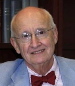 Kenneth M Heilman合作论文数Department of Clinical and Health Psychology, Department of Neurology, College of Medicine, University of Florida9AM Barrett合作论文数7RL Gilmore合作论文数6
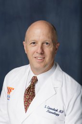 Stephan Eisenschenk合作论文数College of Medicine, University of Florida5
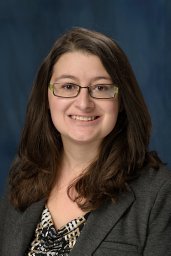 Vasilopoulos Terrie合作论文数Department of Psychiatry and Behavioral Neuroscience, University of Chicago5
 Christoph Seubert合作论文数4LH Lu合作论文数4N Gravenstein合作论文数4RL Schwartz合作论文数4Kaitlyn Melnick合作论文数4
 Lou Ann Cooper合作论文数4
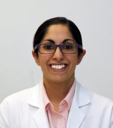 Maryam Rahman合作论文数Department of Neurosurgery, University of Florida4
 Samsun Lampotang合作论文数Department of Anesthesiology, College of Medicine, University of Florida;Department of Biomedical Engineering, College of Engineering, University of Florida;UF Health Shands Experiential Learning Center4Marcia Hodik合作论文数3Chau Destiny F合作论文数3
 Mingzhou Ding合作论文数J. Crayton Pruitt Family Department of Biomedical Engineering, University of Florida3
 Frederick Schmitt合作论文数3David Lizdas合作论文数3Toufic Fakhoury合作论文数3Steven N. Roper合作论文数3David T. R. Berry合作论文数2Daniel S Demos合作论文数2Meriem Bensalem Owen合作论文数2Michael Bewernitz合作论文数2W Travis Johnson合作论文数2JM Anderson合作论文数2
 Matthew Goldman合作论文数Department of Neurosurgery, University of Florida2
 Ashley Ghiaseddin合作论文数2
 Daniela J Conrado合作论文数e‐Quantify LLC2GP Crucian合作论文数2SN Roper合作论文数2
 Edwin Trevathan合作论文数Dept Neurol, Vanderbilt Univ2
 Benjamin C. Warf合作论文数Department of Neurosurgery, Boston Children's Hospital;Harvard Medical School, Harvard University2
 William Alan Friedman合作论文数Lillian S. Wells Department of Neurosurgery, College of Medicine, University of Florida2Sy Sherwin K B合作论文数2
 Hartmut Derendorf合作论文数Department of Pharmaceutics, College of Pharmacy, University of Florida2Dona E. Cragar合作论文数2Matthew L. Cohen合作论文数1CA O'Donovan合作论文数1DC Bergen合作论文数1RK Ristanovic合作论文数1Douglas R. Labar合作论文数1Nina McGrew合作论文数1Greg Murad合作论文数1JI Sirven合作论文数1Michael Gold合作论文数1Jorge A Trejo-Lopez合作论文数1AJ Wilensky合作论文数1




