A challenge posed by model-based formal methods such as Event-B is the validation of the models. This has been recognized and some tools have been created to provide modelers with means to animate models and to explore their behaviour through graphical display. These tools are quite effective on standalone models but lack the ability to connect the model to other external models. CPS systems fall under this category, as well as systems built of components interacting through a communication network. In the context of Jeb, an animation tool for Event-B models based on JavaScript, we explore the possibility of connecting models through Websockets. The paper presents a simple protocol to connect simulations. Using an example inspired by the Lung Ventilator case study, it shows how the implementation expands JeB functionality without modifying its core.
Glutathione transferases are detoxification enzymes with multifaceted roles, including a role in the metabolism and scavenging of nitric oxide (NO) compounds in cells. Here, we explored the ability of Trametes versicolor glutathione transferases (GSTs) from the Omega class (TvGSTOs) to bind metal-nitrosyl compounds. TvGSTOs have been studied previously for their ligandin role and are interesting models to study protein‒ligand interactions. First, we determined the X-ray structure of the TvGSTO3S isoform bound to the dinitrosyl glutathionyl iron complex (DNGIC), a physiological compound involved in the storage of nitric oxide. Our results suggested a different binding mode compared to the one previously described in human GST Pi 1 (GSTP1). Then, we investigated the manner in which TvGSTO3S binds three nonphysiological metal-nitrosyl compounds with different metal cores (iron, ruthenium and osmium). We assayed sodium nitroprusside, a well-studied vasodilator used in cases of hypertensive crises or heart failure. Our results showed that the tested GST can bind metal-nitrosyls at two distinct binding sites. Thermal shift analysis with six isoforms of TvGSTOs identified TvGSTO6S as the best interactant. Using the Griess method, TvGSTO6S was found to improve the release of nitric oxide from sodium nitroprusside in vitro, whereas the effects of human GST alpha 1 (GSTA1) and GSTP1 were moderate. Our results open new structural perspectives for understanding the interactions of glutathione transferases with metal-nitrosyl compounds associated with the biochemical mechanisms of NO uptake/release in biological systems.
The chloroplast is the site of essential reactions in plant cells, including photosynthesis, but also chlorophyll, starch, or amino acid biosynthesis. These metabolic and/or electron transfer reactions inevitably lead to the production of reactive oxygen species that modify the oxidation state of certain proteins and in particular their cysteine residues, causing the formation of disulfide bonds or other reversible redox modifications. In addition, many chloroplastic proteins have evolved such that the appearance of cysteine residues at key positions combined with structural changes allows for light-dependent redox control of their activity. In the chloroplast, the regulation of the redox state of proteins containing reactive cysteine residues is mainly controlled by thioredoxins (TRX). Oxidized TRXs are reduced by ferredoxin-thioredoxin reductase but will oxidize partner proteins when they are not, or only partially, reduced. A NADPH-dependent TRX reductase-TRX chimera referred to as NTRC acts in a complementary manner. In this chapter, we will present the functional properties of both reducing systems and some redox-regulated pathways.
Volume 52 of Advances in Botanical Research "Oxidative stress and redox regulation in plants" edited by Jean-Pierre Jacquot (series Editors Michel Delseny, Jean-Claude Kader) was published in 2009, so now more than 12 years ago. It featured 15 different chapters and more than 60 authors contributed. Volume 52 has covered many aspects of plant physiology including the generation of oxidative stress, the molecular responses to oxidative stress and redox regulation. In general such a volume is initiated about 2 years before final publication and so it has been now 15 years overall. We believe thus that it is now appropriate for the anniversary volume 100 of ABR to summarize what was described in that volume but also to provide a survey of more recent literature showing how the plant redox field has evolved and expanded since.
This chapter is dedicated to the memory of Pierre Gadal a former Series Editor for Advances in Botanical Research. Its aim is to describe the research that has been done in his laboratory covering essentially the period 1970-2000 and provides first-hand testimony about how it has been done. Besides the scientific output concerning CO2 and nitrogen assimilation and their regulation described in this chapter Pierre Gadal has been very influential in plant science in France through his interactions with the University system but also with CNRS and INRA communities. Through the organization of the doctorate school in University of Paris-Sud he has helped train hundreds of students, and more specifically concerning his own research group, more than 10 of his former students have become CNRS researchers, many with high positions and responsibilities, a similar number INRA researchers and Professors and Assistant Professors in the University system. Fourteen different colleagues have contributed to this chapter, each with his own perception, heart and guts, a further testimony to the exceptional human and scientific qualities of Pierre.
The history of the series is described by three successive Editors. Each one reports how he has been approached, his conception of the different volumes and gives an overview of the topics which have been treated. The first period, from creation of the series in 1963 by Professor Reginald Preston to 2006, was covered by Professor James (Jim) Callow who became Editor after Professor Harold Woolhouse, in 1985. During this period, 44 volumes were published, essentially eclectic, covering most of the fields in plant biology. Under James Callow's editorship, the frequency of issues increased up to two/year and a few thematic volumes were published. When he decided to stop in 2006, Dr. Jean Claude Kader and Dr. Michel Delseny were appointed. They maintained a rate of two to three issues/year, alternating eclectic and thematic volumes. They together published 16 volumes and resigned in 2012. Finally, they were replaced by Professors Jean-Pierre Jacquot and Pierre Gadal. They published only thematic volumes and the rate increased to four-five issues/year.
The Gram-positive Bacillus methanolicus shows plasmid-dependent methylotrophy. This facultative ribulose monophosphate (RuMP) cycle methylotroph possesses two fructose bisphosphate aldolases (FBA) with distinct kinetic properties. The chromosomally encoded FBA C is the major glycolytic aldolase. The gene for the major gluconeogenic aldolase FBA P is found on the natural plasmid pBM19 and is induced during methylotrophic growth. The crystal structures of both enzymes were solved at 2.2 Å and 2.0 Å, respectively, and they suggested amino acid residue 51 to be crucial for binding fructose-1,6-bisphosphate (FBP) as substrate and amino acid residue 140 for active site zinc atom coordination. As FBA C and FBA P differed at these positions, site-directed mutagenesis (SDM) was performed to exchange one or both amino acid residues of the respective proteins. The aldol cleavage reaction was negatively affected by the amino acid exchanges that led to a complete loss of glycolytic activity of FBA P . However, both FBA C and FBA P maintained gluconeogenic aldol condensation activity, and the amino acid exchanges improved the catalytic efficiency of the major glycolytic aldolase FBA C in gluconeogenic direction at least 3-fold. These results confirmed the importance of the structural differences between FBA C and FBA P concerning their distinct enzymatic properties. In order to investigate the physiological roles of both aldolases, the expression of their genes was repressed individually by CRISPR interference (CRISPRi). The fba C RNA levels were reduced by CRISPRi, but concomitantly the fba P RNA levels were increased. Vice versa, a similar compensatory increase of the fba C RNA levels was observed when fba P was repressed by CRISPRi. In addition, targeting fba P decreased tkt P RNA levels since both genes are cotranscribed in a bicistronic operon. However, reduced tkt P RNA levels were not compensated for by increased RNA levels of the chromosomal transketolase gene tkt C .
The fluorescence properties of FAD of Thermotoga maritima thioredoxin reductase (TmTR), taken together with the amino acid sequences and structures of similar TRs, are consistent with the interdomain rotation in the catalysis of TmTR. The standard redox potential of FAD of TmTR, –0.230 V, determined by the reactions with 3-acetylpyridine adenine dinucleotide (APAD+/APADH) redox couple, is close to that of E. coli TR. During the reduction of duroquinone with TmTR, the transient formation of neutral FAD semiquinone, and, possibly, FADH2–NAD+ complex was observed. This shows that in spite of obligatory twoelectron (hydride)-transfer between NADH and physiological disulfide oxidants, the FAD cofactor of TmTR may exist under a stable semiquinone form.
The special issue on Thioredoxin and Glutaredoxin systems (http://www [...]
Protein Tyrosine Phosphatases is a book of 280 pages written by Dr Lalima G. Ahuja, who is currently a postdoctoral student at the University of California San Diego in the Department of Pharmacology. Her training is in molecular biophysics and thus she is very well suited for the task. Following a short introductory preface, the book is divided into eight different chapters. The first chapter is mostly historical but also contains more recent data. It recollects the discovery of protein phosphorylation and especially of tyrosine phosphorylation. It describes tyrosine kinases, their structures and their mode of regulation, and then turns its attention to the phosphotyrosine recognition domains in the SH2 and PTB domain containing proteins. The second chapter deals with the classification of protein phosphatases. The author first indicates that there are several types of protein phosphatases which are categorized depending on the nature of the phosphorylated amino acid (either Ser or Thr, or Tyr). Overall there are 15 families of protein phosphatases, 3 of them acting as Ser/Thr phosphatases and the rest as Tyr phosphatases. Amazingly enough, there are more than 100 genes coding for the various protein tyrosine phosphatases (PTPs) in the human genome. This chapter then sets out to describe the relative properties of protein phosphatases especially at the structural level, detailing in particular the fold of the 12 classes of PTPs and the nature of the protein modules that constitute them. The mechanisms of Cysand Asp-based catalysis are also briefly addressed. There is actually some redundancy between this more general chapter and the next five chapters, especially Chapter 3 which details the three-dimensional organization of the various domains. In this chapter a very useful table (Table 3.1) provides a summary of all the PTP proteins of the human genome (two pages are required) and two additional tables detail conserved sequences that have helped classify the various families. Chapter 4 is dedicated to the structure and function of receptor PTPs, and their modular organization and structures are detailed at length. This chapter also addresses their physiological function in signal transduction. Chapter 5 also deals with receptor protein tyrosine phosphatases but focuses on those possessing double domains. Interestingly, it addresses the question of their regulation via the reversible oxidation of cysteine residues. Chapters 6 and 7 detail the non-receptor protein tyrosine phosphatases and not only address their structures, physiological functions and regulation but also their role in cancer and as therapeutic targets. Finally Chapter 8 evaluates the strategies for drug development linked to the PTPs. Overall, after giving a presentation of the field including the necessity for amino acid phosphorylation and dephosphorylation, the book details the organization of the very large families of PTPs – which is not an easy task. I believe that the author has succeeded reasonably well given the complexity of the PTP families. This is actually an amazing task for a postdoctoral student and it certainly reveals high scientific maturity. The literature cited is abundant with overall more than 1000 citations in the book. On the negative side there is some redundancy between the chapters and there are a number of typographical errors, and the quality of the figures (especially of the structures) is not very high, which is unfortunate. It should be mentioned that this book essentially addresses the significance ISSN 2059-7983
From the pioneering work that the Buchanan group started in the late 1960s, we know that the division of photosynthesis into light and dark reactions is inadequate because the activity of a number of chloroplast enzymes, many involved in the Calvin–Benson cycle, is strictly controlled by light; that is, they are activated in the light and deactivated in the dark. An exception is glucose-6-phosphate dehydrogenase, which is regulated in an opposite manner; that is, activation in the dark and deactivation in the light. It took a decade to biochemically dissect the newly identifed redox regulatory pathway responsible for this regulation. Known as the ferredoxin-thioredoxin system, the pathway is composed of three components: ferredoxin, ferredoxin-thioredoxin reductase (FTR), and thioredoxin (Trx) that relay the reducing power generated at photosystem 1 (PSI) to target regulatory enzymes (1, 2). Over the last four decades there have been a number of milestones in the field of redox regulation in plants. The first step was to identify ferredoxin, FTR, and Trx as key components of the regulatory system. We then observed that there were multiple Trxs with differential selectivities in plant eukaryotic cells, and although this seems of little significance now, in the genomic era it was the subject of intense debate at the time. A very important contribution was the elucidation of the 3D structures and catalytic mechanisms of FTRs and Trxs. A lot of effort has also been … [↵][1]1Email: j2p{at}univ-lorraine.fr. [1]: #xref-corresp-1-1
The second volume of the second edition of the book entitled Iron–Sulfur Clusters in Chemistry and Biology, edited by Tracey Rouault and published by de Gruyter, addresses primarily the biochemical properties of iron–sulfur centres (ISCs), their biosynthesis and also, importantly, the dire consequences of some mutations for human diseases. The earlier edition of this book was coordinated by the same editor and the new version has been split into two different volumes. The first one deals primarily with physico-chemical and spectroscopic properties of iron–sulfur centres and iron–sulfur proteins, and it has been subject to an earlier evaluation by the same author (Jacquot, 2017). The second volume, analysed in the present review, contains 17 different chapters, four of them being new additions. A short preface by Tracy Rouault introduces the whole volume. Chapter 1 by Dos Santos and Dean retraces the discovery of the nif operon in Azobacter vinelandii, a nitrogen-fixing species, and the assembly of the metallic cofactors in nitrogenase. The structural features of these metallic centres have been largely discussed in Volume 1. Chapter 2 is a new addition by Barras and colleagues which details the ISC system of bacteria. The third system for ISC incorporation, the suf operon, is described in a third chapter by Wayne Outten who addresses the interesting question as to why suf and ISC coexist in some species such as Escherichia coli. The hypothesis that they are not equally sensitive to stress is obviously of high interest. The next chapter by Mettert et al. describes the properties and phylogeny of E. coli IscR, a cellular sensor of ISC demand, and its binding properties to DNA promoter regions. Chapter 5 by Dos Santos investigates the ISC assembly in Gram-positive bacteria which possess only one assembly system, i.e. clostridia (ISC), actinomycetes (SUF) and bacilli (SUF). In general, it seems that Grampositive bacteria contain fewer client proteins with iron–sulfur centres than their Gramnegative counterparts. Chapter 6 by Pain and Dancis transports us into the eukaryotic world, describing ISC assembly in yeast, essentially via the mitochondrial system. The next chapter by Caryn Outten describes the role of ISCs in the regulation of iron homeostasis in yeast. Moving further into the eukaryotic world, Tracey Rouault details the biogenesis of Fe–S proteins in mammals and the regulatory switches IRP1 and IRP2. This chapter is followed by a new addition by Maio and Rouault dealing with the delivery of ISCs to recipient proteins, detailing in particular the role of chaperone and cochaperone proteins and the importance of the LYR motif in target proteins for the binding to chaperones such as HSC20. Chapter 10 is the largest of them all by Wing Hang Tong with a whopping 600+ references. This analysis details a number of mutations either in ISC-containing proteins or in components of the assembly systems that lead to diseases with severe phenotypes. Table 10.1 of this chapter is very informative about the large number and variety of syndromes associated with mutations of these systems. The next chapter, also an innovation in this volume by Knight and Wilson concentrates on one of these diseases Friedreich ataxia, which results from a defect in frataxin, a protein essential for ISC synthesis and assembly. Then Silke Leimkühler discusses the connections between molybdenum cofactor biosynthesis, ISCs and tRNA thiolation in humans. Basically these reactions rely on cysteine desulfurases of the NFS type and on the formation of persulfides. In Chapter 13 Kerstin Gari draws our attention to the relationship between ISC-containing proteins and genome stability. The bottom line is that ISSN 2059-7983
The sixth edition of Biochemistry and Molecular Biology by edited by Despo Papachristodoulou, Alison Snape, William H. Elliott and Daphne C. Elliott is reviewed.
 Rouhier N.合作论文数Department of Interactions Arbres-Microorganismes, Universite de Lorraine109
 Eric Gelhaye合作论文数Faculté des Sciences, INRA-Université Henri-Poincaré59
 Myroslawa Miginiac-Maslow合作论文数54P Decottignies合作论文数29P Gadal合作论文数26
 Claude Didierjean合作论文数23
 Jeremy Couturier合作论文数Inst Univ France20Peter Schürmann合作论文数17
 Bob B. Buchanan合作论文数Department of Plant & Microbial Biology, College of Natural Resources, University of California, Berkeley16Catherine Corbier合作论文数15
 Kamel Chibani合作论文数Faculté des Sciences, Nancy University15
 Michel Droux合作论文数Laboratoire Mixte Centre;National de Ia Recherche Scientifique;Centre National de Ia Recherche Scientifique;Laboratoire Mixte Centre, Centre National de Ia Recherche Scientifique14
 Melanie Morel-Rouhier合作论文数UMR1136, Université de Lorraine;UMR1136, INRA;INRA, Université de Lorraine12Mariana Stein合作论文数12J M Lancelin合作论文数11
 Stephane D Lemaire合作论文数Lab Biol Mol & Cellulaire Eucaryotes, Univ Paris 0611
 Atif Mashkoor合作论文数LORIA, Nancy Université11Jeanine Souquieres合作论文数l'Universite Nancy211Andre Aubry合作论文数11
 David B. Knaff合作论文数Department of Chemistry & Biochemistry, Texas Tech University10Masakazu Hirasawa合作论文数10
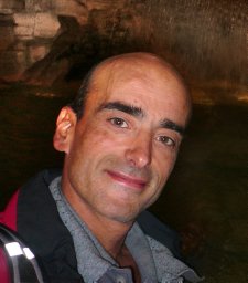 Jose Gualberto合作论文数Institut de Biologie Moléculaire des Plantes, University of Strasbourg10
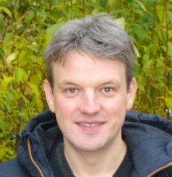 Nicolas Navrot合作论文数Faculté des Sciences BP, Nancy Université Unité Mixte de Recherches 1136 INRA10Jean Vidal合作论文数10Yves Meyer合作论文数9Decottignies Paulette合作论文数8Pierre Gadal合作论文数8Philippe Gérardin合作论文数Laboratoire D'études Et De Recherches Sur Le Matériau Bois, Université Henri Poincaré8Faqing Yang合作论文数8
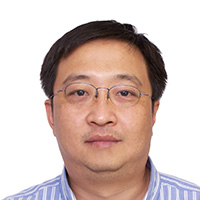 Bin Xia (夏斌)合作论文数College of Chemistry and Molecular Engineering, Peking University8Cenas Narimantas合作论文数7
 Jean-Louis Montastruc合作论文数Faculty of Medicine, University Hospital Toulouse France7
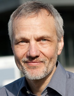 Karl-Josef Dietz合作论文数Faculty of Biology, Bielefeld University;Faculty of Biochemie und Physiologie der Pflanzen, Bielefeld University;Center for Biotechnology, Bielefeld University7
 Alda Navaza合作论文数7
 Jean-Marc Lancelin合作论文数7Isabelle Krimm合作论文数Laboratoire de RMN Biomoléculaire Associé au CNRS, Université Claude Bernard - Lyon 17
 Benjamin Selles合作论文数Unité Mixte de Recherches 1136, INRA-Nancy Université7Claude Crétin合作论文数7
 Ezzedine El Ferjani合作论文数7
 Moez Smiri合作论文数Bio-Physiologie Cellulaires, Faculté des Sciences de Bizerte, 7021 Zarzouna, Tunisie7
 Abdelilah Chaoui合作论文数Bio-Physiologie Cellulaires, Faculté des Sciences de Bizerte, 7021 Zarzouna, Tunisie7S.D. Lemaire合作论文数6Filipe Gama合作论文数6J M Schmitter合作论文数6
 Desiree Gutle合作论文数university of freiburg6
 Francis M. Martin合作论文数Laboratory of Excellence, France's National Research Institute for Agriculture, Food and Environment;Tree-Microbes Interactions Department, France's National Research Institute for Agriculture, Food and Environment6
 Michael Hodges合作论文数CNRS, Institute of Plant Sciences Paris-Saclay, Universite PSud6Pascal Rey合作论文数6Haleh Bagheri合作论文数6




