Background: The treatment of patients who sustain a proximal humerus fracture (PHF) remains controversial. The purpose of this study was to find consensus among experts using a validated iterative process in the treatment of patients after a PHF. Methods: The Neer Circle is an organization of shoulder experts recognized for their service to the American Shoulder and Elbow Surgeons. Consensus among 86 identified experts from this group was sought with a series of surveys using the Delphi process. The first 3 surveys included vignettes with 2-, 3-, and 4-part fractures, under 2 scenarios: (1) a healthy 55-year-old and (2) and a 75-year-old with significant medical comorbidities. Within each vignette, respondents were asked about their preference on computed tomography (CT) use, and whether they would select operative or nonoperative treatment. A final survey was administered to elicit respondent preferences on general treatment approaches regardless of specific vignette characteristics. Results: Consensus was reached on the value of CT scans with 3D reconstructions, age as an important factor in determining treatment, functional demand, fracture pattern, bone quality, and the presence of more significant medical comorbidities, all of which would strongly impact decision making. Experts agreed that medial calcar involvement would have no impact on their decision, and gender and nondominant arm involvement would have a low impact. Consensus was reached in the following scenarios for an operative treatment in a young and healthy patient: 2-part shaft fracture (vignette 3, 98%), 3-part varus fracture (vignette 5, 98%), 3-part valgus fracture (vignette 7, 98%), 4-part fracture (vignette 9, 100%), 4-part dislocation fracture (vignette 11, 98%), and 4-part valgus impacted fractures (vignette 13, 95%); for older unhealthy patients, in 4-part dislocation fractures (vignette 12, 100%). In the remainder of scenarios, there was no consensus reached for preferred treatment, either operative or nonoperative. Finally, there was no consensus on preferred rehabilitation protocols, whether for nonoperative management or postoperative care. Conclusion: In conclusion, this study demonstrates that consensus when managing PHFs is limited to specific scenarios, whereas lack of consensus still exists in others. The presented study advocates nonoperative treatment of PHFs in the sicker patient and surgical treatment methods for 3-part and 4-part fractures in the young, healthy patient. Level of evidence: Level V; Consensus Development Study; Delphi Method (c) 2025 Journal of Shoulder and Elbow Surgery Board of Trustees. All rights are reserved, including those for text and data mining, AI training, and similar technologies.
Background:This study aims to determine the effect of time and imaging modality (three-dimensional (3D) CT vs. 3D magnetic resonance imaging (MRI)) on the surgical procedure indicated for shoulder instability. The hypothesis is there will be no clinical difference in procedure selection between time and imaging modality.Methods:Eleven shoulder surgeons were surveyed with the same ten shoulder instability clinical scenarios at three time points. All time points included history of present illness, musculoskeletal exam, radiographs, and standard two-dimensional MRI. To assess the effect of imaging modality, survey 1 included 3D MRI while survey 2 included a two-dimensional and 3D CT scan. To assess the effect of time, a retest was performed with survey 3 which was identical to survey 2. The outcome measured was whether surgeons made a "major" or "minor" surgical change between surveys.Results:The average major change rate was 14.1% (standard deviation: 7.6%). The average minor change rate was 12.6% (standard deviation: 7.5%). Between survey 1 to the survey 2, the major change rate was 15.2%, compared to 13.1% when going from the second to the third survey (P = .68). The minior change rate between the first and second surveys was 12.1% and between the second to third interview was 13.1% (P = .8).Discussion:The findings suggest that the major factor related to procedural changes was time between reviewing patient information. Furthermore, this study demonstrates that there remains significant intrasurgeon variability in selecting surgical procedures for shoulder instability. Lastly, the findings in this study suggest that 3D MRI is clinically equivalent to 3D CT in guiding shoulder instability surgical management.Conclusion:This study demonstrates that there is significant variability in surgical procedure selection driven by time alone in shoulder instability. Surgical decision making with 3D MRI was similar to 3D CT scans and may be used by surgeons for preoperative planning.
This study aims to determine the effect of time and imaging modality (three-dimensional (3D) CT vs. 3D magnetic resonance imaging (MRI)) on the surgical procedure indicated for shoulder instability. The hypothesis is there will be no clinical difference in procedure selection between time and imaging modality.
BACKGROUND:Full-thickness rotator cuff tears (FTRCTs) represent a common shoulder injury that, if untreated, can progress in size, become increasingly painful, and inhibit function. These lesions are often surgically repaired, with double-row arthroscopic repair often preferred for larger tears. Biological augmentation technologies have been developed to improve rates of postoperative radiographic retear and enhance patient-reported outcomes after surgical FTRCT repair. This study sought to confirm that augmented repair with a bioinductive bovine collagen implant results in favorable retear rates and patient outcomes with follow-up to 2 years.METHODS:A prospective multicenter cohort study was undertaken to determine the efficacy and safety of augmenting single- or double-row arthroscopic repair of FTRCTs with a bioinductive bovine collagen implant. Of 115 adult patients participating, 66 (57.4%) had medium (1-3-cm) tears and 49 (42.6%) had large (3-5-cm) tears. Magnetic resonance imaging and patient-reported outcomes (American Shoulder and Elbow Surgeons Standardized Shoulder Assessment Form [ASES] and Constant-Murley Score [CMS]) were performed and recorded at baseline, 3 months, 1 year, and 2 years.RESULTS:Mean duration of follow-up was 2.1 years (range, 1.5-2.9 years). Between baseline and 2-year follow-up, mean total thickness of the supraspinatus tendon increased by 12.5% for medium tears and by 17.1% for large tears. Radiographic retear was noted in 7 of 61 available patients (11.5%) with medium tears, and in 14 of 40 patients (35.0%) with large tears. In both groups, these tears primarily occurred before the 3-month follow-up visit (13 of 21 [61.9%]). Radiographic retear with the supplemented double-row (DR) repair technique was 13.2% overall (12 of 91 DR patients; 11.3% for medium tears and 15.8% for large tears). The minimal clinically important difference was achieved by >90% of patients with both medium and large tears for both ASES and CMS. There were 2 serious adverse events classified by the treating surgeon as being possibly related to the device and/or procedure (1 case of swelling/drainage and 1 case of intermittent pain). Nine patients (7.8%; 4 medium tears and 5 large tears) required reoperation of the index rotator cuff surgery.CONCLUSION:Final 2-year data from this study confirm that using this implant in augmentation of arthroscopic double-row repair of FTRCTs provides favorable rates of radiographic retear and substantial functional recovery. The relative safety of the device is also further supported.
BackgroundBiologic technologies can potentially augment existing arthroscopic rotator cuff repair to improve retear rates and postoperative outcomes. The purpose of this study was to evaluate healing rates and clinical outcomes of full-thickness rotator cuff repairs augmented with a bioinductive bovine collagen implant.MethodsIn this prospective multicenter study, investigators enrolled 115 patients (mean age, 60.4 years) with full-thickness rotator cuff tears. There were 66 (57.4%) medium (1-3 cm) tears and 49 (42.6%) large (3-5 cm) tears. Eligible patients consisted of those ≥21 years of age with chronic shoulder pain lasting longer than 3 months and unresponsive to conservative therapy. Patients underwent single- or double-row repair augmented with a bioinductive bovine collagen implant. At the baseline, 3 months, and 1 year, magnetic resonance imaging was performed and patients were assessed for American Shoulder and Elbow Surgeons (ASES) Shoulder Score and Constant-Murley Score (CMS). The primary failure end point was retear, classified as any new full-thickness defect observed on magnetic resonance imaging.ResultsThere were 13 retears (11.3%) at 3 months, with an additional 6 (19 total [16.5%]) found at 1 year. In large tears, double-row repair had a significantly lower rate of retear at 3 months (P = .0004) and 1 year (P = .0001) compared with single-row repair. ASES and CMS scores significantly improved between the baseline and 1 year for medium and large tears. At 1 year, the minimally clinically important difference for ASES and CMS was met by 91.7% (95% CI: 84.9-96.1) and 86.4% (95% CI: 78.2-92.4) of patients, respectively. Patients without retear and those <65 years of age had significantly better CMS scores at 1 year when compared with those with retear and those ≥65 years (P < .05). There was no statistically significant difference in outcomes based on treatment of the biceps tendon. Of 9 reported reoperations in the operative shoulder, only 2 were considered potentially related to the collagen implant.ConclusionInterim results from this prospective study indicate a favorable rate of retear relative to the literature and improvement in clinical function at 1 year after adjunctive treatment with the study implant augmenting standard arthroscopic repair techniques.
Background: Current surgical treatment options for partial-thickness tears (eg, takedown and repair, in situ repair) are limited by the degenerative nature of the underlying tendon and may require extensive intervention that can alter the anatomic footprint. The complexity of available techniques to address these issues led to the development of a resorbable collagen implant, which can be used to create a bioinductive repair of partial-thickness tears. Methods: We prospectively enrolled 33 patients with chronic, degenerative, intermediate-grade (n = 12), or high-grade (n = 21) partial-thickness tears (11 articular, 10 bursal, 4 intrasubstance, and 8 hybrid) of the supraspinatus tendon in a multicenter study. After arthroscopic subacromial decompression without a traditional rotator cuff repair, a bioinductive implant was secured over the bursal surface of the tendon. Clinical outcomes were assessed using American Shoulder and Elbow Surgeons (ASES) and Constant-Murley scores (CMS) preoperatively and at 3 months, 1 year, and 2 years postoperatively. Magnetic resonance imaging was performed to assess postoperative tendon healing and thickness at the original tear site. Results: At 2-year follow-up, mean ASES and CMS scores improved both clinically and statistically at 1 and 2 years, compared with baseline, for intermediate- and high-grade tears. There was magnetic resonance imaging evidence of new tissue fill-in within the original baseline tear in 100% of the intermediate-grade tears and 95% of the high-grade tears. In 90.9% of the intermediate-grade tears and 84.2% of the high-grade tears, this new tissue fill-in represented at least an additional 50% of the volume of the initial lesion. From baseline to 2-year follow-up, the mean tendon thickness increased by 1.2 mm (standard deviation, 1.3; P = .012) and 1.8 mm (standard deviation, 2.2; P = .003) in the intermediate- and high-grade tears, respectively. The analysis of tear grade and location revealed no statistically significant difference in the change in mean tendon thickness at any time point. One patient with a high-grade articular lesion demonstrated progression to a full-thickness tear; however, the patient was noncompliant and the injury occurred while shoveling snow 1 month after surgery. Neither tear location nor treatment of bicep pathology affected the ASES or CMS scores at any follow-up point. No serious adverse events related to the implant were reported. Conclusion: Final results from this 2-year prospective study indicate that the use of this resorbable bovine collagen implant for isolated bioinductive repair of intermediate- and high-grade partial-thickness rotator cuff tears of the supraspinatus is safe and effective, regardless of tear grade and location. (C) 2021 Published by Elsevier Inc. on behalf of Journal of Shoulder and Elbow Surgery Board of Trustees.
Anti-EGFR therapy has the potential to increase antitumor immune responses. Therapeutic strategies targeting EGFR and immune checkpoints may benefit patients (pts) with mCRC. We conducted a phase Ib/II clinical trial of cetuximab with pembrolizumab in RAS wild-type (RASwt) mCRC; the primary efficacy results of the trial have been reported (Fountzilas et al ESMO GI 2020). Herein, we present correlative tissue analysis. Tumor biopsies were obtained baseline (pre-Tx) and after 12 weeks (post-Tx); multiplex immunohistochemistry for PD-L1, T-cell exhaustion (TIM3, CTLA4, LAG3) and activation (OX40) markers was performed. T-cells were characterized as partially activated-1 (OX40+/PD-L1-, 1/3 exhaustion markers+); partially activated-2 (OX40+/PD-L1-, 2/3 exhaustion markers+); partially activated-3 (OX40+/PD-L1-, 3/3 exhaustion markers+); exhausted-1 (OX40-/PD-L1-, 1/3 exhaustion markers+); exhausted-2 (OX40-/PD-L1-, 2/3 exhaustion markers+); and exhausted-3 (OX40-/PD-L1-, 3/3 exhaustion markers+). Forty-two pts were enrolled; 14 with matched pre-Tx and post-Tx biopsies, 24 with pre-Tx only. PD-L1 expression was higher in pre-Tx metastasis (M) vs. primary tumor (PT) in both tumor and stromal cells within the microenvironment (TME). The no. of activated T-cells was higher pre-Tx in PT compared to M. The no. of exhausted-2 T-cells in pre-Tx was higher in M vs. PT. The no. of activated T-cells and partially activated-1 T-cells was higher in post-Tx vs. pre-Tx M. There was no change in the no. of exhausted T-cells on post-Tx. PD-L1 decreased on tumor cells and increased in the TME on post-Tx. Temporospatial heterogeneity in the immune landscape of the TME was evident in RASwt mCRC. With the limitation of small sample size, we observed increased PD-L1 expression and exhausted T-cells and decreased activated T-cells in M compared to PT. There was an increase in activated T-cells post-Tx, but the no. of exhausted T-cells was similar, potentially explaining the lack of additive benefit from pembrolizumab. Evaluating the PT in mCRC might not be reflective of immunosuppression in M. Combinations with mAb targeting other T-cell exhaustion pathways may be necessary to improve the antitumor efficacy of the EGFR-based regimens.
Anti-EGFR monoclonal antibody (mAb) therapy can increase EGFR-specific T cells in peripheral blood (Trivedi et al Clin Cancer Res 2016). Thus, therapeutic strategies jointly targeting the EGFR-RAS-MAPK pathway as well as block critical immune checkpoints may be of benefit for patients with metastatic colorectal cancer (mCRC). We conducted a phase Ib/II study of cetuximab, a mAb targeting EGFR, with the anti-PD-1 mAb pembrolizumab in RAS wild-type (RAS-wt) mCRC; the results of the phase Ib part have been previously reported (Boland et al ASCO GI 2018). Here we present the primary efficacy results. Patients with RAS-wt mCRC with at least one prior systemic therapy in the advanced setting were treated in 3-week cycles with cetuximab (400 mg/m2 loading dose followed by 250 mg/m2 weekly) and pembrolizumab (200 mg on day 1). Tumor biopsies were obtained at baseline and on-treatment (C4D1). The primary objectives were to estimate the objective response rate (ORR) by RECIST 1.1 and the 6-month progression-free survival (PFS). We utilized a single-stage version of the bivariate design of Sill et al., 2012. For ORR, H0=0.2 vs. H1=0.4 and 6-month PFS, H0=0.3 vs. H1=0.5, a=0.1, with 38 evaluable patients the study had ≥80% power to detect activity based on ORR alone, ≥80% power to detect activity based on 6-month PFS alone, and ≥97% power if the regimen is active on both endpoints. Forty-two RAS-wt patients were enrolled through October 2019. There were no new safety signals and the combination was well tolerated. Three patients had prior exposure to anti-EGFR therapy. 6-month PFS was 30% (CI: 19%-43%) and ORR was 5%. The median PFS and overall survival (OS) were 4.1 months (95% CI 3.9-6) and 14.9 months (95% CI 8.3-24), respectively. The disease control rate (DCR) was 73%. Thirty percent of patients had their CEA levels decrease >50% and 49% had a decrease in disease burden based on a decrease in the sum of target lesions. An increase in tumor-infiltrating cytotoxic lymphocytes (CTLs, CD3+CD8+) was observed (p=0.035). CTL infiltration was more pronounced in patients with CEA levels that had decreased by >50% vs. not (34% vs. 17%, respectively; p=ns) and those with any decrease in tumor burden vs. not (34% vs. 18%, respectively; p=ns). Although the primary efficacy endpoint was not achieved, cetuximab plus pembrolizumab had modest anti-tumor activity in patients with RAS-wt mCRC. Compared to historic controls of anti-EGFR monotherapy, the PFS results are similar, though OS appears longer. Increased intratumoral CTL infiltration was noted following therapy; this appeared most pronounced in patients who benefited from therapy. Tissue analyses were underpowered owing to the small sample size. Further correlative analysis of tumor and blood specimens is ongoing.
Background: The optimal surgical approach for recurrent anterior shoulder instability remains controversial, particularly in the face of glenoid and/or humeral bone loss. The purpose of this study was to use a contingent-behavior questionnaire (CBQ) to determine which factors drive surgeons to perform bony procedures over soft tissue procedures to address recurrent anterior shoulder instability. Methods: A CBQ survey presented each respondent with 32 clinical vignettes of recurrent shoulder instability that contained 8 patient factors. The factors included (1) age, (2) sex, (3) hand dominance, (4) number of previous dislocations, (5) activity level, (6) generalized laxity, (7) glenoid bone loss, and (8) glenoid track. The survey was distributed to fellowship-trained surgeons in shoulder/elbow or sports medicine. Respondents were asked to recommend either a soft tissue or bone-based procedure, then specifically recommend a type of procedure. Responses were analyzed using a multinomial-logit regression model that quantified the relative importance of the patient characteristics in choosing bony procedures. Results: Seventy orthopedic surgeons completed the survey, 33 were shoulder/elbow fellowship trained and 37 were sports medicine fellowship trained; 52% were in clinical practice similar to 10 years and 48% <10 years; and 95% reported that the shoulder surgery made up at least 25% of their practice. There were 53% from private practice, 33% from academic medicine, and 14% in government settings. Amount of glenoid bone loss was the single most important factor driving surgeons to perform bony procedures over soft tissue procedures, followed by the patient age (19-25 years) and the patient activity level. The number of prior dislocations and glenoid track status did not have a strong influence on respondents' decision making. Twenty-one percent glenoid bone loss was the threshold of bone loss that influenced decision toward a bony procedure. If surgeons performed 10 or more open procedures per year, they were more likely to perform a bony procedure. Conclusion: The factors that drove surgeons to choose bony procedures were the amount of glenoid bone loss with the threshold at 21%, patient age, and their activity demands. Surprisingly, glenoid track status and the number of previous dislocations did not strongly influence surgical treatment decisions. Ten open shoulder procedures a year seems to provide a level of comfort to recommend bony treatment for shoulder instability. (C) 2020 Journal of Shoulder and Elbow Surgery Board of Trustees. All rights reserved.
Clinical studies combining radiation and immunotherapy have shown promising response rates, strengthening efforts to sensitize tumors to immune-mediated attack. Thus, there is an ongoing surge in trials using preconditioning regimens with immunotherapy. Yet, due to scarcity of resected tumors treated in situ with radiotherapy, there has been little investigation of radiation's sole contributions towards local and systemic anti-tumor immunity in patients. Without this access, translational studies have been limited to evaluating circulating immune subsets and systemic remodeling of peripheral T cell receptor repertoires. This constraint has left gaps in how radiation impacts intratumoral responses and whether tumor-resident T cell clones are amplified following treatment. Therefore, to interrogate the immune impact of radiation on the tumor microenvironment and test the hypothesis that radiation initiates local and systemic expansion of tumor-resident clones, we analyzed renal cell carcinoma from patients treated with stereotactic body radiation therapy. Blood and tumor samples were collected from patients prior to nephrectomy for renal cell carcinoma. These samples were compared to samples from patients treated on a 15 patient clinical trial of single fraction stereotactic body radiation therapy delivered 4 weeks prior to nephrectomy. Transcriptomic comparisons were evaluated by bulk RNA sequencing. T cell receptor sequencing monitored repertoires during the 4-week period between SBRT and nephrectomy. Pathway analysis from tumor transcriptomic data showed radiation-specific enrichment of immune-related processes. T cell receptor sequencing revealed increased clonality in radiation-treated tumors. The frequency of identified, tumor-enriched clonotypes was tracked across serial blood samples. Compared to pre-treatment, we observed increased abundance of tumor-enriched clonotypes 2 weeks post-radiation. However, this expansion was not sustained and contracted towards baseline levels 4 weeks after treatment. Altogether, these results indicate robust intratumoral immune remodeling and a window of tumor-resident T cell expansion following radiation that may be leveraged for rational design of combinatorial strategies. Samples from renal cell carcinoma (RCC) patients underwent high-throughput analysis to reveal transcriptional immune activation and increased clonality in irradiated tumors. Analysis across longitudinal blood samples showed tumor-enriched clonotypes undergo phases of peripheral expansion and contraction following radiation. Collectively, these findings demonstrate radiotherapy remodels intratumoral T cell responses and have significant implications for refined sequencing of combination strategies in RCC.
Rotator cuff tears are increasing in frequency in the aging population and are a common issue seen by orthopaedic surgeons. In patients with large, multi-tendon rotator cuff tears or retears, treatment can be challenging. Failure rates of up to 90% have been reported for rotator cuff repair (RCR) of large, multi-tendon tears. Biological augmentation has been an area of interest because of the distinctly different biology of the repaired tendon compared with the native tendon. These biological differences affect the ultimate tensile properties of the repair and may contribute to gap formation and the high failure rate of repairs. RCR with allograft augmentation is a technique that shows potential benefit to healing and preventing retears. Arthroscopic augmentation of RCRs can be challenging. The technique described in this Technical Note illustrates a simple and easily reproducible method for augmenting RCRs with human acellular dermal allograft.
The controversy as to what is the best technique to repair a rotator cuff continues, with single-anchor row versus double-row techniques being highlighted. The literature has presented multiple studies with clinical outcomes being similar, even though double-row linked and transosseous-equivalent repairs have a higher success rate with postoperative imaging. Clinical outcome instruments weigh pain as a major criterion, but strength improvement favors an intact repair. Treatment of chronic rotator cuff tears often yields muscular changes that may compromise the strength-improvement portion of the outcome. Larger tears benefit from additional fixation, and tissue loss continues to require adjustments to the repair strategy. Attempting a repair that emphasizes footprint coverage may over-tension the cuff repair and risk shoulder stiffness and medial failure of the repair. By use of a 3-dimensional spherical attachment surface, a linked infraspinatus repair can be combined with an anteromedial supraspinatus repair to create a lower-tensioned secure repair. Additional grafting methods, including use of the biceps, may provide additional strength to the repair construct.
Surgical repair of the unstable shoulder begins with reattachment of the detached capsulolabral complex. The degree of damage to the glenohumeral articulation can be variable and is often related to the degree of trauma, duration of dislocation, and the number of instability events. There have been many surgical procedures proposed for the treatment of shoulder instability in the athlete, ranging from soft-tissue repair to coracoid transfer or the addition of a bone graft. The arthroscope provides an opportunity to visualize and repair the injured structures, returning the shoulder to maximal range of motion and permitting functional improvement. Indications for arthroscopic anterior stabilization include a first-time dislocation, patients with apprehension following dislocation, and recurrent dislocation and subluxation prior to creating advanced bone loss. If there is advanced bone loss, an augmented repair or a procedure other than arthroscopic stabilization has been recommended. Mobilization of the anterior capsule and fixation to recreate the proper anterior tension will limit translation and potential recurrence of instability. The steps of the arthroscopic anterior stabilization include:Perform examination under anesthesia to identify the directions and degree of humeral translation relative to the glenoid.Position the patient with the shoulder 30° abducted and 20° flexed.Create proper portals, including a posterior viewing portal, dual anterior portals, and accessory portals for suture anchor placement.Perform a diagnostic arthroscopy to determine the damaged structures and how they relate to shoulder positions that may invite future dislocations.Perform capsule and labrum mobilization to permit anatomic relocation of the injured ligament.Place a series of suture anchors along the anterior and inferior glenoid margin.Utilize suture hooks to retrieve the sutures placed through the capsule to advance the capsule superiorly to the glenoid margin.Assess glenoid deficiency and place an autograft anterior to the damaged glenoid rim in selected cases.Tenodese the posterior capsule and infraspinatus to a large Hill-Sachs lesion on the posterosuperior aspect of the humeral head in selected cases.Repair additional labral structures superiorly and posteriorly if they contribute to glenohumeral instability. The anticipated outcome is a return to sport and high-demand activities. Bracing is available, but the internal repair is the most reliable technique to protect the glenohumeral articulation. Additional techniques can be implemented when added trauma has resulted in severe bone loss of the glenoid, humeral head, or anterior capsular structures. A return to high-risk activities can be anticipated in 4 to 7 months.
Superior capsular reconstruction is a powerful tool for the treatment of massive irreparable rotator cuff tears. Several authors have described this evolving technique. Issues of graft sizing, graft passage, graft tensioning, and suture management make this a challenging procedure even in the hands of experienced shoulder surgeons. We describe our arthroscopic technique for superior capsular reconstruction using nonirradiated human acellular dermis. We introduce several techniques for graft passage and tensioning that may help to simplify this challenging procedure and make it more reproducible.
Surgical repair of shoulder instability is challenging, and multiple procedures have been proposed. In an attempt to reduce risk of recurrence following surgical reconstruction, some surgeons have added steps to prior arthroscopic procedures, and other surgeons have selected a bone reinforcement procedure. These additional augmented repair techniques have reduced the risk of postoperative recurrence, but introduced additional risk of complications related to hardware, fixation, and possible need for additional surgery. Surgeons should become familiar with multiple surgical procedures to treat patients with recurrent shoulder instability, and select the appropriate procedure that addresses the demands of the athlete's shoulder and minimize the risk of complication.
Objective: To present recommendations for the diagnosis, management, outcomes, and return to play of athletes with superior labral anterior-posterior (SLAP) injuries. Background: In overhead athletes, SLAP tears are common as either acute or chronic injuries. The clinical guidelines presented here were developed based on a systematic review of the current evidence and the consensus of the writing panel. Clinicians can use these guidelines to inform decision making regarding the diagnosis, acute and long-term conservative and surgical treatment, and expected outcomes of and return-to-play guidelines for athletes with SLAP injuries. Recommendations: Physical examination tests may aid diagnosis; 6 tests are recommended for confirming and 1 test is recommended for ruling out a SLAP lesion. Combinations of tests may be helpful to diagnose SLAP lesions. Clinical trials directly comparing outcomes between surgical and nonoperative management are absent; however, in cohort trials, the reports of function and return-to-sport outcomes are similar for each management approach. Nonoperative management that includes rehabilitation, nonsteroidal anti-inflammatory drugs, and corticosteroid injections is recommended as the first line of treatment. Rehabilitation should address deficits in shoulder internal rotation, total arc of motion, and horizontal-adduction motion, as well as periscapular and glenohumeral muscle strength, endurance, and neuromuscular control. Most researchers have examined the outcomes of surgical management and found high levels of satisfaction and return of shoulder function, but the ability to return to sport varied widely, with 20% to 94% of patients returning to their sport after surgical or nonoperative management. On average, 55% of athletes returned to full participation in prior sports, but overhead athletes had a lower average return of 45%. Additional work is needed to define the criteria for diagnosing and guiding clinical decision making to optimize outcomes and return to play.
Background: Treatment of partial-thickness cuff tears remains controversial. Although conservative therapy may treat symptoms, these defects do not spontaneously heal and conversion to a full-thickness lesion with subsequent repair may alter the tendon footprint. The ability to induce new tissue formation and limit tear progression in intermediate-and high-grade partial-thickness tears without surgical repair may represent a significant advancement in the treatment paradigm for these lesions. Methods: We prospectively enrolled 33 patients with chronic, degenerative, intermediate-grade (n = 12) or high-grade (n = 21) partial-thickness tears (11 articular, 10 bursal, 4 intrasubstance, and 8 hybrid) of the supraspinatus tendon in a multicenter study. Following arthroscopic subacromial decompression without repair, a bioinductive implant was attached over the bursal surface of the tendon. Clinical outcomes were assessed using American Shoulder and Elbow Surgeons and Constant-Murley scores preoperatively and at 3 and 12 months postoperatively. Magnetic resonance imaging was performed to assess postoperative tendon healing and thickness at the original tear site. Results: At 1-year follow-up, clinical scores improved significantly (P<.0001) and the mean tendon thickness increased by 2.0 mm (P<.0001). Magnetic resonance imaging evidence of complete healing was found in 8 patients and a considerable reduction in defect size was shown in 23, whereas 1 lesion remained stable. In 1 noncompliant patient with a high-grade articular lesion, progression to a full-thickness tear occurred while shoveling snow 1 month after surgery. No serious adverse events related to the implant were reported. Conclusions: Arthroscopic implantation of a bioinductive collagen scaffold is a safe and effective treatment for intermediate- to high-grade partial-thickness rotator cuff tears of the supraspinatus tendon. (c) 2017 Journal of Shoulder and Elbow Surgery Board of Trustees. All rights reserved.
I am honored to have served in 2016 as President of the Arthroscopy Association of North America (AANA) and to present to you my Presidential Address. I would like to welcome the audience—the membership and those who are not members but considering joining us. I would also like to welcome our friends to the north, our Canadians, to whom I owe so much in terms of education, background, culture, and of course, friendship. There are many international people in the audience. Over the years, I have been blessed with the opportunity to be able to visit your countries, spend time with you, and gain so much through education, your company, and your friendship. And of course, I would like to welcome the organizational Presidents who have come to the AANA meeting this year. There is a great tradition of Presidents of AANA, and these individuals have really structured the organization as you see it today. Many times, there are events, challenges, and difficulties—“speed bumps,” if you will—that require certain decisions; the past Presidents clearly have created a wonderful organization and made difficult decisions with a tradition of honor, respect, and education (Fig 1). I feel that we are all indebted to these individuals for their contributions. Where is AANA today? We are larger and reach an audience without boundaries, but there are challenges ahead, and I would like to share with you those challenges. Membership now exceeds 4,100, as presented in the membership report. As logic and history defines, if you just continue to do what you have done in the past, it becomes stale and repetitive. The design and the paradigm for learning for students, residents, and fellows need to be recognized and addressed through strategic planning for education in the future. Our mission statement, of course, is to continue medical education and develop surgical skills. We want to strive for competence, performance, and of course, improve our patient outcomes. So if I may, I would like to go over some of the critical issues we have had over the year and talk to you a bit about innovation, similar to that which individuals like Steve Snyder and Marlowe Goble experienced in shaping the design of our specialty and our future. Most realize that “meeting congestion” is an issue, including meetings you attend and possibly many meetings at which you teach. This is a common concern that many of you in the audience have experienced. It used to be that you could go to a meeting here and there, but the problem right now has become the number of meetings that you need to attend and how you can functionally be at home, at your office, and in the audience at a meeting. Consequently, we have competition of dates, competition of venues, and I would like to say that one of the strategic goals of my Presidency was to see whether we could combine society-sponsored meetings to reduce this congestion. Along that path, we have a meeting that is scheduled for September 2016 with the American Shoulder and Elbow Surgeons; they have offered this meeting in the past with the Academy, but instead, we are partnering with them this year in Chicago, combining basically 3 to 4 meetings into one venue, which will include the Orthopaedic Learning Center (OLC) for surgical demonstrations. The Biennial Shoulder and Elbow Meeting will include didactic and a Spanish-speaking OLC experience over the weekend. In addition, we will invite physical therapists and allied health professionals to the meeting and will hopefully set a pattern to see whether these combined meetings can be successful. This is an ambitious endeavor, but it looks very good at this time. Right now, we are an older generation that enjoys attending meetings, meeting with friends, going out to dinner, and learning in this environment. But there is a group of younger surgeons who are graduating from residency and fellowships who clearly have a completely different design on how they learn and get information, called the Internet. The percentage of individuals who actually read a newspaper is in single digits now. The majority go to the Internet, visit their favorite sites, and become educated on the day-to-day events. Like it or not, that is probably the direction that this type of meeting will go. That is not to be in competition with what we do here—because I still love this environment and love to have time to spend with all of you. The truth is that we will need another design for educating our membership, and that will include Internet-based education. We will have webinars, meetings, the case panels enjoyed by many. We can even have interviews. For that matter, we are looking at several different vendors—the G9 is one of the vendors that we have met with during our Board meeting—and it looks promising. I think moving toward that design is inevitable. Members will be able to stay at home to attend and try to be educated in the same way, rather than traveling away from home. Again, that is not to say that this meeting format will go away, but it may change in size, and we should be prepared for this. Surgical Skills work is certainly a big part of our mission. We moved into this large new building in Rosemont a couple of years ago; the new building includes the OLC. New expenses, new people, new equipment, and new partnerships came along with this change. However, thanks to key individuals, including Lise Puckorius, Kevin Plancher, and the committee, we have a design where the surgeons can come one-on-one and they can bring their assistants; this offers a personalized, individual mentorship similar to what Rick Ryu designed probably 3 to 4 years ago, with one-on-one tutorials. This new setting will include surgical demos offered a faculty that has been vetted and rated by the attendees and other faculty. You are constantly being scored both as an educator and to a certain extent as a student, because somewhere along the line—with your Board recertification and as outcomes—your surgical skills will be reviewed as well. It would be wonderful to be able to credential, be tested, and let individuals know where they stand in terms of surgical skills, certainly with the opportunity of designing and demonstrating improvement before and after a laboratory experience. The new AANA textbook series has arrived and is available for purchase. There was an initial series several years ago; this series is the update, with new, additional chapters supported by videos. I would like to thank Rick Ryu for his leadership and serving as a series co-editor, and I would also like to thank the individual textbook editors. When we asked the publisher what our pricing should be on this, it was nearly double. AANA decided on a break-even philosophy—to accommodate both our membership and industry—and so we chose not to go over $99.00 and limit additional documentation to the government. Because of Advamed rules, there is a requirement for industry to report when providing educational materials to physicians, including textbooks. This additional burden could jeopardize the generosity of some industry partners, and by reducing the price, this would put this textbook—or textbooks—in everyone's hands and enrich our learning experience. Arthroscopy has also moved in many directions under the leadership of Jim Lubowtiz. The services and expertise of Matt Provencher and Michael Rossi as the Assistant Editors-in-Chief—collectively with other assisting editors and the reviewers—have made this a world-class journal that is something to be very proud of. Some have termed it “the crown jewel.” Melissa Schmidt is the Managing Editor, and together with the AANA team, this has been a great collaboration. This relationship, defined by congeniality and education, should be rewarding. People question why AANA is in other parts of the world. Part of the answer is for us to share education, because I do not believe that we have all the answers. As Rich Hawkins has quoted, “I'm not even sure we have all the questions, never mind the answers.” That being said, I think that the Journal would be one way to monetize the aspect of our travel and our outreach. When we go to different countries, our international friends can purchase subscription series to our often-quoted “green journal.” There is significant competition with other forms of self-education. We are trying to look at other aspects of teaching, and Arthroscopy Techniques is really the next venue for this organization. In addition to having the written journal, we now have articles and videos submitted to a technique journal, which is an Internet-driven educational medium. The benefits to the authors are as they are with the written journal; they have PubMed Central credit—meaning they are cited—and this will not only be read by orthopaedic surgeons with passwords, but also by other individuals, because the content will be open access, with YouTube being part of the carrier in this endeavor. There are aspects that make certain types of articles difficult to put into print, particularly case reviews and certain techniques, and this offers opportunity to place these types of articles in a well-respected, well-visualized medium, which I believe will be helpful to those residents and fellows in the room. I would envision that surgeons preparing for a case would review and communicate at the scrub sink before they go into the room to take on the next difficult surgical challenge. In addition to the scientific channels, we are embracing industry, and Arthrex would be the first to take on the contract with submitting a certain number of articles and having several of their champions place articles on the ATech channels. The videos and articles are available not only by computer, but also by some of the mobile units, that is, your cell phone and tablets. This organization has always had a great respect for the military. Featured on your podium are active and retired military, so it is no surprise that orthopaedics and the military orthopaedic organizations are one, and both should partner in education. The education of the military surgeons can be somewhat abrupt. They go through their residency, followed by fellowship, and then they are asked to serve. Some of them may be deployed to Afghanistan within months of finishing their training. All of us understand how much value there was in being in practice for a certain amount of time before handling some of the more difficult and serious surgically treated injuries. Military surgeons find themselves in frontline positions with tremendous responsibilities, so AANA has assisted and exchanged experiences to provide additional education to our orthopaedic brotherhood. We have a SOMOS (Society of Military Orthopedic Surgeons) program at OLC, which includes mini-fellowships. The expanding relationship would include military traveling fellows, visiting certain institutions and practices around the country, and spending time with surgeons—whether it is a short period of time, or whatever the program will allow. We also know that the VA (Veterans' Administration) hospitals have been under great scrutiny in terms of the level of care given to the soldiers who have returned. We need to expand these educational opportunities to our military physicians. The number of orthopaedic and sports-related injuries that prevent an individual from going back into active military service is alarming. As dramatic as it is to see some of the most horrific pictures from people coming home, the truth of the matter is that a large percentage of people have become disabled in the military because of injuries that many of you have seen in this conference, including knee and shoulder injuries. Rob Hunter has gone to lobbyists and gained their attention, enlisting their help in trying to gain some financial assistance from the government in this military education. Up until now—and this will likely continue—the majority of this educational benefit is based on volunteer service. Currently, we have asked industry, physicians, and organizations to help fund those meetings. There are many entities involved in this partnership, and with anticipated success, we will have the government help underwrite some of the expenses of getting soldiers and physicians to orthopaedic educational events in the United States; I applaud this effort. Many people in the audience are involved in the recertification process for Board of Orthopaedic Surgeons. There has always been a relationship with the Academy, which has embraced additional assistance in preparation for the Board and has encouraged continued education and competence. That being said, I think it is very important that our organization, as educators, help prepare our surgeons for the recertification examinations. We have created 3 written examinations that help us with this self-assessment; it is a requirement to have at least 20 credits from self-assessment sources of CME, in addition to the ones that you get for attending meetings. Mark Getelman and his committee have created a new examination to help comply with recertification requirements. I would like to applaud Rick Angelo, as he has put together a project that I think is unique and innovative, along with many volunteers: the Copernicus project. This project was designed to create a measurement instrument for surgical skill that is more appropriate than measuring with a multiple-choice examination. This is an educational instrument, as well. You take the examination; you learn; you become a better surgeon. With Rick's direction and assistance from everybody in the audience, you would see how this may impact the credentialing process in the future and be of interest to an organization like ABOS (the American Board of Orthopedic Surgery), as well as perhaps even insurance carriers and hospitals. Our international collaboration cannot be understated. As I said earlier, the opportunity of visiting other countries to lecture and listen is the way we get better. We talked about several orthopaedic procedures in this meeting, and clearly there are some different opinions. I am not sure, as one would say, that either team has gotten it completely right. I think it is the open exchange of information that makes us have our eyes open, our ears listening, and our curiosity piqued to see what is available. ISAKOS (the International Society of Arthroscopy, Knee Surgery, and Orthopedic Sports Medicine) shares our dream of international collaboration, which is why we have visiting nations for our meetings–in this case, Canada is our featured nation. Famous shoulder surgeon Dr. Uhtoff is from Canada; many of us quote him in our papers as an individual who has taught us an awful lot about what can make a rotator cuff heal. The Foundation is very important and vital to our organization. AANA contributors to the Education Foundation are featured on the wall, just as a small demonstration of thanks. Ed Goss played a role with AANA as Executive Director, and now plays a significant role with our Education Foundation. I would suggest that our membership should consider joining the existing contributors to keep our mission healthy and productive. We are doing our best, and I think our Education Foundation certainly deserves to be applauded for its ability to put on meetings and continue to look at what this membership needs to get further and better at what we are supposed to do. Industry is a great partner in all this. I have always enjoyed the industry part of our relationships, not only for what you heard earlier this morning about innovation, but also for the ability to put on meetings and the ability to walk up and to interact with individuals who can help us with new techniques, new equipment, and perhaps familiarize us sometimes with opportunities that would not be so easy to obtain when we go back home and become quite busy. We have traveling fellowships. You might have met some of our traveling fellows who went through the East Coast of the United States; their final stop was this meeting in Boston. The traveling fellows were funded by industry and outside resources, to help defray some of the costs of this important educational project. Through AANA advocacy expertise, you heard critical and instructive comments about trying to protect your income. Our active committee has enlisted and informed many surrounding organizations. In fact, when it comes down to the American Academy of Orthopaedic Surgeons (AAOS), I think they look to the AANA leadership to lead the charge, because AAOS has, at times, been compared with a large battleship trying to turn around, whereas the agility and quickness of AANA can take on certain challenges—for instance, denials of payment and certain things that can affect our ability to treat our patients. The most recent one was the fact that the shoulder was grouped as a single environment and the coding systems were being dismissed and denied, which did not get much attention from CMS (the Centers for Medicare and Medicaid), the major organization that oversees the NCCI (National Correct Coding Initiative). Whether this comes from the White House is not clear, but the efforts of AANA's Louis McIntyre, combined with AAOS leadership, were able to change that decision, which benefits many shoulder surgeons. From a strategic position, our organization helps with reimbursement, not only to challenge denials, but also to “play offense”—to prepare our audience to anticipate changes in our collective future. I do not know how many of you have prepared yourself to take on surgical outcomes in the surgical outcome system. I will warn you that this is 2016. The payment that you were paid before was through an SGR (Sustainable Growth Rate) formula system, and all of these initials can become very confusing to me and maybe to some of you. The SGR formula was the way Medicare and the government reimbursed you. That has gone. In its place is MACRA (Medicare Access and CHIP Reauthorization Act), which includes surgical outcome measurements, unless you deal with bundle payments. So your practice and billing will change in the next 18 months, and if you do not do it differently, you will see substantial changes in your reimbursement. These could exceed a 20% difference, which is not a small change. I would pay attention to these lectures, as they may seem like they are distant, but this is all around the corner. In the next 18 months, start thinking about additional employees in your office and how you are dealing with data collection; if you do not start doing it in the very near future, you are going to find yourself being one of “the stories.” This is why you might look at a surgical outcome system where you can organize results and plot them out for a procedure. This can be compared with other individuals who are submitting data-creating graphics. This is not only an instrument for patients who would like to see how do you do with this procedure or how they will potentially do, but it also applies on a much larger scale. If we combine the surgical outcome system, potentially, with the work of JT Tokish and Richard Hawkins in South Carolina, we may find that we have successfully served the MIPS (Merit-Based Incentive Payment System) requirement in terms of getting it as a national quality review and we may have the opportunity to achieve the Board and the national CMS requirements, not only not being penalized, but being rewarded for our data collection. Let us switch gears a little bit. I would like to talk to you about innovation. You have had an opportunity to be enriched by several individuals at the podium during this meeting: Marlowe Goble, in his talk about the first suture anchor; Dave McGuire, who we sorely miss, who was a Past President and, in addition to his professionalism, was a tremendous innovator, particularly if you start talking about Kurowsaka interference screw fixation; and Dr. James Andrews, who—perhaps more on the technique than development of equipment and implants—clearly has changed the way we perform reparative, minimally invasive procedures on athletes. Ray Thal, for those of you who do not know, has helped AANA design an innovation center. That is a way for individuals in the audience who come up with ideas. If you listened to Steve Snyder's talk a little bit earlier today about having things patented, he provided a mechanism for individuals in the audience to deal with this in an inexpensive way where you can actually, through AANA, be attached to someone who could help you with that patent. Good ideas do not have to be aborted because of time and expense, because of the work of these individuals. I have had an opportunity to be part of innovation. I was a Fellow in London, Ontario, and here, and I was being taught by Rich Hawkins as we were trying to demonstrate quantifying humeral translation. We were just doing translation studies on lax and unstable shoulders, and were able to try to put a name that combined with the previous notoriety of “pivot shift” coming from Canada; we decided that “load-and-shift” would be a good title for this examination that started back during my fellowship year. A bidirectional repair would seem like a new concept. Actually, I reported and wrote on this technique in 1996 while traveling through France, presenting it to improve footprint coverage. At the same time, there were 2 other authors with similar techniques with medial and lateral row fixation, one of whom presented at the same meeting. So this seems to be new, but it maybe 20 years old. There may be good news and bad news about this, but there is no question it certainly has been embraced by surgeons trying to create coverage of a footprint and improving the healing process in an otherwise compromised area. Knotless anchors were not very popular for a bit. It took a while. I received a patent on an early design that is commonly used to capture the suture. I am not sure how this all happened, but recently, I was recognized for this patent with a very close friend of mine from home. Although this recognition was not monetary, I received an award for the knotless anchor and the design and was inducted into the New Jersey Hall of Fame. I shared the podium with some people who created things on the space shuttle, mass communications, fuel, and electronic cars. Tesla was posthumously inducted at the same meeting, and two Nobel laureates were at this meeting at the time. If I can, I will share with you a couple of thoughts and some concerns about new innovations. Resorption of bone graft and exposed hardware from a Latarjet is a concern and a complex surgical dilemma. I think we have to appreciate the significance of complications. Internationally, these things get mentioned, but surgical experience is considered the best way to reduce failure and complications. Some believe that Latarjet is a better way to go because of the likelihood of stabilization, but I do not think that we can underscore enough the fact that these complications can be devastating to a very young population. Hardware can become exposed in certain individuals, and these are difficult operations. I think we need to instruct caution; the pendulum may have pulled back from the arthroscopic techniques a little bit, but I would be cautious in trying to tell you whether we should be embracing some alternative techniques. When considering new technology, we should attend the OLC to perfect our technique and minimize the chances of a poor outcome. As an example, we have the patient with the reverse inverted pair. We are looking from an anterior-superior view at a shoulder with glenoid rim deficiency. We can take a piece of bone from the patient's clavicle and insert it through an anterior portal. With suture anchor fixation, we can use arthroscopic techniques to re-establish the anterior-inferior quadrant. We have radiographic confirmation of the intact glenoid rim. I believe that this will play a role in the recurrent dislocated shoulder. Rotator cuff repairs: you have heard some ideas this morning about novel ways of doing these. I think we are ignoring the fact that we are taking a compromised structure—in most cases, elderly patients' tissue loss—and trying to re-establish a tensionless construct. This is impossible. Your shoulders are under tension with your rotator cuff intact, and we need to recreate the proper degree of tension, so we need to take a longer and harder look at potentially grafting tissue. The patient's long head of the biceps can be used as the tendon extension of the supraspinatus, as it is sewn anteriorly into an anchor, posteriorly into the infraspinatus, and medially into the supraspinatus stump. Additional, exciting types of grafts are also available. We are looking at them closely, and I think by bringing in additional tissue, these can be done in a way that is not so time consuming and expensive. I believe that tissue loss has to be treated differently than just trying to revise or repeat the repair, and I think this may become more routine in our future. Do not let age scare you. I have a patient from Hopewell, NJ, a neighboring town to Princeton. He is 102 years old. I would like to say that follow-up even in community medicine can be pretty good, because at 84 years of age he tore his rotator cuff chopping a tree down in his backyard. We fixed his rotator cuff, and he came in to ask me about his other shoulder. When asked to lift his arms up, he still has pretty good function. I would not be afraid of dealing with older patients, trying to help them with their pathology, and improving their life. Strategic planning is important. This is an aspect of our organization that makes us unique. We need to actually have a structured approach and a structured schedule for developing the educational patterns and ways we deal with our membership to make this an enriching experience. This schedule is put together at our strategic planning meeting, and I applaud John Richmond and others following him in that endeavor, as we try to continue to evolve—just as this program, I think you would admit, is different and unique from other programs before it. This is my opportunity to express appreciation to a special friend and well-respected educator. I am honored to acknowledge my program chairman, Dr. J.T. Tokish. He has done a wonderful job throughout his term as Education Chair. He demonstrates character, which is hard to define, but certainly enviable in a leader. He is to be trusted, he leads in a humble way, and his goal is to make you more successful. He is a friend and respected educator, and you will be seeing a lot of Dr. Tokish in the future. Personally, I was asked about best decisions. I guess the first best decision has already been taken. That happened 31 years ago: marrying my wife, Kathleen. My second best decision involves the Board of Directors, a group of individuals who participated all the time. Their impact on what is going on in your organization is always being demonstrated, well stated, and well heard; they are always interested and always available. It is because of these individuals that you have an organization running as well as it is running. I, at times, deflected many problems to them, and they made the job here at the top quite adaptable. Your AANA office in Rosemont is exceptional. These are a group of dedicated people who have put together and have carried out the Board's wishes (Fig 2). The day-to-day operations need immediate attention and can be difficult for the physician at home. We pass on to them Board decisions; they give us suggestions, and through that, we exchange ideas and formulate a plan. It is these people's efforts that activate the Board decisions. I would like to introduce Laura Downes, who is your new Executive Director this year. Ed Goss, former Executive Director, will continue with the Education Foundation. Through her leadership and bringing to our organization new skills based on her prior experiences, such as introducing strategic planning, Laura will be a great asset to our Arthroscopy Association. Your fellowship never ends. There is no shortage of advice, but I will tell you, there is no shortage of well-meaning individuals who want nothing more than the success of their offspring. I would suggest that you continue to share that type of experience with your instructors. The truth of the matter is that your professional future does not happen without them. Being a mentor plays a special role in the recipient's life. Embrace this role. A lot of things come along as benefits of being in this organization, including forming relationships. I have many, many, many friends in the audience now. One group of individuals formed together somewhere back in…actually, during Dave McGuire's presidency. This friendship, referred to in 2008 as the “Rat Pack” of the AANA organization, has been well centered in the culture of AANA over the past decade (Fig 3). You need to continue to develop relationships. You need to continue to watch after them and do not assume that even if you have not been in communication with people for years, things just pick up when you can. Keep working on relationships. This is one of the major motivations for me in terms of coming to meetings; I owe a great deal to my friends. You cannot lecture and travel around the country unless certain things are looked after at home. In this particular case, I work in a small college town. Princeton Orthopaedic Associates, as was stated earlier, has grown in size, number, and reach. If your practice at home is doing well, then you have more opportunity to travel and offer education outside of your home city. If things are difficult there, you do not have this opportunity. So make sure you become famous at home before you try to do something on the road! Our brand new hospital is about 2 years old. I get a lot of walking because there is a single row of rooms from one end to the other, but exercise is good for most of us, and again, having hospitals in your partnership scheme is extremely important. I actually have partnerships with surgery centers, with the hospital being my other partner. Keep an eye on things, but the truth of the matter is that these types of relationships should not be alien to you. I think your future is that you need to increase your reach and increase your contact with these types of relationships. Kids are great. They bring you back, and they make it all real and important. I know I should have been at a few more events, but I have seen my children become palm trees in plays during elementary school. It is not uncommon for me to tell the operating room, “I'm buying pizza, I'll back in 40 minutes.” You try to do what you can to keep your family engaged in what you do, and you engaged in what they do. Richard Hawkins started off this meeting by talking about family and the journey. David Epstein talks about your DNA versus your environment. Clearly, my DNA perhaps should have pushed me further and higher, because the individuals in my family other than myself are quite accomplished in many ways. The honesty, the relationships, and the encouragement along the way have made a major impact and clearly define why I am who I am. I get to travel with Kathleen; in addition to accomplishing a “selfie” yearbook that you would not be able to lift and carry, she is a great partner in life. She has unselfishly been not only interested, but also a part of the journey with me, and I could not ask for a better partner. The journey takes many changes and alternate paths, some of them good and some of them bad. Some of them could not be here today, but are here in spirit. We love our friends and family and are there for them, in good times and sad times. Gaining a patient's trust is an important feature that surgeons develop. For example, an individual from Princeton comes in, and you try to say what can we do. Life can be tough at times and we need to be able to do the best we can. As surgeons, we can offer treatment combining preparation and opportunity. This individual basically has one limb; his other limb is paralyzed from childhood neurologic disorder. And you have that opportunity to change his life, as he has an injury to his healthy side. And in this case, do not ever underestimate a patient who trusts you. Do not take that trust for granted. I leave you in good hands. John Richmond, your next President, is an exceptional individual—talented and organized, with great communication skills—and he will serve you well. I am hoping that we did not leave him with any major burden, but we will be by his side, or just behind him, should he need any assistance with some of the projects that are ongoing, which is often the story in this position. So from my bride Kathleen and I: ciao, thank you, and enjoy the rest of your meeting. I appreciate the opportunity of serving as your President. Thank you. Download .pdf (.28 MB) Help with pdf files ICMJE Author Disclosure Forms
 Bushnell Brandon D合作论文数5Ho Charles P合作论文数4Joseph Aisner合作论文数4
 William N. Levine合作论文数Center for Shoulder, Elbow and Sports Medicine, Columbia University Medical Center4
 Brett Douglas Owens合作论文数Department of Orthopaedics, Alpert Medical School, Brown University;The Sports Injury Research Lab, Alpert Medical School, Brown University;Cartilage Research Lab, Alpert Medical School, Brown University;Department of Surgery, F. Edward Hebert School of Medicine, Uniformed Services University of Health Sciences;Department of Orthopaedic Surgery, Alpert Medical School, Brown University4Provencher Matthew T合作论文数3Kaar Scott G合作论文数School of Medicine, St Louis University3
 Jonathan Braman合作论文数3Garrigues Grant E合作论文数3Riboh Jonathan C合作论文数3
 Brian C Lau合作论文数Center for Sports Medicine, University of Pittsburgh Medical Center3Macdonald Peter S合作论文数3Richard K.N. Ryu合作论文数3Sameer H. Nagda合作论文数3
 Anthony Miniaci合作论文数3
 Patrick J. McMahon合作论文数McGowan Institute for Regenerative Medicine, University of Pittsburgh;Department of Bioengineering, University of Pittsburgh;UPMC Department of Orthopedics3Jovan Laskovski合作论文数2Howard W Harris合作论文数2K. Wang合作论文数2J. Muhitch合作论文数2C. Fountzilas合作论文数2Alexandra Paul合作论文数2Frances Cuomo合作论文数2Patrick M Connor合作论文数2AHG PATERSON合作论文数2JH VONROENN合作论文数2
 David Bajor合作论文数2
 Sarbajit Mukherjee合作论文数Roswell Park Comprehensive Cancer Center2Steven L Bokshan合作论文数2
 Marc Hellerstein合作论文数Department of Nutritional Sciences & Toxicology, College of Natural Resources, University of California, Berkeley2Schlegel Theodore F合作论文数2
 Joshua S Dines合作论文数2
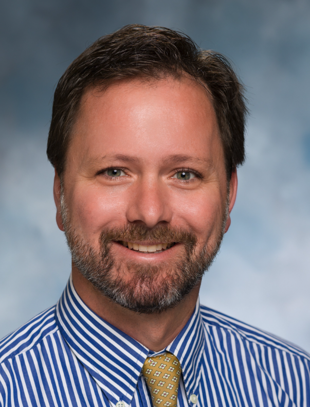 Patrick Mckay Boland合作论文数Department of Medicine, Roswell Park Cancer Institute2
 Richard L Angelo合作论文数2Larry D. Field合作论文数2Felix H. Savoie合作论文数2
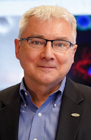 Pawel Kalinski合作论文数Department of Immunology, Roswell Park Comprehensive Cancer Center2Nikhil N. Verma合作论文数2Julia M. Cruz合作论文数2
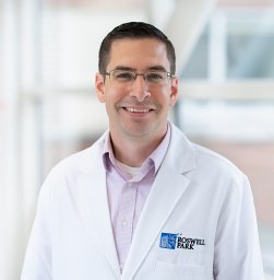 Jason B. Muhitch合作论文数Department of Immunology, Roswell Park Cancer Institute2Thomas Schwaab合作论文数2
 Renuka Iyer合作论文数Roswell Park Cancer Institute2Scott W. Trenhaile合作论文数2
 Joseph A. Abboud合作论文数Pennsylvania Hospital, University of Pennsylvania Health System2
 Joaquin Sanchez Sotelo合作论文数Division of Shoulder and Elbow Surgery, Department of Orthopedic Surgery, Mayo Clinic2
 Anand Murthi合作论文数2Charles A. Coltman合作论文数2Mark A. Mighell合作论文数2Joel Saltzman合作论文数2




