9518 Background: MPNSTs are rare soft tissue tumors (STS) that are relatively resistant to chemotherapy. 26% have epidermal growth factor receptor (EGFR) amplification. In preclinical studies, NF1/p53 murine MPNSTs in vitro are stimulated by EGF and inhibited by EGFR inhibitors. We proposed to assess response rate (RR) to erlotinib in adult patients with unresectable or metastatic MPNST. Methods: Patients (pts) were required to have measurable disease, Zubrod performance status (PS) <3, adequate organ function, no prior EGFR therapy, and centrally reviewed confirmation of diagnosis. Treatment was erlotinib 150 mg by mouth daily, in continuous 28 day cycles. Disease evaluation was performed every 2 cycles. Results: 24 pts enrolled in 22 months, from 13 institutions. 20 pts were deemed eligible. Median age was 45.3 years; 50% had neurofibromatosis. At enrollment, 15 had a PS of 0–1, 18 had metastatic disease, and 19 had unresectable disease. Pts were heavily pretreated: 9 pts had ≥ 2 prior regimens, 6 pts had 1 prior regimen, and 4 pts had no prior chemotherapy (1 unknown). 19 of 20 pts were assessable for toxicity: 6 pts had grade 3 toxicities; only 1 was hematologic (anemia). Mild rash (in 15 pts) and fatigue (in 8 pts) were the most common side effects. 20 pts were evaluable for response: 1 had stable disease after first evaluation and 19 had no response. Median progression-free survival was 2 months. 14 patients have died; median overall survival was 4 months. Because no objective responses were observed in the first stage of the study, it was closed to further accrual. Conclusions: We were able to accrue pts with a rare STS to a phase II histology specific study through a single adult cooperative group. In spite of preclinical promise of EGFR inhibition, erlotinib does not appear to be active in MPNSTs. To further understand the role of EGFR in MPNST, we are assessing EGFR expression immunohistochemically and performing quantitative PCR for genes altered by EGFR stimulation/inhibition on pt tumors. No significant financial relationships to disclose.
Benign neurofibromas and malignant peripheral nerve sheath tumors are serious complications of neurofibromatosis type 1. The epidermal growth factor receptor is not expressed by normal Schwann cells, yet is overexpressed in subpopulations of Nf1 mutant Schwann cells. We evaluated the role of EGFR in Schwann cell tumorigenesis. Expression of EGFR in transgenic mouse Schwann cells elicited features of neurofibromas: Schwann cell hyperplasia, excess collagen, mast cell accumulation, and progressive dissociation of non-myelin-forming Schwann cells from axons. Mating EGFR transgenic mice to Nf1 hemizygotes did not enhance this phenotype. Genetic reduction of EGFR in Nf1(+/-);p53(+/-) mice that develop sarcomas significantly improved survival. Thus, gain- and loss-of-function experiments support the relevance of EGFR to peripheral nerve tumor formation.
Individuals with tuberous sclerosis complex (TSC) exhibit a variety of neurologic abnormalities, including mental retardation, epilepsy, and autism. Examination of human TSC brains demonstrate dysplastic astrocytes and neurons, areas of abnormal neuronal migration (tubers), and hamartomatous growths, termed subependymal nodules, which can progress to subependymal giant cell astrocytomas (SEGA). Previous studies have suggested that these neuropathologic features may result from abnormal neuroglial cell differentiation. In an effort to provide support for this hypothesis and to identify specific markers of aberrant neuroglial cell differentiation in TSC, we employed gene expression profiling on Tsc1 conditional knockout ( Tsc1 GFAP CKO) mouse astrocytes. We identified several transcripts implicated in central nervous system development that are differentially expressed in Tsc1 −/− astrocytes compared to wild‐type astrocytes. We validated the differential expression of select transcripts on the protein level both in primary cultures of Tsc1 −/− astrocytes and in Tsc1 GFAP CKO mouse brains. Moreover, we show that these markers are also differentially expressed within cortical tubers, but not in adjacent normal tissue from TSC patient brains. This study provides supportive evidence for a developmental defect in neuroglial cell differentiation relevant to the genesis of TSC nervous system pathology and underscores the utility of mouse modeling for understanding the molecular pathogenesis of human disease. © 2004 Wiley‐Liss, Inc.
Background: Angiofibromas occur sporadically, and they develop in most patients with tuberous sclerosis complex (TSC), which is associated with alterations of the tumor suppressor genes TSC1 or TSC2. Loss of tuberin, the protein product of TSC2, has been shown in the interstitial fibroblast compartment of TSC-associated angiofibromas. It is unclear whether there is also a loss of hamartin, the product of TSC1 in TSC-associated and sporadic angiofibromas.Methods: The expression of hamartin and tuberin was analyzed by immunohistochemistry in 59 TSC-associated and 12 sporadic angiofibromas using affinity-purified antibodies.Results: Loss of expression of both tuberin and hamartin was detected in 14 angiofibromas, loss of only tuberin in three, and loss of only hamartin in four TSC-associated angiofibromas; but there was no loss in the sporadic angiofibromas. Only the interstitial cells, but not the vascular cells, showed a loss of expression of tuberin or hamartin.Conclusions: Loss of tuberin or hamartin occurred in a minority of the TSC-linked angiofibromas, but not in the sporadic angiofibromas. The absence of both tuberin and hamartin in some of the tumors suggests that the stability of tuberin and hamartin, which are believed to form an active complex in vivo , is negatively affected by the absence of either of the partners.
We investigated the effects of overexpression of the tuberous sclerosis-1 and -2 (TSC1/2) gene products (hamartin and tuberin, respectively) in the human kidney epithelial cell line 293 with an inducible expression system. As we had observed previously in fibroblasts, 293 cells overexpressing hamartin and/or tuberin grew more slowly in vitro. However, here we also observed that the 293 overexpressing cells underwent a dramatic morphological change in which groups of cells formed compact clusters. The overexpressing cells also displayed decreased dissociation and increased reaggregation in vitro. These changes were found to be associated with an increased level of E-cadherin, which is known to regulate cell-cell interactions in epithelial cells, and of its binding partner beta-catenin. Consistent with the role of E-cadherin in these effects, we found that the observed changes in 293 cell morphology, dissociation, and adhesion were calcium-dependent, and were reproduced by overexpression of E-cadherin. In contrast, overexpression of TSC1 in rat embryo fibroblasts, which lack E-cadherin, failed to elicit the same changes as in 293 cells. We conclude that the hamartin/tuberin complex exerted a direct effect on the morphology and adhesive properties of 293 cells through regulation of the level and/or activity of cellular E-cadherin/beta-catenin.
Loss of axonal contact characterizes Schwann cells in benign and malignant peripheral nerve sheath tumors (MPNST) from neurofibromatosis type 1 (NF1) patients. Tumor Schwann cells demonstrate NF1 mutations, elevated Ras activity, and aberrant epidermal growth factor receptor (EGFR) expression. Using cDNA microarrays, we found that brain lipid binding protein (BLBP) is elevated in an EGFR-positive subpopulation of Nf1 mutant mouse Schwann cells (Nf1(-/-) TXF) that grows away from axons; BLBP expression was not affected by farnesyltransferase inhibitor, an inhibitor of H-Ras. BLBP was also detected in EGFR-positive cell lines derived from Nf1:p53 double mutant mice and human MPNST. BLBP expression was induced in normal Schwann cells following transfection with EGFR but not H-Ras12V. Furthermore, EGFR-mediated BLBP expression was not inhibited by dominant-negative H-Ras, indicating that BLBP expression is downstream of Ras-independent EGFR signaling. BLBP-blocking antibodies enabled process outgrowth from Nf1(-/-) TXF cells and restored interaction with axons, without affecting cell proliferation or migration. Following injury, BLBP expression was induced in normal sciatic nerves when nonmyelinating Schwann cells remodeled their processes. These data suggest that BLBP, stimulated by Ras-independent pathways, regulates Schwann cell-axon interactions in normal peripheral nerve and peripheral nerve tumors.
BACKGROUND:Patients affected with tuberous sclerosis complex (TSC) are prone to the development of multiple benign tumors of the skin and other organs. Tuberin, the protein product of the tuberous-sclerosis-complex-2 tumor suppressor gene (TSC2) has been shown to inhibit cell proliferation. In TSC associated kidney tumors and sporadic brain tumors the loss/reduction of tuberin has been shown. METHODS:Specimens of nine squamous cell carcinomas (SCC) and five basal cell carcinomas (BCC) from patients without TSC and six biopsies of connective tissue nevi (CTN) of patients with TSC were obtained. Specimens were analyzed by immunoblotting for the expression of tuberin. RESULTS:Absent or reduced levels of tuberin were detected in the dermal parts of three of six shagreen patches, two of five BCC, and four of nine SCC. CONCLUSIONS:In tumors/hamartomas of patients with TSC the complete loss of TSC2 and tuberin is a mechanism which could be shown for CTN, thereby excluding the possibility of haploinsufficiency of TSC2. In a substantial number of cutaneous BCC and SCC the loss or downregulation of tuberin seems to be epigenetic, as alterations of TSC2 are not known in these tumors. The absence or reduction of tuberin might contribute to their proliferation.
The human disease neurofibromatosis type 1 (NF1) is caused by mutations in the NF1 gene, and is characterized by the formation of benign and malignant tumors of the peripheral nervous system. We have shown previously that aberrant expression of the epidermal growth factor receptor (EGFR) is a common feature of human NF1-related tumor development in humans and in NF1 animal models. One recent approach taken to investigate the changes associated with NF1 tumor formation is the development of the Nf1:p53 mouse tumor model. Here, we examined a series of tumor cell lines derived from Nf1:p53 mice for their expression of EGFR family members. Immunoblotting analyses revealed that 23 of the 24 cell lines examined express the EGFR, and 24 of 24 express the related tyrosine kinase erbB2, whereas erbB3 was detected in only 6 of 24. All of the cell lines expressing EGFR responded to epidermal growth factor (EGF) by activation of the downstream signaling pathways, mitogen-activated protein (MAP)/extracellular signal-regulated kinase kinase/MAP kinase, and phosphatidylinositol 3'-kinase (PI3k)/AKT. Growth of the cell lines was greatly stimulated by EGF in vitro and could be blocked by an antagonist of the EGFR. In addition, inhibition of the PI3k pathway potently inhibited the EGF-dependent growth of these cell lines, whereas inhibition of the MAP/extracellular signal-regulated kinase kinase/MAP kinase pathway had more limited effects. We conclude that EGFR expression is a common feature of the Nf1:p53 tumor cell lines and that inhibition of this molecule or its downstream target PI3k, may be useful in the treatment of NF1-related malignancies.
Session III 8 1tissues, but at tenfold lower levels than in prostate.The only prostate cancer cell line that expresses 1B10 is an androgen-sensitive cell line, LNCaP, suggesting that the gene might be androgen-regulated.Cloning of the full-length cDNA has so far resulted in 4.6 kb of the transcript.The 3′ end of the coding region is significantly homologous to a prostate-specific transmembrane protein, STEAP, but the rest of the sequence does not show homology to any known gene.Cloning of the 5′ end of the cDNA and functional studies are now in progress.
Background: Tuberous sclerosis complex (TSC) is an autosomal dominantly inherited disorder associated with an alteration of the TSC2 tumor suppressor gene which encodes for the protein product tuberin. The disease is characterized by the development of hamartomas, e.g. cutaneous angiofibromas which consist of vascular cells, interstitial cells, and normal components of the skin. The Eker rat model, an animal model of inherited cancer, has been shown to carry a mutation of TSC2.Methods: Immunohistochemical analyses of human angiofibromas were performed using antibodies directed against tuberin and angiogenic growth factors. Proliferation of human dermal microvascular endothelial cells (HDMEC) was determined after incubation with the supernatants of TSC2 (+/+) and TSC2 (-/-) rat embryonic fibroblasts (REF) that were derived from the Eker strain.Results: Loss of the expression of tuberin was observed in the interstitial cells of 13 of 39 angiofibromas. The expression of tuberin was retained in the vascular cells. In all analyzed angiofibromas, the angiogenic factors bFGF, PD-ECGF, VEGF and angiogenin were detected in the interstitial cells and/or vascular cells. Expression of PDGF-B and TGF-beta1 was weak. Tissue culture supernatants from TSC2 (-/-) REF stimulated the growth of HDMEC significantly more than supernatants from TSC2 (+/+) REF.Conclusion: A functional loss of tuberin may stimulate vascular growth.
Critical functions of hamartin and tuberin, encoded by the TSC1 and TSC2 genes, are likely to be closely linked. The proteins interact directly with one another and mutations affecting either gene result in the tuberous sclerosis phenotype. However, the regions of hamartin and tuberin that interact have not been well defined, and the relationship between their interaction and the pathogenesis of tuberous sclerosis has not been explored. To address these issues a series of hamartin and tuberin constructs were used to assay for interaction in the yeast two-hybrid system. Hamartin (amino acids 302-430) and tuberin (amino acids 1-418) interacted strongly with one another. A region of tuberin encoding a putative coiled-coil (amino acids 346-371) was necessary but not sufficient to mediate the interaction with hamartin, as more N-terminal residues were also required. A region of hamartin (amino acids 719-998) predicted to encode coiled-coils was capable of oligermerization but was not important for the interaction with tuberin. Subtle, non-truncating mutations identified in patients with tuberous sclerosis and located within the putative binding regions of hamartin (N198_F199delinsI;593-595delACT) or tuberin (G294E and I365del), abolished or dramatically reduced interaction of the proteins as assessed by yeast two-hybrid assays and by co-immunoprecipitation of the full-length proteins from Cos7 cells. In contrast, three non-pathogenic missense polymorphisms of tuberin (R261W, M286V, R367Q) in the same region as the disease-causing TSC2 mutations did not. These results indicate a requirement for interaction in critical growth suppressing functions of hamartin and tuberin.
We report here that overexpression of the tuberous sclerosis-1 (TSC1) gene product hamartin results in the inhibition of growth, as well as changes in cell morphology. Growth inhibition was associated with an increase in the endogenous level of the product of the tuberous sclerosis-2 (TSC2) gene, tuberin. As overexpression of tuberin inhibits cell growth, and hamartin is known to bind tuberin, these results suggested that hamartin stabilizes tuberin and this contributes to the inhibition of cell growth. Indeed, transient transfection of TSC1 increased the endogenous level of tuberin, and transient co-transfection of TSC1 with TSC2 resulted in higher tuberin levels. The stabilization was explained by the finding that tuberin is highly ubiquitinated in cells, while the fraction of tuberin that is bound to hamartin is not ubiquitinated. Co-expression of tuberin stabilized hamartin, which is weakly ubiquitinated, in transiently transfected cells. The amino-terminal two-thirds of tuberin was responsible for its ubiquitination and for stabilization of hamartin. A mutant of tuberin from a patient missense mutation of TSC2 was also highly ubiquitinated, and was unable to stabilize hamartin. We conclude that hamartin is a growth inhibitory protein whose biological effect is likely dependent on its interaction with tuberin.
We have found that EGF-R expression is associated with the development of the Schwann cell-derived tumors characteristic of neurofibromatosis type 1 (NF1) and in animal models of this disease. This is surprising, because Schwann cells normally lack EGF-R and respond to ligands other than EGF. Nevertheless, immunoblotting, Northern analysis, and immunohistochemistry revealed that each of 3 malignant peripheral nerve sheath tumor (MPNST) cell lines from NF1 patients expressed the EGF-R, as did 7 of 7 other primary MPNSTs, a non-NF1 MPNST cell line, and the S100(+) cells from each of 9 benign neurofibromas. Furthermore, transformed derivatives of Schwann cells from NF1(-/-) mouse embryos also expressed the EGF-R. All of the cells or cell lines expressing EGF-R responded to EGF by activation of downstream signaling pathways. Thus, EGF-R expression may play an important role in NF1 tumorigenesis and Schwann cell transformation. Consistent with this hypothesis, growth of NF1 MPNST lines and the transformed NF1(-/-) mouse embryo Schwann cells was greatly stimulated by EGF in vitro and could be blocked by agents that antagonize EGF-R function.
ABSTRACT The full-length versions of the Ras-specific exchange factors Ras-GRF1 (GRF1) and Ras-GRF2 (GRF2), which are expressed in brain and a restricted number of other organs, possess an ionomycin-dependent activation of Erk mitogen-activated protein kinase activity in 293T cells (C. L. Farnsworth et al., Nature 376:524–527, 1995; N. P. Fam et al., Mol. Cell. Biol. 17:1396–1406, 1996). Each GRF protein contains a Dbl homology (DH) domain. A yeast two-hybrid screen was used to identify polypeptides that associate with the DH domain of GRF1. In this screen, a positive cDNA clone from a human brain cDNA library was isolated which consisted of the GRF2 DH domain and its adjacent ilimaquinone domain. Deletion analysis verified that the two-hybrid interaction required only the DH domains, and mutation of Leu-263 to Gln (L263Q) in the N terminus of the GRF1 DH domain abolished the two-hybrid interaction, while a cluster of more C-terminally located mutations in the DH domain did not eliminate the interaction. Oligomers between GRF1 and GRF2 were detected in a rat brain extract, and forced expression of GRF1 and GRF2 in cultured mammalian cells formed homo- and hetero-oligomers. Introduction of the L263Q mutation in GRF1 led to a protein that was deficient in oligomer formation, while GRF1 containing the DH cluster mutations formed homo-oligomers with an efficiency similar to that of wild type. Compared to wild-type GRF1, the focus-forming activity on NIH 3T3 cells of the GRF1 DH cluster mutant was reduced, while the L263Q mutant was inactive. Both mutants were impaired in their ability to mediate ionomycin-dependent Erk activity in 293T cells. In the absence of ionomycin, 293T cells expressing wild-type GRF1 contained much higher levels of Ras-GTP than control cells; the increase in Erk activity induced by ionomycin in the GRF1-expressing cells also induced a concomitant increase in Raf kinase activity, but without a further increase in the level Ras-GTP. We conclude that GRF1 and GRF2 can form homo- and hetero-oligomers via their DH domains, that mutational inactivation of oligomer formation by GRF1 is associated with impaired biological and signaling activities, and that in 293T cells GRF1 mediates at least two pathways for Raf activation: one a constitutive signal that is mainly Ras-dependent, and one an ionomycin-induced signal that cooperates with the constitutive signal without further augmenting the level of GTP-Ras.
The murine t complex on chromosome 17 contains a number of homozygous lethal and semi-lethal mutations that disrupt development of the mouse embryo. We recently characterized an embryonic lethality in the rat that results from a germ-line mutation in the tuberous sclerosis 2 (Tsc-2) tumour suppressor gene (the Eker mutation). Remarkably, mouse embryos homozygous for tw8 mutation display cranial defects reminiscent of those observed in rat embryos homozygous for the Eker mutation. To determine whether the Tsc-2 gene, which is in the t complex, is mutated in tw8 or other t haplotypes, we characterized this gene in a series of t haplotype mice. Four Tsc-2 polymorphisms were identified: three in the coding region and one intronic that appeared to be common to all t haplotypes analysed. No evidence was found to argue that the Tsc-2 gene is altered in tw8 haplotype mice. However, in the tw5 haplotype we found a G to T mutation in Tsc-2 that was present only in this t haplotype. In contrast to other polymorphisms within the Tsc-2 coding region which did not result in amino acid changes in Tsc-2 gene product tuberin, this mutation substituted a phenylalanine for a conserved cysteine in tw5 tuberin. Within the t complex, the Tsc-2 gene and the putative tw5 locus appeared to map to different positions, complicating identification of Tsc-2 as a candidate for the tw5 locus and suggesting that the G to T mutation in the Tsc-2 gene may have arisen independently of the tw5 functional mutation.
Cortical dysplasia (CD) broadly defines a complex cerebral malformative lesion associated clinically with intractable, pharmacoresistant epilepsy (including infantile spasms), especially in infants and children. In CD, the spectrum of structural brain abnormalities includes (at a minimum) neuronal dyslamination and (in severe cases) neuronal cytomegaly with cytoskeletal alterations and the presence of gemistocyte-like `balloon cells'. In some CD variants, the neuropathological features are essentially indistinguishable from those of a tuber of tuberous sclerosis (TSC). Two genes associated with the autosomal dominant, multi-system disorder TSC have recently been cloned: TSC2 (on chromosome 16p13.3) encodes the protein tuberin and TSC1 (on 9q34) encodes hamartin. Tuberin has been immunolocalized to neurons and possibly astrocytes in normal brain and CD/TSC tubers, and is widely expressed in normal viscera; loss of heterozygosity and tissue culture studies suggest it functions as a growth suppressor. The TSC1 gene has been cloned within the last year and hamartin as yet has no well-defined cellular function, though its protein product may also function as a growth suppressor. This article focuses on the cellular pathogenesis of CD and TSC brain lesions and how the two may be biologically related. Studies of how TSC1 and TSC2 function in normal and dysplastic cerebral neocortex may provide a paradigm for understanding the neurobiology of other genes that determine epilepsy-associated cerebral malformations (e.g. lissencephaly, double cortex).
Individuals affected with tuberous sclerosis complex (TSC) develop several benign and malignant tumors at increased frequency, including astrocytomas. Tuberin, the protein product of the tuberous sclerosis complex-2 (TSC2) tumor suppressor gene, has been shown to directly inhibit cell growth and is expressed at high levels in normal central nervous system neurons and astrocytes. To determine whether TSC2 RNA and protein are reduced in astrocytomas from individuals without tuberous sclerosis, reverse transcriptase-polymerase chain reaction and immunoblotting analyses were performed on 49 adult astrocytomas, 10 pediatric astrocytomas, and 13 ependymomas. Eighteen of 40 (45%) high-grade (World Health Organization [WHO] grade III/IV) astrocytomas and 4 of 8 (50%) adult low-grade (WHO grade II) astrocytomas demonstrated reduced or absent TSC2 expression, including 1 giant cell astrocytoma, whereas none of the 10 pediatric low-grade astrocytomas analyzed showed a reduction in TSC2 expression. Reduced or absent tuberin was observed in 2 of 6 (33%) ependymomas analyzed. These data demonstrate, for the first time, that reduced or absent TSC2 expression may represent one of the critical genetic events associated with the development of sporadic adult, but not pediatric, astrocytomas.
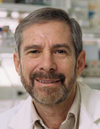 Douglas R. Lowy合作论文数Laboratory of Cellular Oncology, National Cancer Institute, National Institutes of Health18
 R Wienecke合作论文数17William C. Vass合作论文数16Alexander G. Papageorge合作论文数National Cancer Institute, National Institutes of Health9
 Nancy Ratner合作论文数Cincinnati Children's Hospital Medical Center/University of Cincinnati College of Medicine8Zhang K E合作论文数6
 Mark R. Johnson合作论文数Chelsea and Westminster Hospital;Department of Metabolism, Digestion and Reproduction, Faculty of Medicine, Imperial College London6Yeung Raymond S合作论文数5Berthe M. Willumsen合作论文数University Institute of Microbiology5J C Maize合作论文数4
 Harry V. Vinters合作论文数Brain Research Institute, David Geffen School of Medicine, University of California, Los Angeles;Mental Retardation Research Center, David Geffen School of Medicine, University of California, Los Angeles;Mary S. Easton Center for Alzheimer’s Research and Care, David Geffen School of Medicine, University of California, Los Angeles4Matthias Volkenandt合作论文数3Fackler I合作论文数3
 David H. Gutmann合作论文数Departments of Neurology, Neurosurgery, Pediatrics, and Genetics, School of Medicine, Washington University in St. Louis;St. Louis Children's Hospital;Barnes-Jewish Hospital3Richard Braverman合作论文数3Shyra Miller合作论文数3Jeremy P. Cheadle合作论文数3Julian R. Sampson合作论文数Molecular Neurogenetics Unit, Massachusetts General Hospital;and Institute of Medical Genetics, University of Wales College of Medicine;and Institute of Medical Genetics, University of Wales College of Medicine3HA Kim合作论文数2TA Rizvi合作论文数2DR LOWY合作论文数2PA Nguyen-Vu合作论文数2C Kerfoot合作论文数2Mb Valentine合作论文数2H Cen合作论文数2Hongzhen Li合作论文数2Shaowei Li合作论文数2
 A. Thomas Look合作论文数Department of Pediatric Oncology, Dana-Farber Cancer Institute, Inc.;Department of Pediatrics, Harvard Medical School, Harvard University;Children’s Hospital Boston2
 Lee Parry合作论文数2
 Giovanni Benvenuto合作论文数Centro di Endocrinologia ed Oncologia Sperimentale `G. Salvatore'of the Consiglio Nazionale delle Ricerche, Università degli Studi di Napoli `Federico II'2Julie Maynard合作论文数2Kristine S. Vogel合作论文数2GF XU合作论文数1J Degunzburg合作论文数1D W Stacey合作论文数1K. R. Monk合作论文数1Mg Norman合作论文数1Michael W. Johnson合作论文数1R L Heideman合作论文数1A Rust合作论文数1PB Crino合作论文数1Christopher Kerfoot合作论文数1Thorsten Rosenbaum合作论文数1J-F Cordier合作论文数1MA Marchionni合作论文数1Clara C. Chen合作论文数1AK Hodges合作论文数1Gh Xiao合作论文数1




