Currently, the incorrect judgment of burn depth remains common even among experienced surgeons. Contributing to this problem are change in burn appearance throughout the first week requiring periodic evaluation until a confident diagnosis can be made. To overcome these issues, we investigated the feasibility of an artificial intelligence algorithm trained with multispectral images of burn injuries to predict burn depth rapidly and accurately, including burns of indeterminate depth. In a feasibility study, 406 multispectral images of burns were collected within 72 hours of injury and then serially for up to 7 days. Simultaneously, the subject's clinician indicated whether the burn was of indeterminate depth. The final depth of burned regions within images were agreed upon by a panel of burn practitioners using biopsies and 21-day healing assessments as reference standards. We compared three convolutional neural network architectures and an ensemble in their capability to automatically highlight areas of nonhealing burn regions within images. The top algorithm was the ensemble with 81% sensitivity, 100% specificity, and 97% positive predictive value (PPV). Its sensitivity and PPV were found to increase in a sigmoid shape during the first week postburn, with the inflection point at day 2.5. Additionally, when burns were labeled as indeterminate, the algorithm's sensitivity, specificity, PPV, and negative predictive value were: 70%, 100%, 97%, and 100%. These results suggest multispectral imaging combined with artificial intelligence is feasible for detecting nonhealing burn tissue and could play an important role in aiding the earlier diagnosis of indeterminate burns.
BACKGROUND:Exposing a healthy wound bed for skin grafting is an important step during burn surgery to ensure graft take and maintain good functional outcomes. Currently, the removal of non-viable tissue in the burn wound bed during excision is determined by expert clinician judgment. Using a porcine model of tangential burn excision, we investigated the effectiveness of an intraoperative multispectral imaging device combined with artificial intelligence to aid clinician judgment for the excision of non-viable tissue. METHODS:Multispectral imaging data was obtained from serial tangential excisions of thermal burn injuries and used to train a deep learning algorithm to identify the presence and location of non-viable tissue in the wound bed. Following algorithm development, we studied the ability of two surgeons to estimate wound bed viability, both unaided and aided by the imaging device. RESULTS:The deep learning algorithm was 87% accurate in identifying the viability of a burn wound bed. When paired with the surgeons, this device significantly improved their abilities to determine the viability of the wound bed by 25% (p = 0.03). Each time a surgeon changed their decision after seeing the AI model output, it was always a change from an incorrect decision to excise more tissue to a correct decision to stop excision. CONCLUSION:This study provides insight into the feasibility of image-guided burn excision, its effect on surgeon decision making, and suggests further investigation of a real-time imaging system for burn surgery could reduce over-excision of burn wounds.
Objective Prediction of amputation wound healing is challenging due to the multifactorial nature of critical limb ischemia and lack of objective assessment tools. Up to one-third of amputations require revision to a more proximal level within 1 year. We tested a novel wound imaging system to predict amputation wound healing at initial evaluation. Methods Patients planned to undergo amputation due to critical limb ischemia were prospectively enrolled. Clinicians evaluated the patients in traditional fashion, and all clinical decisions for amputation level were determined by the clinician's judgement. Multispectral images of the lower extremity were obtained preoperatively using a novel wound imaging system. Clinicians were blinded to the machine analysis. A standardized wound healing assessment was performed on postoperative day 30 by physical exam to determine whether the amputation site achieved complete healing. If operative revision or higher level of amputation was required, this was undertaken based solely upon the provider's clinical judgement. A machine learning algorithm combining the multispectral imaging data with patient clinical risk factors was trained and tested using cross-validation to measure the wound imaging system's accuracy of predicting amputation wound healing. Results A total of 22 patients undergoing 25 amputations (10 toe, five transmetatarsal, eight below-knee, and two above-knee amputations) were enrolled. Eleven amputations (44%) were non-healing after 30 days. The machine learning algorithm had 91% sensitivity and 86% specificity for prediction of non-healing amputation sites (area under curve, 0.89). Conclusions This pilot study suggests that a machine learning algorithm combining multispectral wound imaging with patient clinical risk factors may improve prediction of amputation wound healing and therefore decrease the need for reoperation and incidence of delayed healing. We propose that this, in turn, may offer significant cost savings to the patient and health system in addition to decreasing length of stay for patients.
Assessing severity of burns based on current clinical methods is difficult but critical in assuring appropriate and expedient burn care. We are continuing to develop a novel device which uses multispectral imaging (MSI) and artificial intelligence (AI) algorithms to aide in determination of burn severity and healing potential. Previously, we reported interim results of this proof-of-concept (POC) case series at the Southern Regional Burn Conference in 2018. We now present final results of this clinical study and demonstrate feasibility for training AI algorithms to predict non-healing burn. Subjects with various burn severities were enrolled within 72 hours of burn and imaged daily with our device up to 7 days post burn. True severity of burns in each image was determined using healing assessments at day 21 post-burn and punch biopsies obtained at time of surgery. The accuracy of the imaging device to identify and differentiate healing and non-healing burn tissue in first, second, and third degree burn injuries was analyzed. Using the accuracy results, we trained an AI algorithm to identify non-healing burn tissue. Evaluation of biopsy utilization in informing the truthing method for burns was also evaluated. Data were collected from 25 subjects with 43 total burns and 303 images. The AI algorithm was trained and achieved a 90.0% sensitivity and 90.2% specificity in predicting non-healing burn tissue using cross validation. Example images for a non-healing burn on the anterior left thigh of an 86-year-old male taken 4 days post-burn injury appear in the attached figure. The AI algorithm trained on 25 subjects had an area under the curve (AUC) of 0.965, compared to an AUC of 0.850 for 10 subjects. Future work is focused on initiating a multi-center study to collect a larger representative burn database used to further train our AI algorithm. This study shows results from a completed POC study for developing an aid to burn assessments. Performance of AI algorithms trained on the data collected using our MSI device gives high confidence that clinically relevant predictions of burn healing potential are feasible using this technology.
Machine learning techniques have been recently applied for discriminating between Viable and Non-Viable tissues in animal wounds, to help surgeons to identify areas that need to be excised in the process of burn debridement. However, the presence of outliers in the training data set can degrade the performance of that discrimination. This paper presents an outlier removal technique based on the Mahalanobis distance to improve the accuracy detection of Non-Viable skin in human injuries. The iteratively application of this technique improves the accuracy results of the Non-Viable skin in a 13.6% when applying K-fold cross-validation.
Non-invasive optical imaging techniques have been recently proposed for distinguishing between different types of tissue in burns generated in porcine models. These techniques are designed to assist surgeons during the process of burn debridement, to identify regions requiring excision and their appropriate excision depth. This paper presents a machine learning tool for discriminating between Viable and Non- Viable tissues in human injuries. This tool merges a supervised (QDA) with an unsupervised (k-means clustering) classification algorithms. This combination improves the Non-Viable tissue detection in 23.7% with respect to a simple QDA classifier.
Accuracy of burn depth assessment depends on expertise and can be as low as 64%, even for skilled practitioners. Imaging devices to classify burn depth, including laser Doppler imaging, multispectral imaging (MSI), and thermography, have been studied to address this issue. The objective of this study was to determine if a MSI device, previously developed in an animal burn model, could translate to clinical burns. We present current results of this ongoing proof-of-concept (POC) clinical study, including study design and initial burn detection accuracy. In an IRB-approved study, data were collected from subjects with 1 , superficial 2, deep 2 , and 3 thermal burns. Subjects were imaged following consent and daily for up to 7 days post injury. At imaging timepoints, the MSI device was used to collect images across the visible and near-IR spectrum. True severity of burn injuries, or ground truth, was determined using 21 day healing assessments or pathology for burns that required excision. Using MSI data and ground truth, we trained two deep learning algorithms to identify pixels in the image that represened non-healing burn tissue. A a fully connected convolutional neural network (CNN) and a fully convolutional neural network (SegNet) were trained to segment non-healing burn pixels from other pixels in the MSI images. Accuracy, sensitivity, and specificity of these algorithms ability to identify non-healing burns on the current study subjects were calculated using cross-validation (CV). Average accuracy was 75 ± 0.3% (fig. 1), and this accuracy increased as we collect more images. Additionally, classified output images could be processed in less than one second using the SegNet algorithm. Results from two deep learning algorithms on initial POC study subjects obtained using leave-one-out CV. Obtaining MSI images with the described clinical study design was feasible. The MSI images contained sufficient information to classify non-healing burn tissue as accurately as a skilled practitioner, and could provide these results rapidly. These preliminary results are promising, and future work will be aimed toward collecting more data to identify sources of variability and to increase algorithm accuracy. This study shows results from an ongoing POC study for developing an MSI device to aid in burn depth assessments. Eventually, the device could assist in EDs that lack specialized burn care and as an aid to burn surgery.
Noncontact photoplethysmography (PPG) has been studied as a method to provide low-cost, noninvasive, two-dimensional blood oxygenation measurements and medical imaging for a variety of near-surface pathologies. To evaluate this technology in a laboratory setting, dynamic tissue phantoms were developed with tunable parameters that mimic physiologic properties of the skin, including blood vessel volume change, pulse wave frequency, and tissue scattering and absorption. Tissue phantoms were generated using an elastic tubing to represent a blood vessel where the luminal volume could be modulated with a pulsatile fluid flow. The blood was mimicked with a scattering and absorbing motility standard, and the tissue with a gelatin-lipid emulsion hydrogel. A noncontact PPG imaging system was then evaluated using the phantoms. Noncontact PPG imaging accurately identified pulse frequency, and PPG signals from these phantoms suggest that the phantoms can be used to evaluate noncontact PPG imaging systems. Such information may be valuable to the development of future PPG imaging systems.
Taking a solution for a clinical unmet need from a mere idea to a profitable medical device company is a long and complex process. After developing a prototype solution, the physician-inventor must quickly file a patent to protect his or her intellectual property. After the patent is secured, the first major business decision arrives: should the inventor sell the patent or maintain ownership? If the inventor decides to maintain ownership, he or she will face a series of hurdles from obtaining additional funding to device development, and ultimately, commercialization and marketing of the product. Although this process is daunting at first glance, and physicians certainly face unique challenges in this endeavor, clinicians are uniquely and strategically positioned to identify clinical unmet needs and, therefore, have the ability to fundamentally transform the way we treat our patients.
Significance: Burn assessments, including extent and severity, are some of the most critical diagnoses in burn care, and many recently developed imaging techniques may have the potential to improve the accuracy of these evaluations. Recent Advances: Optical devices, telemedicine, and high-frequency ultrasound are among the highlights in recent burn imaging advancements. We present another promising technology, multispectral imaging (MSI), which also has the potential to impact current medical practice in burn care, among a variety of other specialties. Critical Issues: At this time, it is still a matter of debate as to why there is no consensus on the use of technology to assist burn assessments in the United States. Fortunately, the availability of techniques does not appear to be a limitation. However, the selection of appropriate imaging technology to augment the provision of burn care can be difficult for clinicians to navigate. There are many technologies available, but a comprehensive review summarizing the tissue characteristics measured by each technology in light of aiding clinicians in selecting the proper device is missing. This would be especially valuable for the nonburn specialists who encounter burn injuries. Future Directions: The questions of when burn assessment devices are useful to the burn team, how the various imaging devices work, and where the various burn imaging technologies fit into the spectrum of burn care will continue to be addressed. Technologies that can image a large surface area quickly, such as thermography or laser speckle imaging, may be suitable for initial burn assessment and triage. In the setting of presurgical planning, ultrasound or optical microscopy techniques, including optical coherence tomography, may prove useful. MSI, which actually has origins in burn care, may ultimately meet a high number of requirements for burn assessment in routine clinical use.
Burn excision, a difficult technique owing to the training required to identify the extent and depth of injury, will benefit from a tool that can cue the surgeon as to where and how much to resect. We explored two rapid and noninvasive optical imaging techniques in their ability to identify burn tissue from the viable wound bed using an animal model of tangential burn excision. Photoplethysmography (PPG) imaging and multispectral imaging (MSI) were used to image the initial, intermediate, and final stages of burn excision of a deep partial-thickness burn. PPG imaging maps blood flow in the skin's microcirculation, and MSI collects the tissue reflectance spectrum in visible and infrared wavelengths of light to classify tissue based on a reference library. A porcine deep partial-thickness burn model was generated and serial tangential excision accomplished with an electric dermatome set to 1.0 mm depth. Excised eschar was stained with hematoxylin and eosin to determine the extent of burn remaining at each excision depth. We confirmed that the PPG imaging device showed significantly less blood flow where burn tissue was present, and the MSI method could delineate burn tissue in the wound bed from the viable wound bed. These results were confirmed independently by a histological analysis. We found these devices can identify the proper depth of excision, and their images could cue a surgeon as to the preparedness of the wound bed for grafting. These image outputs are expected to facilitate clinical judgment in the operating room.
Burn debridement is a challenging technique that requires significant skill to identify regions requiring excision and appropriate excision depth. A machine learning tool is being developed in order to assist surgeons by providing a quantitative assessment of burn-injured tissue. Three noninvasive optical imaging techniques capable of distinguishing between four kinds of tissue-healthy skin, viable wound bed, deep burn, and shallow burn-during serial burn debridement in a porcine model are presented in this paper. The combination of all three techniques considerably improves the accuracy of tissue classification, from 0.42 to almost 0.77.
Introduction: Multispectral imaging (MSI) is an optical technique that measures specific wavelengths of light reflected from wound site tissue to determine the severity of burn wounds. A rapid MSI device to measure burn depth and guide debridement will improve clinical decision making and diagnoses.Methodology: We used a porcine burn model to study partial thickness burns of varying severity. We made eight 4 x 4 cm burns on the dorsum of one minipig. Four burns were studied intact, and four burns underwent serial tangential excision. We imaged the burn sites with 400-1000 nm wavelengths.Results: Histology confirmed that we achieved various partial thickness burns. Analysis of spectral images show that MSI detects significant variations in the spectral profiles of healthy tissue, superficial partial thickness burns, and deep partial thickness burns. The absorbance spectra of 515, 542, 629, and 669 nm were the most accurate in distinguishing superficial from deep partial thickness burns, while the absorbance spectra of 972 nm was the most accurate in guiding the debridement process.Conclusion: The ability to distinguish between partial thickness burns of varying severity to assess whether a patient requires surgery could be improved with an MSI device in a clinical setting. (C) 2015 Elsevier Ltd and ISBI. All rights reserved.
In this issue of the Journal, Henn and colleagues1 from Washington University in St Louis present a novel method to quantify and localize regional left ventricle (LV) contractile function in coronary artery disease by means of cardiac magnetic resonance imaging with radiofrequency tissue tagging. They have developed a mechanism by which the quantified LV function can be compared against a normalized standard to determine the presence and severity of an individual patient's pathologic contractile dysfunction.
Repairing defective cardiac cells is important towards improving heart function. Due to the frequency and severity of ischemic heart disease, management of patients featuring this type of cardiac failure receives significant interest. Previously we discovered that Thymosin β4 (TB4), a 43 amino-acid secreted actin sequestering peptide, is beneficial for myocardial cell survival and coronary re-growth after infarction in adult mammals. Considering the regenerative potential of full-length TB4 in the heart, and that minimal structural variations alter TB4's influence on actin assembly and cell movement, we investigated how various TB4 domains affect cardiac cell behavior and post-ischemic mammalian heart function. We synthesized 17 domain combinations of full-length TB4 and analyzed their impact on embryonic cardiac cells in vitro, and after cardiac infarction in vivo. We discovered the domains of TB4 affect cardiac cell behavior distinctly. We revealed TB4 specific C-terminal tetrapeptide, AGES, increases embryonic cardiac cell migration and myocyte beating in culture, and improves adult mammalian heart function following ischemia. Investigating the molecular background and mechanism we discovered systemic injection of AGES enhances early myocyte survival by activating Akt-mediated signaling mechanisms, increases coronary vessel growth and inhibits inflammation in mice and pigs. Biodistribution analyses revealed cardiomyocytes uptake AGES efficiently in vitro and in vivo projecting a potential independent clinical utilization for the tetrapeptide. Our comprehensive domain investigations also suggest, preservation and/or restoration of cardiomyocyte communication is a target of TB4 and AGES, and critical to improve post-ischemic heart function in pigs. In summary, we identified the C-terminal four amino-acid variable end of TB4 as the essential and responsible domain for the molecule's full benefits in the hypoxic heart. Additionally, we introduced AGES as a novel, systemically applicable drug candidate to aid cardiac infarction in adult mammals.
Multispectral imaging (MSI) was implemented to develop a burn tissue classification device to assist burn surgeons in planning and performing debridement surgery. To build a classification model via machine learning, training data accurately representing the burn tissue was needed, but assigning raw MSI data to appropriate tissue classes is prone to error. We hypothesized that removing outliers from the training dataset would improve classification accuracy. A swine burn model was developed to build an MSI training database and study an algorithm’s burn tissue classification abilities. After the ground-truth database was generated, we developed a multistage method based on Z -test and univariate analysis to detect and remove outliers from the training dataset. Using 10-fold cross validation, we compared the algorithm’s accuracy when trained with and without the presence of outliers. The outlier detection and removal method reduced the variance of the training data. Test accuracy was improved from 63% to 76%, matching the accuracy of clinical judgment of expert burn surgeons, the current gold standard in burn injury assessment. Given that there are few surgeons and facilities specializing in burn care, this technology may improve the standard of burn care for patients without access to specialized facilities.
See related article on pages 3180-8. See related article on pages 3180-8. In this issue of the Journal of Thoracic and Cardiovascular Surgery (JTCVS), Luo and colleagues1Luo J. Weaver M.S. Dennis J.E. Whalen E. Laflamme M.A. Allen M.D. Targeting survival pathways to create infarct-spanning bridges of human embryonic stem cell-derived cardiomyocytes.J Thorac Cardiovasc Surg. 2014; 148: 3180-3188Abstract Full Text Full Text PDF PubMed Scopus (6) Google Scholar have demonstrated a novel approach to transplantation of human embryonic stem cell grafts within infarcted myocardium in an animal model. We commend their emphasis on transplanting stem cell grafts as directionally oriented, infarct-spanning bands. Often missing from basic science laboratories is the understanding of the importance of these macroscopic helical Torrent-Guasp myocardial bands2Torrent-Guasp F. Ballester M. Buckberg G.D. Carreras F. Flotats A. Carrio I. et al.Spatial orientation of the ventricular muscle band: physiologic contribution and surgical implications.J Thorac Cardiovasc Surg. 2001; 122: 389-392Abstract Full Text Full Text PDF PubMed Scopus (242) Google Scholar that allow for torsional deformation of the left ventricle during systole and, thus, appropriate ventricular function. By submitting their work to JTCVS, Luo and colleagues recognize that cardiac surgeons, who handle the human heart on a near daily basis, can best appreciate the importance of the heart's macro-architecture for effective cardiac function. Their demonstrated effects of CoPP preconditioning represent an important step forward in our understanding of the basic mechanisms and principles underlying regenerative stem cell therapy. This bench-top research is crucial to assist clinicians in achieving better clinical outcomes with cardiac stem cell therapy. In light of this success, however, it is essential to recognize the often-frustrating discordance between results of promising basic science research and more humbling clinical trials over the last 15 years of investigation into stem cell therapy for cardiac disease. Extensive research efforts have investigated the potential for stem cell–mediated cardiac tissue regeneration in the treatment of cardiovascular disease.3Malliaras K. Marban E. Cardiac cell therapy: where we’ve been, where we are, and where we should be headed.Br Med Bull. 2011; 98: 161-185Crossref PubMed Scopus (159) Google Scholar Early basic science reports suggested enormous therapeutic potential,4Tomita S. Li R. Weisel R.D. Mickle D.A.G. Kim E. Sakai T. et al.Autologous transplantation of bone marrow cells improves damaged heart function.Circulation. 1999; 100: II-247-II-256Crossref Google Scholar but results from clinical trials have been disappointing to date. Basic investigators have employed various kinds of stem cells: embryonic stem cells (ESCs), induced pluripotent stem cells (iPSCs), and adult progenitor cells (including skeletal myoblasts, bone marrow mononuclear cells [BMMNCs], and cardiac stem cells [CSCs]). So far, however, completed clinical trials have only reported transplantation of the various adult progenitor cells. In fact, a casual review of the clinical trials database (clinicaltrials.gov/ct2/home) shows that the overwhelming majority of trials has employed or will employ adult progenitor stem cells rather than truly pluripotent ESCs or iPSCs. An early clinical trial of skeletal myoblasts (Myoblast Autologous Grafting in Ischemic Cardiomyopathy [MAGIC]5Menasche P. Alfieri O. Janssens S. McKenna W. Reichenspurner H. Trinquart L. et al.The Myoblast Autologous Grafting in Ischemic Cardiomyopathy (MAGIC) trial: the first randomized placebo-controlled study of myoblast transplantation.Circulation. 2008; 117: 1189-1200Crossref PubMed Scopus (762) Google Scholar) was discontinued prematurely after both a lack of efficacy and an increased risk for arrhythmias in patients receiving stem cell therapy were demonstrated. A more recent skeletal myoblast trial (ie, SEISMIC6Duckers H.J. Houtgraaf J. Hehrlein C. Schofer J. Waltenberger J. Gershlick A. et al.Final results of a phase IIa, randomized, open-label trial to evaluate the percutaneous intramyocardial transplantation of autologous skeletal myoblasts in congestive heart failure patients: the SEISMIC trial.EuroIntervention. 2011; 6: 805-812Crossref PubMed Scopus (89) Google Scholar) did not reveal any effect of stem cell therapy on the left-ventricular ejection fraction (LVEF). Although BMMNCs have been the most commonly employed cell type for stem cell therapy trials (eg, REPAIR-AMI,7Assmus B. Rolf A. Erbs S. Elsässer A. Haberbosch W. Hambrecht R. et al.REPAIR-AMI InvestigatorsClinical outcome 2 years after intracoronary administration of bone marrow-derived progenitor cells in acute myocardial infarction.Circ Heart Fail. 2010; 3: 89-96Crossref PubMed Scopus (241) Google Scholar TIME,8Traverse J.H. Henry T.D. Pepine C.J. Willerson J.T. Zhao D.X. Ellis S.G. et al.Effect of the use and timing of bone marrow mononuclear cell delivery on left ventricular function after acute myocardial infarction: the TIME randomized trial.J Am Med Assoc. 2012; 308: 2380-2389Crossref PubMed Scopus (311) Google Scholar POSEIDON9Hare J.M. Fishman J.E. Gerstenblith G. Velazquez D.L.D. Zambrano J.P. Suncion V.Y. et al.Comparison of allogeneic vs autologous bone marrow-derived mesenchymal stem cells delivered by transendocardial injection in patients with ischemic cardiomyopathy: the POSEIDON randomized trial.J Am Med Assoc. 2012; 308: 2369-2379Crossref PubMed Scopus (882) Google Scholar), these investigations have also yielded underwhelming results. Meta-analyses of BMMNC trials for various cardiac disease indications have demonstrated only minimal increases in LVEF across the board (Table 110Fisher S.A. Dorée C. Brunskill S.J. Mathur A. Martin-Rendon E. Bone marrow stem cell treatment for ischemic heart disease in patients with no option of revascularization: a systemic review and meta-analysis.PLoS One. 2013; 8: e64669Crossref PubMed Scopus (78) Google Scholar, 11Kandala J. Upadhyay G.A. Pokushalov E. Wu S. Drachman D.E. Singh J.P. Meta-analysis of stem cell therapy in chronic ischemic cardiomyopathy.Am J Cardiol. 2013; 112: 217-225Abstract Full Text Full Text PDF PubMed Scopus (73) Google Scholar, 12Delewi R. Hirsch A. Tijssen J.G. Schächinger V. Wojakowski W. Roncalli J. et al.Impact of intracoronary bone marrow cell therapy on left ventricular function in the setting of ST-segment elevation myocardial infarction: a collaborative meta-analysis.Eur Heart J. 2014; 35: 989-998Crossref PubMed Scopus (111) Google Scholar). Two trials implementing CSCs (SCIPIO13Bolli R. Chugh A.R. D'Amario D. Loughran J.H. Stoddard M.F. Ikram S. et al.Cardiac stem cells in patients with ischaemic cardiomyopathy (SCIPIO): initial results of a randomized phase 1 trial.Lancet. 2011; 378: 1847-1857Abstract Full Text Full Text PDF PubMed Scopus (1105) Google Scholar and CADUCEUS14Makkar R.R. Smith R.R. Cheng K.E. Malliaras K. Thomson L.E. Berman D. et al.Intra-coronary cardiosphere-derived cells for heart regeneration after myocardial infarction (CADUCEUS): a prospective, randomised phase 1 trial.Lancet. 2012; 379: 895-904Abstract Full Text Full Text PDF PubMed Scopus (1120) Google Scholar) were ineffective in improving meaningful clinical outcomes. Additional trials with adult progenitor cells are currently ongoing, including the ATHENA trial,15Cytori Therapeutics. Safety and feasibility trial of adipose-derived regenerative cells in the treatment of chronic myocardial ischemia. Available at: http://www.clinicaltrials.gov/ct2/show/NCT01556022?term=cytori&rank=3. Accessed October 7, 2014.Google Scholar which is utilizing adipose-derived regenerative cells for transplantation.16Zuk P.A. Zhu M. Ashjian P. De Ugarte D.A. Huang J.I. Mizuno H. et al.Human adipose tissue is a source of multipotent stem cells.Mol Biol Cell. 2002; 13: 4279-4295Crossref PubMed Scopus (5458) Google ScholarTable 1Meta-analyses of bone marrow cell therapy for cardiac disease, encompassing 3 major indications10Fisher S.A. Dorée C. Brunskill S.J. Mathur A. Martin-Rendon E. Bone marrow stem cell treatment for ischemic heart disease in patients with no option of revascularization: a systemic review and meta-analysis.PLoS One. 2013; 8: e64669Crossref PubMed Scopus (78) Google Scholar, 11Kandala J. Upadhyay G.A. Pokushalov E. Wu S. Drachman D.E. Singh J.P. Meta-analysis of stem cell therapy in chronic ischemic cardiomyopathy.Am J Cardiol. 2013; 112: 217-225Abstract Full Text Full Text PDF PubMed Scopus (73) Google Scholar, 12Delewi R. Hirsch A. Tijssen J.G. Schächinger V. Wojakowski W. Roncalli J. et al.Impact of intracoronary bone marrow cell therapy on left ventricular function in the setting of ST-segment elevation myocardial infarction: a collaborative meta-analysis.Eur Heart J. 2014; 35: 989-998Crossref PubMed Scopus (111) Google ScholarStudyIndicationNo. of trialsNo. of patientsΔ LVEF (%)NotesFisher et al (2013)10Fisher S.A. Dorée C. Brunskill S.J. Mathur A. Martin-Rendon E. Bone marrow stem cell treatment for ischemic heart disease in patients with no option of revascularization: a systemic review and meta-analysis.PLoS One. 2013; 8: e64669Crossref PubMed Scopus (78) Google ScholarRefractory angina or HF9659+3.47 ± 1.59Reduced mortality with cell therapy (RR = 0.33, 0.17-0.65)Kandala et al (2013)11Kandala J. Upadhyay G.A. Pokushalov E. Wu S. Drachman D.E. Singh J.P. Meta-analysis of stem cell therapy in chronic ischemic cardiomyopathy.Am J Cardiol. 2013; 112: 217-225Abstract Full Text Full Text PDF PubMed Scopus (73) Google ScholarChronic heart failure10519+4.48 ± 2.05Intramyocardial injection may be superior to intracoronary infusion in patients with LV systolic dysfunctionDelewi et al (2014)12Delewi R. Hirsch A. Tijssen J.G. Schächinger V. Wojakowski W. Roncalli J. et al.Impact of intracoronary bone marrow cell therapy on left ventricular function in the setting of ST-segment elevation myocardial infarction: a collaborative meta-analysis.Eur Heart J. 2014; 35: 989-998Crossref PubMed Scopus (111) Google ScholarAcute STEMI161641+2.55 ± 0.72Improvement better in young patients (<55 y) and patients with low LVEF (<40% baseline)LVEF, Left ventricular ejection fraction; HF, heart failure; RR, relative risk; LV, left ventricle; STEMI, ST-segment elevation myocardial infarction. Open table in a new tab LVEF, Left ventricular ejection fraction; HF, heart failure; RR, relative risk; LV, left ventricle; STEMI, ST-segment elevation myocardial infarction. After this surge of clinical trials employing adult progenitor stem cells yielded largely disappointing results, research efforts have been wisely redirected from the clinic back to the laboratory. In this effort to better understand the underlying principles of cardiac stem cell therapy, basic scientists and clinicians have learned from the mistakes of prior trials and sought solutions to challenging questions in the field, but many questions remain about basic mechanisms of stem cell therapy in cardiac regeneration.17Malliaras K. Kreke M. Marban E. The stuttering progress of cell therapy for heart disease.Clin Pharmacol Ther. 2011; 90: 532-541Crossref PubMed Scopus (72) Google Scholar Garbern and Lee18Garbern J.C. Lee R.T. Cardiac stem cell therapy and the promise of heart regeneration.Cell Stem Cell. 2013; 12: 689-698Abstract Full Text Full Text PDF PubMed Scopus (293) Google Scholar have recently outlined several of these questions expertly, 4 of which we highlight below:1.What is the precise mechanism of action by which stem cell transplantation may demonstrate clinical efficacy? Perhaps the most intuitive explanation for the therapeutic effects of stem cell transplantation is what we call the Cardiomyocyte Replacement Theory. According to this theory, transplanted stem cells differentiate into functional cardiomyocytes that integrate into the existing myocardial architecture to increase cardiac function through additional mechanical force. Nevertheless, data have consistently indicated that the vast majority of transplanted adult progenitor stem cells do not persist in their new ischemic environment for a prolonged period of time. Long-term engraftment rates have been shown to be <1%. These rates are so low because 90% of delivered cells are washed out of the heart within 24 hours, and approximately 90% of any remaining cells die within the first week of transplant.3Malliaras K. Marban E. Cardiac cell therapy: where we’ve been, where we are, and where we should be headed.Br Med Bull. 2011; 98: 161-185Crossref PubMed Scopus (159) Google Scholar, 17Malliaras K. Kreke M. Marban E. The stuttering progress of cell therapy for heart disease.Clin Pharmacol Ther. 2011; 90: 532-541Crossref PubMed Scopus (72) Google Scholar In fact, the beneficial effects of stem cell transplantation often paradoxically outlast the survival of transplanted stem cells in the ischemic heart tissue. Therefore, a second theory, which emphasizes unspecified paracrine effects of transplanted stem cells, has recently gained traction.19Williams A.R. Hare J.M. Mesenchymal stem cells: biology, pathophysiology, translational findings, and therapeutic implications for cardiac disease.Circ Res. 2011; 109: 923-940Crossref PubMed Scopus (685) Google Scholar This theory, which we call the Cardiomyocyte Repair Theory, suggests that trophic factors released by stem cells stimulate existing cardiomyocytes, leading to an increase in cardiac function. It remains to be determined which specific factors are actually released by stem cells or how they promote beneficial cardiac effects. Proposed mechanisms of the Cardiomyocyte Repair Theory include stimulation of endogenous cardiac progenitor cell populations, enhanced tissue repair, improved angiogenesis, and matrix remodeling to reduce fibrosis.20Prowse A.B.J. Timmins N.E. Yau T.M. Li R. Weisel R.D. Keller G. et al.Transforming the promise of pluripotent stem cell derived cardiomyocytes to a therapy: challenges and solutions for clinical trials.Can J Cardiol. 2014; 30: 1335-1349Abstract Full Text Full Text PDF PubMed Scopus (23) Google Scholar Of note, a consensus18Garbern J.C. Lee R.T. Cardiac stem cell therapy and the promise of heart regeneration.Cell Stem Cell. 2013; 12: 689-698Abstract Full Text Full Text PDF PubMed Scopus (293) Google Scholar has recently emerged that the mammalian heart is capable of creating a limited number of cardiac cells after birth and that some vertebrates, including neonatal mice, can regenerate the myocardium after experimentally induced ischemia.21Porello E.R. Mahmoud A.I. Simpson E. Hill J.A. Richardson J.A. Olson E.N. et al.Transient regenerative potential of the neonatal mouse heart.Science. 2011; 331: 1078-1080Crossref PubMed Scopus (1664) Google Scholar Various mechanisms have been proposed to explain how cardiomyocytes may innately regenerate following injury, as summarized in Figure 1. These include a signaling cascade that activates dormant progenitor cells, the proliferation of existing cardiomyocytes, activation of the epicardium, or the de-differentiation of cardiomyocytes into progenitor cells that then proliferate.18Garbern J.C. Lee R.T. Cardiac stem cell therapy and the promise of heart regeneration.Cell Stem Cell. 2013; 12: 689-698Abstract Full Text Full Text PDF PubMed Scopus (293) Google Scholar The proposed paracrine effects of stem cell therapy likely promote some or all of these innate pathways to increase function in the face of cardiac disease. The authors of this editorial favor the Cardiomyocyte Repair Theory as compared to the Cardiomyocyte Replacement Theory to explain the effects demonstrated after transplantation of adult progenitor stem cells. In fact, with various other colleagues, 2 of the authors have previously demonstrated important findings, involving identification of both potential trophic factors and mechanisms of repair, that lend credence to the paracrine cardiac repair paradigm. With Bock-Marquette and colleagues,22Bock-Marquette I. Saxena A. White M.D. DiMaio J.M. Srivastava D. Thymosin β4 activates integrin-linked kinase and promotes cardiac cell migration, survival and cardiac repair.Nature. 2004; 432: 466-472Crossref PubMed Scopus (570) Google Scholar, 23Bock-Marquette I. Shrivastava S. Pipes G.C. Thatcher J.E. Blystone A. Shelton J.M. et al.Thymosin β4 mediated PKC activation is essential to initiate the embryonic coronary developmental program and epicardial progenitor cell activation in adult mice in vivo.J Mol Cell Cardiol. 2009; 46: 728-738Abstract Full Text Full Text PDF PubMed Scopus (119) Google Scholar DiMaio and Thatcher identified a trophic factor, thymosin β4, that promotes cardiomyocyte migration, survival, and repair. In addition, thymosin β4 is capable of organwide activation of the embryonic coronary developmental program in the adult mammalian heart after systemic administration. With Huang, Olson, and colleagues,24Huang G.N. Thatcher J.E. McAnally J. Kong Y. Qi X. Tan W. et al.C/EBP transcription factors mediate epicardial activation during heart development and injury.Science. 2012; 338: 1599-1603Crossref PubMed Scopus (154) Google Scholar DiMaio and Thatcher have also outlined how activation of the epicardium by specific transcription factors plays a role in myocardial repair following ischemic injury. Therefore, we believe it is reasonable to suggest that increases in cardiac function in animal models following stem cell transplantation may be due to paracrine effects on the endogenous cardiomyocyte population rather than cardiomyocyte replacement via stem cell differentiation processes.2.Which cell type is best suited for transplantation therapy in cardiac disease? Although the cardiomyocyte repair theory is gaining wide acceptance as the proper explanation of the results in successful basic science investigations employing adult progenitor stem cells, the next generation of stem cell trials will likely involve transplantation of ESCs and iPSCs.20Prowse A.B.J. Timmins N.E. Yau T.M. Li R. Weisel R.D. Keller G. et al.Transforming the promise of pluripotent stem cell derived cardiomyocytes to a therapy: challenges and solutions for clinical trials.Can J Cardiol. 2014; 30: 1335-1349Abstract Full Text Full Text PDF PubMed Scopus (23) Google Scholar In theory, ESCs and iPSCs could be better suited for stem cell therapy because of their ability to fully differentiate into functional cardiomyocytes as compared to the more limited, less versatile cell lines used in the clinical trials previously discussed. Thus, in addition to paracrine effects, future stem cells employed in regenerative therapy may have greater potential to persist as directionally oriented, infarct-spanning bands that will improve the mechanical performance of the left ventricle in systole.20Prowse A.B.J. Timmins N.E. Yau T.M. Li R. Weisel R.D. Keller G. et al.Transforming the promise of pluripotent stem cell derived cardiomyocytes to a therapy: challenges and solutions for clinical trials.Can J Cardiol. 2014; 30: 1335-1349Abstract Full Text Full Text PDF PubMed Scopus (23) Google Scholar In other words, these truly pluripotent cells could promote therapeutic effects via mechanisms uniting the Cardiomyocyte Regeneration and Cardiomyocyte Repair Theories. Many questions remain about how truly pluripotent cells can be optimized in stem cell therapy. The ethical concerns involving ESCs may ultimately preclude their utility in the clinical setting. iPSCs circumvent this ethical controversy, but the development of a cost-effective protocol to create sufficient quantities of iPSCs for clinical therapy is currently a challenge.18Garbern J.C. Lee R.T. Cardiac stem cell therapy and the promise of heart regeneration.Cell Stem Cell. 2013; 12: 689-698Abstract Full Text Full Text PDF PubMed Scopus (293) Google Scholar Even given a sufficient quantity of pluripotent stem cells, further investigations must determine in what state these cells should be transplanted.20Prowse A.B.J. Timmins N.E. Yau T.M. Li R. Weisel R.D. Keller G. et al.Transforming the promise of pluripotent stem cell derived cardiomyocytes to a therapy: challenges and solutions for clinical trials.Can J Cardiol. 2014; 30: 1335-1349Abstract Full Text Full Text PDF PubMed Scopus (23) Google Scholar Theoretically, pluripotent cells could be delivered as cardiomyocyte progenitors, fetal-like cardiomyocytes, or adult-like cardiomyocytes. Furthermore, these cells could differentiate to become ventricular, atrial, or nodal cells, or even some combination of all 3. Much work still remains before it can be determined which option(s) may produce the greatest clinical efficacy.3.What is the optimal delivery method for transplantation of stem cells? Several delivery mechanisms have been previously employed including intramyocardial (epicardial or transendocardial), intracoronary, retrograde coronary sinus, and systemic intravenous. Each method has significant advantages and disadvantages as described by Dib et al.25Dib N. Khawaja H. Varner S. McCarthy M. Campbell A. Cell therapy for cardiovascular disease: a comparison of methods of delivery.J Cardiovasc Transl Res. 2011; 4: 177-181Crossref PubMed Scopus (73) Google Scholar In exchange for increased accuracy of delivery, the most invasive delivery routes carry a risk of perforation, arrhythmia, or embolus of cells and/or the viscous delivery suspension. On the other hand, poor cell homing and retention limit the effectiveness of less invasive procedures and require the delivery of an increased number of cells to achieve a comparable therapeutic benefit. A summary of the various delivery methods is presented in Table 2. No consensus has been reached upon how best to deliver stem cells to damaged cardiac tissue,18Garbern J.C. Lee R.T. Cardiac stem cell therapy and the promise of heart regeneration.Cell Stem Cell. 2013; 12: 689-698Abstract Full Text Full Text PDF PubMed Scopus (293) Google Scholar and it is likely that different methods may ultimately prove to be superior depending on the particular indication for stem cell therapy. For example, intracoronary delivery is the preferred route of transplant in patients suffering acute myocardial infarct.17Malliaras K. Kreke M. Marban E. The stuttering progress of cell therapy for heart disease.Clin Pharmacol Ther. 2011; 90: 532-541Crossref PubMed Scopus (72) Google Scholar, 25Dib N. Khawaja H. Varner S. McCarthy M. Campbell A. Cell therapy for cardiovascular disease: a comparison of methods of delivery.J Cardiovasc Transl Res. 2011; 4: 177-181Crossref PubMed Scopus (73) Google Scholar Again, we commend Luo and colleagues1Luo J. Weaver M.S. Dennis J.E. Whalen E. Laflamme M.A. Allen M.D. Targeting survival pathways to create infarct-spanning bridges of human embryonic stem cell-derived cardiomyocytes.J Thorac Cardiovasc Surg. 2014; 148: 3180-3188Abstract Full Text Full Text PDF PubMed Scopus (6) Google Scholar for addressing lingering questions about cell delivery by employing an injectable delivery method designed to regenerate Torrent-Gausp myocardial bands. In particular, their attempt to replicate the physiologic architecture of the human heart via stem cell transplantation represents a novel solution for optimal delivery.4.What adjuvants to cell delivery will maximize the benefits of stem cell therapy?Table 2Advantages and disadvantages of the various stem cell delivery methods24Huang G.N. Thatcher J.E. McAnally J. Kong Y. Qi X. Tan W. et al.C/EBP transcription factors mediate epicardial activation during heart development and injury.Science. 2012; 338: 1599-1603Crossref PubMed Scopus (154) Google ScholarMethodLeast invasiveCell retentionLow risk∗Common risks include perforation of the ventricular wall, cardiac arrhythmia secondary to local tissue inflammation, or embolus of the stem cells and/or viscous delivery suspension.Clinical experienceNotesEpicardial○●◔◑Delivery via mini-thoracotomy or with open heart procedureTransendocardial◑◕◔◑Requires delivery of percutaneous catheter to left ventricleIntracoronary◕◑◑●Preferred method during acute MIRetrograde coronary sinus◔◑◑◔Optimal in cases of severe coronary stenosisIntravenous●○●◔Very low retention due to cell trapping in lungsMI, Myocardial infarction.∗ Common risks include perforation of the ventricular wall, cardiac arrhythmia secondary to local tissue inflammation, or embolus of the stem cells and/or viscous delivery suspension. Open table in a new tab MI, Myocardial infarction. Recent investigations have demonstrated many methods to quantitatively assess cell engraftment and survival following delivery in the laboratory.26Terrovitis J.V. Smith R.R. Marbán E. Assessment and optimization of cell engraftment after transplantation into the heart.Circ Res. 2010; 106: 479-494Crossref PubMed Scopus (251) Google Scholar Experiments have consistently revealed that both engraftment and survival rates are extremely low regardless of the cell type or delivery mechanism implemented. Both cardiac blood flow, which washes away stem cells after intracoronary delivery, and myocardial contraction, which accentuates leakage of cells from an injection site, limit cell retention.26Terrovitis J.V. Smith R.R. Marbán E. Assessment and optimization of cell engraftment after transplantation into the heart.Circ Res. 2010; 106: 479-494Crossref PubMed Scopus (251) Google Scholar In addition, ischemic conditions severely inhibit the survival of any cells that successfully engraft by 24 hours posttransplant, as at least 90% of engrafted cells die within the first week. These factors limit total cell survival following transplant to <1% overall.3Malliaras K. Marban E. Cardiac cell therapy: where we’ve been, where we are, and where we should be headed.Br Med Bull. 2011; 98: 161-185Crossref PubMed Scopus (159) Google Scholar, 17Malliaras K. Kreke M. Marban E. The stuttering progress of cell therapy for heart disease.Clin Pharmacol Ther. 2011; 90: 532-541Crossref PubMed Scopus (72) Google Scholar Reasonable estimates suggest that approximately 1 billion cardiomyocytes are required to completely replace all of the cells from the working myocardium that is lost during a myocardial infarction.27Mummery C.L. Zhang J. Ng E.S. Elliott D.A. Elefanty A.G. Kam T.J. Differentiation of human ES and iPS cells to cardiomyocytes: a methods overview.Circ Res. 2012; 111: 344-358Crossref PubMed Scopus (509) Google Scholar At current cell survival rates, stem cell therapy would require the transfer of up to 1 trillion cells (roughly equivalent to 3% of the total number of cells in the human body) to each patient in order to completely replete the devastated cell population. Therefore, various methods of tissue engineering have been investigated as adjuvant support to improve the persistence of stem cells following transplant. By increasing the viability of stem cells, these methods would allow for fewer stem cells to be transplanted while concurrently bettering clinical outcomes. Examples include cell co-culturing, biomaterial formulation, and genetic engineering of cells.17Malliaras K. Kreke M. Marban E. The stuttering progress of cell therapy for heart disease.Clin Pharmacol Ther. 2011; 90: 532-541Crossref PubMed Scopus (72) Google Scholar Cell co-culturing involves pretreating stem cells with various cofactors that induce favorable cell-signaling pathways to increase cell survival and/or homing accuracy following transplantation. Biomaterials, in which cells are engrafted, may be applied as epicardial patches that increase cell retention and survival via mechanical scaffolding to limit cell washout or controlled released of biologic agents to support cells in ischemic environments.28Menasché P. How close are we to using stem cells in routine cardiac therapy?.Can J Cardiol. 2014; 30: 1265-1269Abstract Full Text Full Text PDF PubMed Scopus (2) Google Scholar Many attempts at genetic engineering of cells prior to delivery have been reported, but several concerns about this manner of adjunctive therapy have been raised.26Terrovitis J.V. Smith R.R. Marbán E. Assessment and optimization of cell engraftment after transplantation into the heart.Circ Res. 2010; 106: 479-494Crossref PubMed Scopus (251) Google Scholar Specifically, how can we ensure genetic manipulation increases cell survival without interfering with proper myocardial function or without introducing a significant risk of oncogenesis? Further research is needed to elucidate which adjuvant therapies can be optimally combined with stem cell transplantation to increase cardiac function. In addition to tissue engineering of stem cells, other techniques to promote cardiac repair have been proposed. These include the direct delivery of paracrine factors isolated from stem cell colonies and the reprogramming of noncardiomyocytes into cardiomyocytes using growth factors or microRNA assays as described elsewhere.18Garbern J.C. Lee R.T. Cardiac stem cell therapy and the promise of heart regeneration.Cell Stem Cell. 2013; 12: 689-698Abstract Full Text Full Text PDF PubMed Scopus (293) Google Scholar Figure 2 provides a summary of these approaches to cardiac regeneration therapy. Ultimately, all of these methods likely promote some combination of the innate cardiac repair pathways summarized above. To these questions, which are currently under investigation in the laboratory, we add our own, which cannot be resolved with basic science research alone: How can clinical trials best assess stem cell therapy in a clinically significant manner? Most completed trials have published outcomes by employing metrics such as changes in LVEF, left ventricular end-diastolic volume, and/or left ventricular end-systolic volume. Many of these trials have shown statistically significant, but clinically disappointing, improvements in these metrics. We wonder, moreover, how meaningful these metrics are to clinicians and their patients, especially given the operator-depended variability inherent in acquiring such measurements in addition to the physiologic variability of measurements of the same patient under different loading conditions. We believe that more clinically relevant outcomes, such as Vo2max and cardiac perfusion, must receive greater attention in future clinical trials. Other useful parameters include heart failure and angina classifications, quality of life metrics, and physical exercise performance.10Fisher S.A. Dorée C. Brunskill S.J. Mathur A. Martin-Rendon E. Bone marrow stem cell treatment for ischemic heart disease in patients with no option of revascularization: a systemic review and meta-analysis.PLoS One. 2013; 8: e64669Crossref PubMed Scopus (78) Google Scholar Of course, we cannot lose sight of the most clinically significant outcomes, morbidity and mortality, as stem cell therapy becomes more widespread. Ultimately, the initial promise of stem cell transplantation research has not yielded as much success as initially hoped during the first round of clinical trials. More basic science investigation is required to elucidate the specific mechanism(s) by which transplanted stem cells promote cardiac regeneration and/or repair, which stem cell type is most effective, and how cells can be optimally delivered and engineered. As our understanding of stem cell therapy increases, it becomes more likely that clinical trials can produce truly meaningful results with implications for clinical practice. Another wave of stem cell clinical trials is fast approaching, as the NIH Heart, Lung, and Blood Institute teams with the Cardiothoracic Surgical Trials Network (CTSN) to fund and perform this research in multicentered trials.29Ascheim D.D. Gelijns A.C. Goldstein D. Moye L.A. Smedira N. Lee S. et al.Mesenchymal precursor cells as adjunctive therapy in recipients of contemporary LVADs.Circulation. 2014; 129: 2287-2296Crossref PubMed Scopus (123) Google Scholar We remain cautiously optimistic that further investigations will yield more promising results than the first round of clinical trials, but we emphasize the tremendous challenges yet to be overcome before stem cell therapy can be a realistic therapeutic option for clinicians and their patients. Targeting survival pathways to create infarct-spanning bridges of human embryonic stem cell–derived cardiomyocytesThe Journal of Thoracic and Cardiovascular SurgeryVol. 148Issue 6PreviewGenerating myocyte grafts that bridge across infarcts could maximize their functional impact and best utilize small numbers of stem cells. To date, however, graft survival within acute infarcts has not been feasible. To enhance intrainfarct graft viability, human embryonic stem cell–derived cardiomyocytes (hESC-CMs) were pretreated before implantation with cobalt protoporphyrin (CoPP), a pharmacologic inducer of cytoprotective heme oxygenase-1. Full-Text PDF Open Archive
Non-contact photoplethysmography (PPG) has been studied as a method to provide low-cost and non-invasive medical imaging for a variety of near-surface pathologies and two dimensional blood oxygenation measurements. Dynamic tissue phantoms were developed to evaluate this technology in a laboratory setting. The purpose of these phantoms was to generate a tissue model with tunable parameters including: blood vessel volume change; pulse wave frequency; and optical scattering and absorption parameters. A non-contact PPG imaging system was evaluated on this model and compared against laser Doppler imaging (LDI) and a traditional pulse oximeter. Results indicate non-contact PPG accurately identifies pulse frequency and appears to identify signals from optically dense phantoms with significantly higher detection thresholds than LDI.
The epicardium encapsulates the heart and functions as a source of multipotent progenitor cells and paracrine factors essential for cardiac development and repair. Injury of the adult heart results in reactivation of epicardial progenitor cells, which reengages a developmental gene program, but the underlying transcriptional basis has not been delineated. We established a mouse embryonic heart organ culture and gene expression system that facilitated the identification of epicardial enhancers activated during heart development and injury. Epicardial activation of these enhancers depends on a combinatorial transcriptional code centered on C/EBP, HOX, MEIS, and GRAINYHEAD families of transcription factors. Furthermore, disruption of C/EBP signaling in the adult epicardium reduced injury-induced neutrophil infiltration and improved cardiac function. These findings reveal a transcriptional basis for epicardial progenitor cell activation during heart development and injury, providing a platform for enhancing cardioprotection and regeneration.
 DiMaio J Michael合作论文数24
 John J. Squiers合作论文数11
 Wensheng Fan合作论文数8
 Darlene King合作论文数6
 Eric Olson合作论文数Department of Molecular Biology, University of Texas Southwestern Medical Center6
 Faliu Yi合作论文数5Kevin Plant合作论文数5Yang Lu合作论文数4
 Jose Martinez合作论文数4Eric W. Sellke合作论文数4Robert D. Gerard合作论文数3
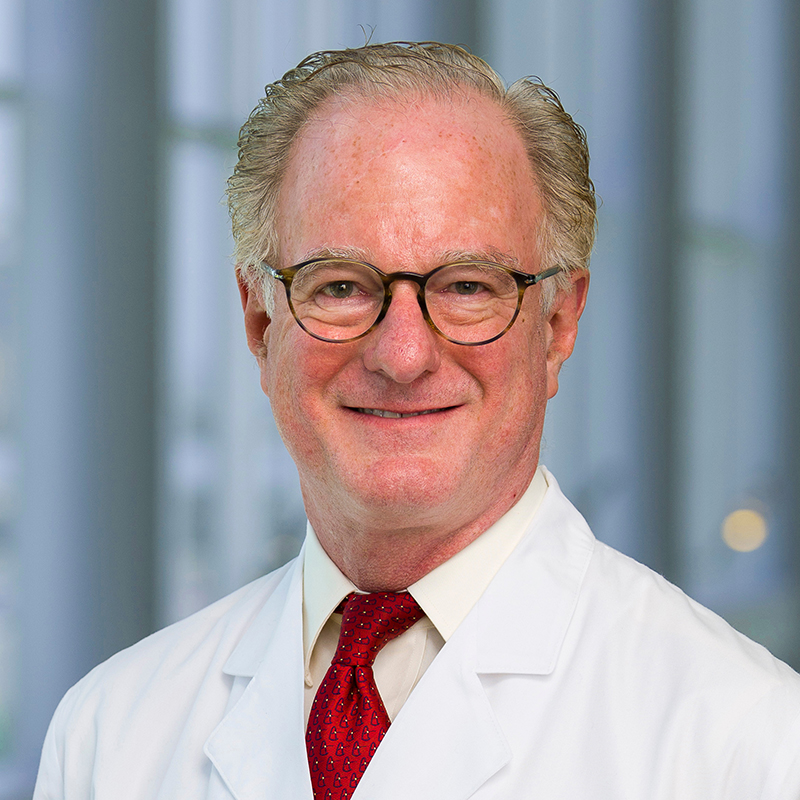 Joseph Hill合作论文数Department of Internal Medicine, University of Texas Southwestern Medical Center;Division of Cardiology, University of Texas Southwestern Medical Center;Department of Internal Medicine, Parkland Hospital;Department of Internal Medicine, Zale University Hospital3
 Weizhi Li (李伟志)合作论文数3
 Weirong Mo合作论文数3
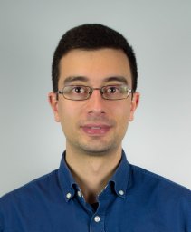 Juan Heredia Juesas合作论文数Department of Electrical Engineering, University of Oviedo Gijón3
 John M. Shelton合作论文数The University of Texas Southwestern Medical Center3Jason Dwight合作论文数2Katherine Graham合作论文数2Xu U. Zhang合作论文数2rachit mohan合作论文数2
 Lillian B Sutherland合作论文数2
 Amatruda James F合作论文数Dept Pediat, Univ Texas SW Med Ctr Dallas2Kong Yongli合作论文数2
 Shuai Yu合作论文数2Tan Wei合作论文数2McCall Brian合作论文数Department of Bioengineering, Rice University2Rhonda Bassel-Duby合作论文数2Xiaoxia Qi合作论文数2
 Ildiko Bock-Marquette合作论文数2
 James Hill Holmes合作论文数School of Medicine, Wake Forest University2
 Guo N. Huang合作论文数2Santwana Shrivastava合作论文数2Kelley A. Hutcheson合作论文数2
 Ajay Singh合作论文数1Andrew J Applewhite合作论文数1
 Jeffrey E Carter合作论文数1Robert C Siska合作论文数1Ronald Baxter合作论文数1Gabor Faskerti合作论文数1J E Carter合作论文数1
 Gordon Teg Pipes合作论文数1Ikenna Nwafor合作论文数1
 Shimeng Du合作论文数1
 Brittany A. Potz合作论文数1
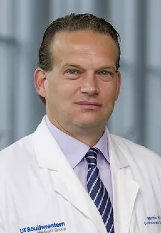 Matthias Peltz合作论文数Department of Cardiovascular & Thoracic Surgery, UT Southwestern Medical Center1
 Yulin Wang (王于琳)合作论文数Corresponding author.1Hinkel Rabea合作论文数Klinikum Grosshadern, Univ Munich1
 Eva van Rooij合作论文数Department of Cardiology, UMC Utrecht1
 Kanick Stephen C合作论文数Dartmouth Coll1




