This article has been retracted. Please see the Retraction Notice for more detail: https://doi.org/10.1186/s12967-021-02852-z
Despite encouraging results reported with regards to Platelet-rich plasma (PRP) application in osteoarthritis (OA) knee, still critical issues like conclusive structural evidence of its efficacy, standard dose and good manual method of preparation to obtain high yield remains unanswered. Present study is an attempt to optimise the dose and concentration of therapeutic PRP and its correlation with structural, physiologic efficacy with a new manual method of PRP preparation. A total of one hundred and fifty patients were randomized to receive either PRP (10 billion platelets) or hyaluronic acid (HA; 4 ml; 75 patients in each group) and followed up till 1 year. An addition of filtration step with 1 µm filter in manual PRP processing improved platelet recovery upto 90%. Significant improvements in WOMAC (51.94 ± 7.35 vs. 57.33 ± 8.92; P < 0.001), IKDC scores (62.8 ± 6.24 vs 52.7 ± 6.39; P < 0.001), 6-min pain free walking distance (+ 120 vs. + 4; P < 0.001) persisted in PRP compared to HA group at 1 year. Significant decline IL-6 and TNF-α levels observed in PRP group (P < 0.05) compared to HA at 1 month. Study demonstrated that an absolute count of 10 billion platelets is crucial in a PRP formulation to have long sustained chondroprotective effect upto one year in moderate knee OA.
Total Knee Arthroplasty (TKA) is one of the ‘last resort’ prosthetic treatments for patients with acute arthritis or other degenerative knee debilities. The primary objective of this non-ablative modality is to alleviate chronic pain and reinstate functionality of the impaired limb. Total knee arthroplasty has seen successive evolutionary changes from ivory implants with plaster of paris to cobalt chromium implants assisted by computerized navigation systems. The evolutionary changes are aimed at improving the prosthesis design, surgical approach, effects and recuperation. Major and minor ethnic differences also must be considered to achieve an optimal rate of success. In this review, we discuss on some of the critical factors that help achieve favorable outcomes while conducting total knee arthroplasty. We have also identified the morphological differences in tibial and patellar structures of the knee that interfere in the prosthetic treatment.
Background: Osteoarthritis (OA) causes substantial physical disability that limits a person's ability to indulge in daily activities. Non-steroidal anti-inflammatory drugs provide only minimal symptomatic benefit and are associated with mild to severe side effects, thus limiting its continuous use. This being the case, new alternate healing strategies with minimal adverse effects are being explored scientifically. In our study we have attempted to evaluate the clinical efficacy of a single dose of Platelet Rich Plasma (PRP) in treating osteoarthritis. Our primary objectives are:i) To optimise the correct dose of PRP preparation to achieve therapeutic relief in knee OAii) To identify the therapeutic efficacy of PRP in selected patients suffering from symptomatic primary knee OA through Randomized Clinical Trials and analyse the possible outcome using MRI analysis.Methods: A randomized, double-blind, 12-month, placebo-controlled study was conducted in 100 outpatients. PRP was prepared from the blood drawn from the subjects and administered intra-articularly guided by ultrasound. The degree of pain was assessed using WOMAC scores, the International Knee Documentation Committee score (IKDC) and six-minute pain free walking distance. The joint space width and articular cartilage thickness was evaluated through x-ray, ultrasonagraphy and MRI.Result: PRP with absolute count of approximately 100 billion platelets brings out significant therapeutic relief. The WOMAC scores showed reduction from 54.7 at baseline to 28.8 at one month, and 29.9 at 3 months showing efficacy of PRP over placebo. Improvement was evident in one month scores of IKDC tests (Baseline- 53.6; 1 month - 76.9) and VAS (Baseline - 5.8; 1 month - 2.46) followed by slight decline at 3, 6-month and one-year scores. Similarly, the pain-free distance covered during a 6-min walk was significantly improved at one month in both the groups (PRP-146 ft.; placebo-122 ft; p value <0.001). No change is seen on MRI and Joint space width.Conclusion: 7 times Concentration and absolute count of 100 billion platelets is crucial in a PRP formulation to be therapeutically effective in alleviating symptoms in moderate knee OA. The results indicate prevention of structural modification in the PRP group and better chondroprotective effects compared to placebo group.Trial Registration: This study has been registered in the US Clinical Trial Registry (U.S. National Library of Medicine) with Trial registration no. 04198467. Date of registration: December 13, 2019. (Retrospectively registered) URL - https://clinicaltrials.gov/ct2/show/NCT04198467?term=04198467&draw=2&rank=1
Objective: This article presents a proof of concept study on the efficacy of autologous treatment with adipose tissue stromal cells and bone marrow stem cells on patients with mild Alzheimer’s disease.Methods: Eligible patients were selected on the basis of SPECT test, NINCDS-ADRDA criteria and specific inclusion and exclusion criteria. Cognitive status of the patients was assessed before and after autologous intrathecal administration of stem cells through different assessment tests. Neurological status was determined using PET scan, SPECT imaging and CSF protein analysis.Results: Steady improvement in the cognitive capability of patients was observed in the first six months of treatment. Later, there was a decline in the neurological ability which was revealed in the cognitive assessments. PET scan results were not changed.Conclusion: In mild AD patients, the effect was obvious although short lived. The enhancement of the general status of the patients revealed the therapeutic potential of the therapy. Future large-scale clinical trials with more study subjects to determine the safety and efficacy of the therapy in different clinical settings are warranted.
OBJECTIVE:To report the safety and therapeutic effectiveness of application of concentrated bone marrow aspirate in three bedridden patients with weakness in both legs, and monitor potential improvement in neurological outcomes.DESIGN:Case report. Intervention: Five infusions of 3x10⁸ mononuclear cells were administrated with 12 week intervals. Bone marrow (240ML) were obtained from the posterior superior iliac spine and Bone marrow mononuclear cells were enriched by standard manual close method under aseptic condition.RESULTS:During the follow-up study of one year after stem cell implantation, the conditions of all three patients were improved and were confirmed by physical assessment, muscle charting and Electromyography (EMG). One year after stem cell implantation patients who were bedridden before treatment could sit without support and walk with support up to 200 feet at a stretch.CONCLUSION:The local application of a cocktail of regenerative cell population found in an MNC fraction of bone marrow was safe and effective in improving quality of life and muscle strength in ALS patients. This case opens the need for further investigations on Autogenic stem cell transplant therapies for MND disease.
Autism is a neural disorder presenting in the early developmental period, usually in the first 2 years of life. It is characterized by persistent deficits in social communication and social interaction, restricted and repetitive patterns of behavior, interests, or activities, and causes clinically significant impairment in social, occupational, or other important areas of functioning.Cellular therapy is an advanced approach to treat disorders where current therapies do not offer a cure or efficient symptom relief. Herein we present the data of ten autistic patients who were enrolled in this study utilizing stem cells. All patients were scored on ISAA and WeeFIM scales before and at 3, 6, 12 or 24 months following the injection of stem cells. The ISAA scores of the patients improved with the injection, and no adverse effects were noted. We report promising results in this small pilot study using autologous Bone Marrow Aspirate Concentrate (BMAC) injected intrathecally into these ten enrolled autistic patients.
INTRODUCTION:Human mesenchymal stem cells from bone marrow (hMSCs) have broad therapeutic potential. These cells can be are readily isolated from bone marrow by their property to adhere to tissue culture treated culture wares. However, the proliferation rates and other properties of the cells gradually change during expansion. This study aims to validate the protocol of isolation and differentiation of hMSCs from bone marrow for therapeutic applications.METHODS:Sixty ml of bone marrow was extracted from 5 patients and MSCs were isolated. These were characterized by Flow Cytometry, CFU assay and were differentiated into bone, fat cells and neurocytes.RESULTS:The cells were having healthy morphology. These were positive for the markers CD105, CD90 and CD73 and negative for CD45, CD34 and HLA-DR. The cells could differentiate into fat, bone and neural cells.CONCLUSION:MSCs from the bone marrow were isolated and differentiated. These cells were morphologically healthy and passed CFU assay. The cells exhibited differentiation potential into bone, fat and neural tissue. These cells can be used in therapeutic applications.
BACKGROUND:Stem cell therapy is a promising treatment for cerebral palsy, which refers to a category of brain diseases that are associated with chronic motor disability in children. Autologous bone marrow stem cells may be a better cell source and have been studied for the treatment of cerebral palsy because of their functions in tissue repair and the regulation of immunological processes.METHODS:To assess autologous marrow stem cells as a novel treatment for patients with moderate-to-severe cerebral palsy, a total of 10 cerebral palsy patients were enrolled in this clinical study with 24 months follow-up. A total of 10 cerebral palsy patients received autologous bone marrow cells transplantation (4.5 × 10⁸ mononuclear cells; 90% viability) into the subarachnoid cavity and rehabilitation.RESULTS:We recorded the gross motor function measurement scores, manual ability function measurement score, and adverse events up to 24 months post-treatment. The gross motor function measurement scores were significantly higher at month 6 post-treatment compared with the baseline scores and were stable up to 24 months follow-up. The increase in manual ability and communication function measurement scores at 6 months were not significant when compared to the baseline score. All the 10 patients survived and none of the patients experienced any serious adverse events or complications.CONCLUSION:Our results indicated that bone marrow derived MNCs are safe and effective for the treatment of motor deficits related to cerebral palsy. Further randomized clinical trials are necessary to establish the efficacy of this procedure.
INTRODUCTION:Spinal cord injury is a traumatic neurological condition which makes the patient disable. Its management still remains challenging but advancements in the regenerative medicine have changed the approach of treating this serious debilitating condition of the central nervous system. Cell based therapies can restore function in spinal cord injury by replacing the lost neural tissue. These therapies also rejuvenate the existing intact neurons by facilitating remyelination and by repairing and reducing progressive tissue damage and scarring.METHODS:Autologous bone marrow stem cells were collected from the patients. 5 ml of the processed sample was injected back into the patients via lumbar puncture at L1/L2 level. The bone marrow harvesting and administration was repeated every 4 weeks 3 times (12 weeks).RESULTS:Significant improvements were noticed following the injections into the patients with the duration of injury less than 6 months. ASIA grade improvements were observed in 6 out of 10 patients. VTC and walking, at least with the support, was restored in eight patients. Bladder control and sexual functions improved in three and five patients respectively. Eight patients exhibited decreased spasticity.DISCUSSION:We believe that autologous bone marrow stem cells contributed towards the neuroplaticity and/or paracrine effect due to which we observed the considerable improvements in the conditions of the patients.CONCLUSION:This preliminary proof of patient improvement reinforces the potential of autologous bone marrow stem cell treatment in the patients suffering from Spinal Cord Injury. Although the results are encouraging further studies are needed to substantiate the claims.
 Himanshu Bansal合作论文数8
 Prasad S Koka合作论文数6Anupama Agrawal合作论文数5Sundell I Birgitta合作论文数5anupama bansal合作论文数3Iustin Preoteasa合作论文数3Lipi Singh合作论文数2Sathya Meonah St合作论文数2diwaker agarwal合作论文数1
 Poonam Verma合作论文数Birbal Sahni Institute of Palaeosciences1Kristin Comella合作论文数1
 David Wilson合作论文数Department of Mechanical Engineering, Faculty of Applied Science, The University of British Columbia1
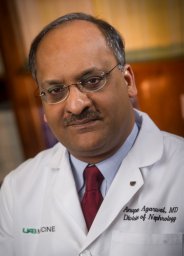 Anupam Agarwal合作论文数Harvard-Thorndike Laboratory and the Department of Medicine, Beth Israel Deaconess Medical Center and Harvard Medical School;Cardiovascular and Metabolic Division, GlaxoSmithKline Pharmaceuticals1
 Thomas Ichim合作论文数Venvalo Group1




