The role of acoustic cavitation in various surface cleaning disciplines is important. However, the physical mechanisms underlying acoustic cavitation-induced surface cleansing are poorly understood. This is due to the combination of microscopic and ultrashort timescales associated with the dynamics of acoustic cavitation bubbles. Here, we have precisely controlled single-bubble cavitation in both space and time. Ultrasonic excitation leads to the cavitation of generated single bubbles. A synchronous ultrafast photomicrographic system simultaneously records the dynamics of single acoustic cavitation bubbles (SACBs) and the cleaning process of the nearby surface in liquids with varying viscosities. Finally, we analysed the correlation between bubble dynamics and surface cleaning situations. The differences in the typical dynamic characteristics of the bubbles during collapse in liquids with varying viscosities reveal two main mechanisms underlying surface cleaning by acoustic cavitation, which are respective the Laplace pressure during the bubble's movement and liquid jets during bubble collapse. Our study provides a better physical understanding of the ultrasonic cleaning process based on acoustic cavitation, and will help to optimize and facilitate the applications of surface cleaning, especially for the cleaning of substrates with tightly attached dirt.
This paper reports a facile fabrication method of polymeric chip with about five-fold increasement in reaction area by in-situ constructing porous microreactor array, which is therefore beneficial for low-cost, high throughput and high capacity de novo DNA synthesis. Microwell arrays were laser-engraved on polytetrafluoroethylene (PTFE) substrate, filled with sugar and then casted with polydimethylsiloxane (PDMS). After sugar removed, porous PDMS (pPDMS) arrays would be incorporated easily. A 40-mer oligonucleotides were successfully synthesized on the pPDMS array and validated by fluorescent hybridization experiments. With the high reaction sites provided by pPDMS, the yield of oligo is about four times higher than the pristine planar PDMS with the same footprint. In addition, the porosity of pPDMS can be easily adjusted to meet different yield requirements.
It remains a great challenge to construct nanostructures and modify two-dimensional (2D) nanomaterials on the cylindrical surface of a micro-scale biosensor to enhance its performance. In the paper, a novel fiber surface plasmon resonance (SPR) biosensor was proposed to enhance the sensitivity of glucose monitoring by forming nanostructures and coating graphene on the gold film. Ultrasonic cavitation was used for the first time to form nanostructures on the cylindrical gold film surface, thus inducing local surface plasmon resonance (LSPR). To enhance the adsorption of glucose molecules, a liquid transfer method was innovatively proposed to cover the surface of the cylindrical roughened gold film of the biosensor with multilayer chemical vapor deposition (CVD) graphene. According to the experimental results, the SPR sensor treated with ultrasonic cavitation for 15 min and modified with two-layer graphene achieved top sensitivity, exhibiting about 3.57 times enhancement compared to the untreated one. This result verifies the sensitivity enhancement effect of both methods, ultrasonic cavitation to construct nanostructures and the liquid transfer of graphene, which may also be applied to construct nanostructures and modify 2D nanomaterials on arbitrary shapes of micro-scale biosensors.
In recent years, DNA-based biosensors have shown great potential as the candidate of the next generation biomedical detection device due to their robust chemical properties and customizable biosensing functions. Compared with the conventional biosensors, the DNA-based biosensors have advantages such as wider detection targets, more durable lifetime, and lower production cost. Additionally, the ingenious DNA structures can control the signal conduction near the biosensor surface, which could significantly improve the performance of biosensors. In order to show a big picture of the DNA biosensor's advantages, this article reviews the background knowledge and recent advances of DNA-based biosensors, including the functional DNA strands-based biosensors, DNA hybridization-based biosensors, and DNA templated biosensors. Then, the challenges and future directions of DNA-based biosensors are discussed and proposed.
A novel micro surface plasmon resonance (SPR) sensor with multiple sensing areas was designed and fabricated, which realized the joint detection of various early liver cancer markers simultaneously. The SPR sensor has two sensing areas constructed by different metal layers to realize the separation of resonance peaks, which makes it possible to detect multiple substances by one single sensor. Then the two sensing regions were modified by "monoclonal antibody - AFP - polyclonal antibody" and "DNA probe - miRNA-125b - S9.6 antibody" respectively. These two sandwich structures enhance the specific detection of Alpha-fetoprotein (AFP) and miRNA-125b. The feasibility of the micro SPR sensor for joint diagnosis of early liver cancer markers was verified.
Fiber-based techniques make it possible to implant a miniaturized and flexible surface plasmon resonance (SPR) sensor into the human body for glucose detection. However, the miniaturization of fiber SPR sensors results in low sensitivity compared with traditional prism-type SPR sensors due to limited sensing area. In this paper, we proposed a D-shaped fiber SPR sensor with a composite nanostructure of molybdenum disulfide (MoS2)-graphene to improve the sensor sensitivity. Compared with the traditional cylindrical fiber, the planar sensing area on the side-polished fiber makes it easier to modify two-dimensional materials. Chemical vapor deposition (CVD) graphene and CVD MoS2 were modified on the sensor surface to obtain the MoS2-graphene composite nanostructure. π-π stacking interactions were used to modify pyrene-1-boronic acid (PBA) on the graphene. The excellent photoelectric properties of the MoS2-graphene composite nanostructure and the ability of PBA to specifically bind glucose molecules improved the glucose detection performance of the SPR sensor. The results show that specific detection of glucose was realized and that the highest sensitivity was achieved with three-layer MoS2 and monolayer graphene.
This paper reports a flexible precise volume sensor with metal-on-polyimide (PI) electrodes to substitute for the peripheral ration pump of a microfluidic system, thus beneficial for integration and miniaturization. This in-channel volume sensor consists of multi-electrode pairs, and it can perform volume measurement of the fluid flowing through microchannels by testing the resistance variation of the electrode pairs, which makes the device possible to help automatically control the sample volume in the mixing and reacting processes inside a microfluidic chip without peripheral ration pumps. The electrode pairs of the sensor are fabricated on flexible PI surface directly by inkjet printing. Then, the electrodes with the PI substrate are transferred and sandwiched by a polydimethylsiloxane (PDMS) substrate layer and a PDMS channel layer to form the flexible precise volume sensor. This method overcomes the challenge of patterning metals on PDMS and the sandwiched PDMS–PI–PDMS structure is beneficial for integration with other PDMS-based microfluidic chips. The effects of electrode-tip shapes and numbers of the electrode pairs are also investigated. A novel calculating method is proposed to obtain more precise results when different numbers of electrode pairs are used in different situations. According to the experimental results, the more electrode pairs are used in the same spacing, the better measurement precision can be obtained. The volume sensor with optimized electrode-tip and multi-electrode pairs can detect the fluid volume in nanolitre scales with the relative error of < 0.8%. This work exhibits the potential to form a total lab-on-a-chip without peripheral ration pumps.
This paper proposes a non-invasive method to research on the dynamic behaviors of a micrometer scale air-bubble near a rigid boundary in ultrasound field. As the rapid change and tiny size of the acoustic bubble near rigid wall, it is very hard to record the dynamic behaviors of acoustic bubbles, which is one of the key fundamental problems in the application of ultrasonic cavitation. In this paper, a new method combining microscope, high speed photography and synchronous technology is proposed to noninvasively and accurately record the dynamic behaviors of a single bubble in ultrasound field. By this method, a single air bubble in tiny size can be created in water whose inner composition is almost the same as acoustic cavitation bubbles, and the relative position of the generated bubble near rigid wall can be controlled. In the experiment, the temporal evolution of the bubble is recorded by the high-speed camera at 300,000 frames per second, as well as the corresponding data, such as volume of the bubble and the velocity of the farthermost point on the bubble from the rigid boundary, is recorded. Results are demonstrated for a single bubble generated over a rigid wall under a certain standoff distance (1.85) in an ultrasound field with frequency of 20.47 kHz. The results show that the dynamics of a single bubble near the rigid wall in ultrasonic field can be divided into four parts: oscillation, movement, collapse and rebound. The maximum velocity of the bubble boundary, which is due to the formation of the high speed liquid jet, is around 11.3 m/s. Furthermore, the dynamic behaviors of a single bubble in the experiment of this paper have a good agreement with the corresponding numerical results.
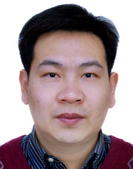 Dachao Li (栗大超)合作论文数School of Precision Instrument and Opto-electronics Engineering, Tianjin University9
 Haixia Yu (于海霞)合作论文数School of Precision Instrument and Optoelectronics Engineering, Tianjin University5
 Xiao Su合作论文数3Zhihua Pu合作论文数3
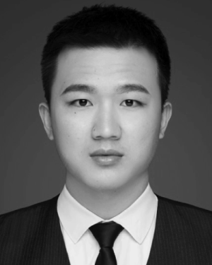 Hao Wu (吴昊)合作论文数2Penghao Zhang合作论文数2Hao Zheng合作论文数2
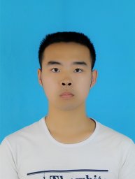 Xingguo Zhang合作论文数State Key Laboratory of Precision Measuring Technology and Instruments, Tianjin University2
 Xiaochen Lai (赖笑辰)合作论文数School of Artificial Intelligence, Nanjing University of Information Science & Technology2
 Ridong Wang (王日东)合作论文数School of Precision Instrument and Opto-electronics Engineering, Tianjin University;Laboratory of Digital-Intelligent Sensing Chips and Instruments, Tianjin University2Chengcheng Li合作论文数1Yu Hua合作论文数1Bingyu Lu合作论文数1Yanyun Fan合作论文数1Hailong Chen合作论文数1
 Cheng Zhou合作论文数1
 Xiaoping Li (李小平)合作论文数School of Precision Instrument and Opto-electronics Engineering, Tianjin University1
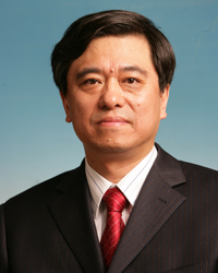 Kexin Xu (徐可欣)合作论文数School of Precision Instrument and Opto-Electronics Engineering, Tianjin University1
 Claus-Dieter Ohl合作论文数Department Soft Matter, Faculty of Natural Sciences, Otto-von-Guericke University, Magdeburg;Institute for Physics, Otto-von-Guericke University, Magdeburg1




