Reactivation of endogenous retroviruses (ERVs) has been proposed to be involved in aging. However, the mechanism of reactivation and contribution to aging and age-associated diseases is largely unexplored. In this study, we identified a subclass of ERVs reactivated in senescent cells (termed senescence-associated ERVs (SA-ERVs)). These SA-ERVs can be bidirectional transcriptionally activated by activating transcription factor 3 (ATF3) to generate double-stranded RNAs (dsRNAs), which activate the RIG-I/MDA5-MAVS signaling pathway and trigger a type I interferon (IFN-I) response in senescent fibroblasts. Consistently, we found a concerted increased expression of ATF3 and SA-ERVs and enhanced IFN-I response in several tissues of healthy aged individuals and patients with Hutchinson-Gilford progeria syndrome. Moreover, we observed an accumulation of dsRNAs derived from SA-ERVs and higher levels of IFNβ in blood of aged individuals. Together, these results reveal a previously unknown mechanism for reactivation of SA-ERVs by ATF3 and illustrate SA-ERVs as an important component and hallmark of aging.
Telomeres, at the ends of chromosomes, protect chromosomes from fusion and preserve genomic stability. However, the molecular mechanisms underlying telomere attrition-induced genome instability remain to be understood. We systematically analyzed the expression of retrotransposons and performed genomic sequencing of different cell and tissue types with telomeres of varying lengths due to telomerase deficiency. We found that critically short telomeres altered retrotransposon activity to promote genomic instability in mouse embryonic stem cells, as evidenced by elevated numbers of single nucleotide variants, indels and copy number variations (CNVs). Transpositions of retrotransposons such as LINE1 resulting from the short telomeres can also be found in these genomes with elevated number of mutations and CNVs. Retrotransposon activation is linked to increased chromatin accessibility, and reduced heterochromatin abundance correlates with short telomeres. Re-elongation of telomeres upon recovery of telomerase partly represses retrotransposons and heterochromatin accumulation. Together, our findings suggest a potential mechanism by which telomeres maintain genomic stability by suppressing chromatin accessibility and retrotransposon activity.
Ubiquitin-fold modifier 1 (UFM1) is a recently identified ubiquitin-like posttranslational modification with important biological functions. However, the regulatory mechanisms governing UFM1 modification of target proteins (UFMylation) and the cellular processes controlled by UFMylation remain largely unknown. It has been previously shown that a UFM1-specific protease (UFSP2) mediates the maturation of the UFM1 precursor and drives the de-UFMylation reaction. Furthermore, it has long been thought that UFSP1, an ortholog of UFSP2, is inactive in many organisms, including human, because it lacks an apparent protease domain when translated from the canonical start codon (445AUG). Here, we demonstrate using the combination of site-directed mutagenesis, CRISPR/Cas9-mediated genome editing, and mass spectrometry approaches that translation of human UFSP1 initiates from an upstream near-cognate codon, 217CUG, via eukaryotic translation initiation factor eIF2A-mediated translational initiation rather than from the annotated 445AUG, revealing the presence of a catalytic protease domain containing a Cys active site. Moreover, we show that both UFSP1 and UFSP2 mediate maturation of UFM1 and de-UFMylation of target proteins. This study demonstrates that human UFSP1 functions as an active UFM1-specific protease, thus contributing to our understanding of the UFMylation/de-UFMylation process.
Telomerase plays a pivotal role in tumorigenesis by both telomere-dependent and telomere-independent activities, although the underlying mechanisms are not completely understood. Using single-sample gene set enrichment analysis (ssGSEA) across 9,264 tumour samples, we observe that expression of telomerase reverse transcriptase (TERT) is closely associated with immunosuppressive signatures. We demonstrate that TERT can activate a subclass of endogenous retroviruses (ERVs) independent of its telomerase activity to form double-stranded RNAs (dsRNAs), which are sensed by the RIG-1/MDA5-MAVS signalling pathway and trigger interferon signalling in cancer cells. Furthermore, we show that TERT-induced ERV/interferon signalling stimulates the expression of chemokines, including CXCL10, which induces the infiltration of suppressive T-cell populations with increased percentage of CD4+ and FOXP3+ cells. These data reveal an unanticipated role for telomerase as a transcriptional activator of ERVs and provide strong evidence that TERT-mediated ERV/interferon signalling contributes to immune suppression in tumours.
Ufmylation (UFM1 modification) is a newly identified ubiquitin-like modification system involved in numerous cellular processes. However, the regulatory mechanisms and biological functions of this modification remain mostly unknown. We have recently reported that Ufmylation family genes have frequent somatic copy number alterations in human cancer including melanoma, suggesting involvement of Ufmylation in skin function and disease. UFL1 is the only known Ufmylation E3-like ligase. In this study, we generated the skin-specific Ufl1 knockout mice and show that ablation of Ufl1 caused epidermal thickening, pigmentation and shortened life span. RNA-Seq analysis indicated that Ufl1 deletion resulted in upregulation of the genes involved in melanin biosynthesis. Mechanistically, we found that Endothelin-1 (ET-1) is a novel substrate of Ufmylation and this modification regulates ET-1 stability, and thereby deletion of Ufl1 upregulates the expression and secretion of ET-1, which in turn results in up-regulation of genes in melanin biosynthesis and skin pigmentation. Our findings establish the role of Ufl1 in skin pigmentation through Ufmylation modification of ET-1 and provide opportunities for therapeutic intervention of skin diseases.
Endogenous retroviruses (ERVs), deriving from exogenous retroviral infections of germ line cells occurred millions of years ago, represent ~8% of human genome. Most ERVs are highly inactivated because of the accumulation of mutations, insertions, deletions, and/or truncations. However, it is becoming increasingly apparent that ERVs influence host biology through genetic and epigenetic mechanisms under particular physiological and pathological conditions, which provide both beneficial and deleterious effects for the host. For instance, certain ERVs expression is essential for human embryonic development. Whereas abnormal activation of ERVs was found to be involved in numbers of human diseases, such as cancer and neurodegenerative diseases. Therefore, understanding the mechanisms of regulation of ERVs would provide insights into the role of ERVs in health and diseases. Here, we provide an overview of mechanisms of transcriptional regulation of ERVs and their dysregulation in human diseases.
The ubiquitin and ubiquitin-like protein modifications contribute to functional changes of substrate proteins and thus regulate a range of biological processes including cancer.1 Ubiquitin-fold modifier 1 (UFM1) is a newly identified ubiquitin-like modification with essential biological functions.2, 3 Like ubiquitination, UFM1 modification (UFMylation) is catalyzed by the dynamic E1–E2–E3 enzymatic reaction. Unlike ubiquitination which encompasses several E1s and different E2s (>50) and E3s (>600), only one of each enzyme, UBA5 (E1-like), UFC1 (E2-like), and UFL1 (E3-like), has been identified for the UFMylation reaction.2, 3 The proteases UFM1-specific cysteine proteases 1 (UFSP1) and 2 (UFSP2) execute the maturation of the UFM1 precursor and de-UFMylation process.2, 3 In addition, the satellite components DDRGK1 and CDK5RAP3 are considered as key regulators of the UFMylation system.3-5 Recent studies suggest that UFMylation plays critical roles in diverse cellular processes.3 However, the roles of UFMylation in tumorigenesis have not been systematically explored. In this study, we comprehensively analysed the genomic alterations of these eight UFMylation family genes across the Cancer Genome Atlas (TCGA) data cohort. (Data S1). We performed GISTIC2.0 analysis and identified a total of 55 recurrent and focal somatic copy number alterations (SCNA) events in UFMylation family genes across the whole TCGA cohort with 33 cancer types (Figure 1A). Among the UFMylation genes, UFSP2 was frequently deleted in 14 cancer types. We calculated the frequencies of copy number gain or loss of UFMylation genes in each cancer type (Figure 1B). We found UFSP2 (31%), UFM1 (31%) and UFL1 (28%) showed the highest average frequency for copy number loss, whereas UFC1 (34%), UFSP1 (34%) and DDRGK1 (30%) had the highest average alteration frequency for copy number gain (Figure 1B). Similar SCNAs patterns of the UFMylation genes were observed by using the ICGC Data Portal (Figure S1 and Data S2). Interestingly, we found that the UFSP2 copy number was mainly heterozygous loss in tumours and cancer cell lines (Figure S2), and homozygous deletions were rarely detected, indicating a critical biological function and possible haploinsufficiency of the UFSP2 gene (Figure 1A and Data S3). To further explore the SCNA patterns of UFMylation genes, we calculated the high-level alteration of SCNAs and found that 11.08% of TCGA samples have high-level copy number alterations in at least one of the eight genes. Noteworthy, the high-level amplifications and deletions of UFMylation genes appeared to be occurred in a pattern of mutually exclusive (Figure 1C), suggesting UFMylation genes share common biological functions. However, we noticed that most of UFMylation genes exhibit low frequencies of somatic mutations (<5%) and transcript fusions (<.1%) (Figure S3 and Data S4–S7). Gene expression profiles analysed based on the RNA sequencing data from TCGA database (Data S8 and S9) indicate that UFMylation genes were ubiquitously expressed (Figure S4). Although most UFMylation genes generally have low mutation frequencies in cancers, we found that UFSP2 is frequently mutated in colon adenocarcinoma and uterine corpus endometrial carcinoma. The high recurrent copy number loss and frequent somatic mutations of UFSP2 suggest that UFSP2 may function as a tumour suppressor. It has been previously demonstrated that knockdown UFSP2 promoted breast cancer cell growth and tumour formation, suggesting that UFSP2 is a tumour suppressor in breast cancer.6 In addition, we found that the levels of UFSP2 mRNA were significantly lower in 11 cancer types (Figure S5A). Using human tissue microarrays, we confirmed that the expression of UFSP2 was significantly reduced in cancer tissues (Figures 2A and S6). To further validate the potential tumour suppressor function of UFSP2 in colon cancer, we examined the effects of UFSP2 knockdown on cell growth of colon cancer cells HT29 and HCT116, and observed that knockdown UFSP2 expression significantly promoted growth rates of colon cancer cells and its anchorage-independent cell growth. (Figure 2B–G). Significantly, knockdown UFSP2 expression significantly promoted the growth of xenograft tumours from UFSP2 depleted HT29 cells (Figure 2H–J). In addition, we observed that total UFMyaltion levels were increased in both UFSP2 depleted cells and xenograft tumours (Figure S7). These findings suggested that genomic alterations of UFSP2 were associated with the functional involvement of UFMylation in human colon cancer. Furthermore, our GSEA analysis indicates that loss of UFSP2 is mainly associated with the pathways in DNA replication, cell cycle, spliceosome, ribosome and mismatch repair (Figure S5C–D and Data S10). This is in line with previous reports that the UFMylation is critically involved in DNA damage, cell cycle, and ribosome protein modification.3, 4, 7, 8 We confirmed that knockdown UFSP2 increased the expression of some marker genes in DNA replication (PCNA and MCM2), cell cycle (CDK4 and CCND1), and ribosome protein (RPL26) (Figure S5E). It has been reported that MCM2, CDK4, and PCNA are interacted with UFM1 and are potential targets of the UFMylation.7 Thus, we speculate that UFSP2 may contribute to the tumorigenesis by modulating key regulators of cell cycle, DNA replication or protein biogenesis through Ufmylation modification. Together, our experimental findings revealed that knockdown UFSP2 expression significantly promotes the growth of colon cancer cells both in vitro and in vivo, suggesting that UFSP2 is a potential tumour suppressor in colon cancers. Given that UFMylation genes exhibit a high frequency of SCNA with a mutually exclusive pattern in common adult cancers, this suggests that involvement of UFMylation in cancer may be cell type or tissue specific. In line with this, it has been previously reported that CDK5RAP3 may function as a tumour suppressor9 in HNSCC and as an oncogene in liver cancer.10 In summary, we found that UFMylation family genes have a high frequency of SCNAs. Especially, UFSP2 is recurrently and focally deletion in a total of 14 human common cancers with the highest alteration score. We further demonstrated that depleted UFSP2 expression significantly promotes the growth of tumour cells both in vitro and in vivo, suggesting that UFSP2 may function as a tumour suppressor in colon cancers. Our integrated genomic analysis and functional studies provide insights in understanding this new post-translational modifier in cancer and potential therapeutic opportunities. We are grateful to Dr. Shuo Mou for bioinformatics analysis assistance, and Dr. Yongkang Zou for helpful discussions. This work was supported by grants from the National Natural Science Foundation of China (31730020), and the Hangzhou Science and Technology Bureau (20182014B01). Please note: The publisher is not responsible for the content or functionality of any supporting information supplied by the authors. Any queries (other than missing content) should be directed to the corresponding author for the article.
Human endogenous retroviruses (HERVs) represent ∼8% of human genome, deriving from exogenous retroviral infections of germ line cells occurred millions of years ago and being inherited by the offspring in a Mendelian fashion. Most of HERVs are nonprotein-coding because of the accumulation of mutations, insertions, deletions, and/or truncations. It has been long thought that HERVs were "junk DNA". However, it is now known that HERVs are involved in various biological processes through encoding proteins, acting as promoters/enhancers, or lncRNAs to affect human health and disease. In this review, we summarized recent findings about HERVs, with implications in embryonic development, pluripotency, cancer, aging, and neurodegenerative diseases.
The UFMylation modification is a novel ubiquitin-like conjugation system, consisting of UBA5 (E1), UFC1 (E2), UFL1 (E3), and the conjugating molecule UFM1. Deficiency in this modification leads to embryonic lethality in mice and diseases in humans. However, the function of UFL1 is poorly characterized. Studies on Ufl1 conditional knockout mice have demonstrated that the deletion of Ufl1 in cardiomyocytes and in intestinal epithelial cells causes heart failure and increases susceptibility to experimentally induced colitis, respectively, suggesting an essential role of UFL1 in the maintenance of the homeostasis in these organs. Yet, its physiological function in other tissues and organs remains completely unknown. In this study, we generate the nephron tubules specific Ufl1 knockout mice and find that the absence of Ufl1 in renal tubular results in kidney atrophy and interstitial fibrosis. In addition, Ufl1 deficiency causes the activation of unfolded protein response and cell apoptosis, which may be responsible for the kidney atrophy and interstitial fibrosis. Collectively, our results have demonstrated the crucial role of UFL1 in regulating kidney function and maintenance of endoplasmic reticulum homeostasis, providing another layer of understanding kidney atrophy.
Telomerase plays a pivotal role in tumorigenesis by maintaining telomere homeostasis, a hallmark of cancer. However, the mechanisms by which telomerase is reactivated or upregulated during tumorigenesis remain incompletely understood. Here, we report that the Hippo pathway effector Yes-associated protein (YAP) regulates the expression of human telomerase reverse transcriptase (hTERT). Ectopic expression or physiological activation of YAP increases hTERT expression, whereas knockdown of YAP decreases the expression of hTERT. YAP binds to the hTERT promoter through interaction with the TEA domain family transcription factors and activates hTERT transcription. Furthermore, sustained YAP hyperactivation promotes telomerase activity and extends telomere length, with increased hTERT expression. In addition, we show that hTERT expression is positively correlated with YAP activation in human liver cancer tissues. Together, our results demonstrate that YAP promotes hTERT expression, which could contribute to tumor progression.
p53 is the most intensively studied tumour suppressor 1 . The regulation of p53 homeostasis is essential for its tumour-suppressive function 2 , 3 . Although p53 is regulated by an array of post-translational modifications, both during normal homeostasis and in stress-induced responses 2 – 4 , how p53 maintains its homeostasis remains unclear. UFMylation is a recently identified ubiquitin-like modification with essential biological functions 5 – 7 . Deficiency in this modification leads to embryonic lethality in mice and disease in humans 8 – 12 . Here, we report that p53 can be covalently modified by UFM1 and that this modification stabilizes p53 by antagonizing its ubiquitination and proteasome degradation. Mechanistically, UFL1, the UFM1 ligase 6 , competes with MDM2 to bind to p53 for its stabilization. Depletion of UFL1 or DDRGK1, the critical regulator of UFMylation 6 , 13 , decreases p53 stability and in turn promotes cell growth and tumour formation in vivo. Clinically, UFL1 and DDRGK1 expression are downregulated and positively correlated with levels of p53 in a high percentage of renal cell carcinomas. Our results identify UFMylation as a crucial post-translational modification for maintenance of p53 stability and tumour-suppressive function, and point to UFMylation as a promising therapeutic target in cancer.
Pigs are proposed to be suitable large animal models for test of the efficacy and safety of induced pluripotent stem cells (iPSCs) for stem cell therapy, but authentic pig ES/iPS cell lines with germline competence are rarely produced. The pathways or signaling underlying the defective competent pig iPSCs remain poorly understood. By improving induction conditions using various small chemicals, we generated pig iPSCs that exhibited high pluripotency and differentiation capacity that can contribute to chimeras. However, their potency was reduced with increasing passages by teratoma formation test, and correlated with declined expression levels of Rex1, an important marker for naïve state. By RNA-sequencing analysis, genes related to WNT signaling were upregulated and MAPK signaling and TGFβ pathways downregulated in pig iPSCs compared to fibroblasts, but they were abnormally expressed during passages. Notably, pathways involving in DNA repair and replication were upregulated at early passage, but downregulated in iPSCs during prolonged passage in cluster with fibroblasts. Our data suggests that reduced DNA repair and replication capacity links to the instability of pig iPSCs. Targeting these pathways may facilitate generation of truly pluripotent pig iPSCs, with implication in translational studies.
Inadequate silencing of exogenous genes represents a major obstacle to complete epigenetic reprogramming of porcine-induced pluripotent stem cells (piPSCs) by conventional pluripotency transcription factors (OSKM). We tested the hypothesis that epigenetic modification by active DNA or histone demethylation or by inhibition of histone deacetylase would enhance reprogramming and exogenous gene silencing in piPSCs. piPSCs induced by OSKM in combination with epigenetic factors, specifically Ten-Eleven Translocation (Tet1 or Tet3) or lysine (K)-specific demethylase 3A (Kdm3a), expressed higher levels of Rex1 and other genes representing naive state and exhibited more open chromatin status, compared with those of OSKM controls. Tet1 also improved differentiation capacity. Conversion with inhibitors of histone deacetylases (HDACi), NaB, TSA, or VPA, further increased Rex1 expression, while decreasing expression of exogenous genes. piPSCs induced by Tet1+OSKM followed by conversion with HDACi show high pluripotency. Together, epigenetic modifiers enhance generation of piPSCs and reduce their reliance on exogenous genes.
Mouse embryonic stem cell (ESC) cultures exhibit a heterogeneous mixture of metastable cells sporadically entering the 2-cell (2C)-embryo-like state, critical for ESC potency. One of 2-cell genes, Zscan4, has been shown to be responsible for telomere maintenance, genomic stability and pluripotency of mouse ESCs. Functions of other 2C-genes in ESCs remain elusive. Here we show that 2C-genes Tcstv1 and Tcstv3 play a role in regulation of telomere lengths. Overexpression or knockdown Tcstv1 and Tcstv3 does not immediately affect proliferation, pluripotency and differentiation in vitro of ESCs. However, ectopic expression of Tcstv1 or Tcstv3 results in telomere elongation, whereas Tcstv1/3 knockdown shortens telomeres of ESCs. Overexpression of Tcstv1 or Tcstv3 does not alter telomere stability. Furthermore, Tcstv1 can increase Zscan4 protein levels and telomere recombination by telomere sister chromatid exchange (T-SCE). Depletion of Tcstv1/3 reduces Zscan4 protein levels. Together, Tcstv1 and Tcstv3 are involved in telomere maintenance that is required for long-term self-renewal of mouse ESCs. Our data also suggests that Tcstv1/3 may co-operate and stabilize Zscan4 protein but the molecular bases remain to be determined.
Various types of somatic cells can be reprogrammed to induced pluripotent stem (iPS) cells. Somatic stem cells may generate iPS cells more efficiently than do differentiated cells. We show that granulosa cells exhibit characteristic of somatic stem cells and can be reprogrammed to iPS cells more efficiently or with few factors. Here, we describe generation of mouse and pig iPS cells from granulosa cells with high efficiency.
Jnk1-deficient mice manifest disrupted anterior commissure formation and loss of axonal and dendritic microtubule integrity. However, the mechanisms and the specific stages underlying the developmental defects remain to be elucidated. Here, we report the generation of Jnk1-deficient (Jnk1 KO) iPSCs from Jnk1 KO mouse tail-tip fibroblasts (TTFs) for modeling the neural disease development. The efficiency in the early induction of iPSCs was higher from Jnk1 KO fibroblasts than that of wild-type (WT) fibroblasts. These Jnk1 KO iPSCs exhibited pluripotent stem cell properties and had the ability of differentiation into general three embryonic germ layers in vitro and in vivo. However, Jnk1 KO iPSCs showed reduced capacity in neural differentiation in the spontaneous differentiation by embryoid body (EB) formation. Notably, by directed lineage differentiation, Jnk1 KO iPSCs specifically exhibited an impaired ability to differentiate into early stage neural precursors. Furthermore, the neuroepitheliums generated from Jnk1 KO iPSCs appeared smaller, indicative of neural stem cell developmental defects, as demonstrated by teratoma tests in vivo. These data suggest that Jnk1 deï¬ciency inhibits the development of neural stem cells/precursors and provide insights to further understanding the complex pathogenic mechanisms of JNK1-related neural diseases.
Telomere attrition and genomic instability are associated with organism aging. Concerns still exist regarding telomere length resetting in cloned embryos and ntES cells, and possibilities of premature aging of cloned animals achieved by somatic cell nuclear transfer (SCNT). Trichostatin A (TSA), a histone deacetylase inhibitor, effectively improves the developmental competence of cloned embryos and animals, and recently contributes to successful generation of human ntES cells by SCNT. To test the function of TSA on resetting telomere length, we analyzed telomeres in cloned blastocysts and pigs following treatment of SCNT embryos with TSA. Here, we show that telomeres of cloned pigs generated by standard SCNT methods are not effectively restored, compared with those of donor cells, however TSA significantly increases telomere lengths in cloned pigs. Telomeres elongate in cloned porcine embryos during early cleavage from one-cell to four-cell stages. Notably, TSA facilitates telomere lengthening of cloned embryos mainly at morula-blastocyst stages. Knockdown of pTert by shRNA in donor cells reduces telomerase activity in cloned blastocysts but does not abrogate telomere elongation in the TSA-treated embryos (p > 0.05). However, genes associated with recombination or telomerase-independent mechanism of alternative lengthening of telomeres (ALT) Rad50 and BLM show increased expression in TSA-treated embryos. These data suggest that TSA may promote telomere elongation of cloned porcine embryos by ALT. Together, TSA can elongate telomeres in cloned embryos and piglets, and this could be one of the mechanisms underlying improved development of cloned embryos and animals treated with TSA.
Female germline or oogonial stem cells transiently residing in fetal ovaries are analogous to the spermatogonial stem cells or germline stem cells (GSCs) in adult testes where GSCs and meiosis continuously renew. Oocytes can be generated in vitro from embryonic stem cells and induced pluripotent stem cells, but the existence of GSCs and neo-oogenesis in adult mammalian ovaries is less clear. Preliminary findings of GSCs and neo-oogenesis in mice and humans have not been consistently reproducible. Monkeys provide the most relevant model of human ovarian biology. We searched for GSCs and neo-meiosis in ovaries of adult monkeys at various ages, and compared them with GSCs from adult monkey testis, which are characterized by cytoplasmic staining for the germ cell marker DAZL and nuclear expression of the proliferative markers PCNA and KI67, and pluripotency-associated genes LIN28 and SOX2, and lack of nuclear LAMIN A, a marker for cell differentiation. Early meiocytes undergo homologous pairing at prophase I distinguished by synaptonemal complex lateral filaments with telomere perinuclear distribution. By exhaustive searching using comprehensive experimental approaches, we show that proliferative GSCs and neo-meiocytes by these specific criteria were undetectable in adult mouse and monkey ovaries. However, we found proliferative nongermline somatic stem cells that do not express LAMIN A and germ cell markers in the adult ovaries, notably in the cortex and granulosa cells of growing follicles. These data support the paradigm that adult ovaries do not undergo germ cell renewal, which may contribute significantly to ovarian senescence that occurs with age.
Various types of somatic cells can be reprogrammed to induced pluripotent stem (iPS) cells. Somatic stem cells exhibit enhanced reprogramming efficiency by fewer factors, in contrast to fully differentiated cells. Nuclear LaminA is highly expressed in differentiated cells, and stem cells are characterized by the absence of LaminA. Granulosa cells (GCs) and cumulus cells in the ovarian follicles effectively and firstly generated cloned mice by somatic cell nuclear transfer, and these cells lack LaminA expression. We tested the hypothesis that GCs could be effectively used to generate iPS cells with fewer factors. We show that iPS cells are generated from GCs at high efficiency even with only two factors, Oct4 and Sox2, like the iPS cells generated using four Yamanaka factors. These iPS cells show pluripotency in vitro and in vivo, as evidenced by high expression of pluripotency-associated genes, Oct4, Nanog, and SSEA-1, differentiation into three embryonic germ layers by embryoid body formation and teratoma tests, as well as high efficient generation of chimeras. Moreover, the exogenous genes are effectively silenced in these iPS cells. These data provide additional evidence in supporting the notion that reduced expression of LaminA and stem cells can improve the reprogramming efficiency to pluripotency.
 Lin Liu (刘林)合作论文数School of Life Sciences, Nankai University12
 Yusheng Cong (丛羽生)合作论文数School of Basic Medical Sciences, Hangzhou Normal University10
 Miao Wang合作论文数8
 Lu Xu合作论文数6
 Junzhi Zhou合作论文数School of Basic Medical Sciences, Guangdong Medical University5Qian Liang合作论文数5Xiaoying Wu合作论文数4Fu Haifeng合作论文数4
 Kai Liu合作论文数4
 Xiaoying Ye (叶孝颖)合作论文数College of Life Sciences, Nankai University4Guangzhen Ji合作论文数3Jiang Liu合作论文数Institute of Aging Research, School of Medicine, Hangzhou Normal University, Hangzhou, Zhejiang 310036, China.3Xiaohe Ma合作论文数3
 Liu Mengyuan (刘梦媛)合作论文数3
 Di Guan合作论文数3
 Na Liu (刘娜)合作论文数School of Medicine, Nankai University3
 Lei Wang (王磊)合作论文数School of Statistics and Data Science, Nankai University3
 Zhonghua Liu (刘忠华)合作论文数College of Animal Scienceand Technology, Northeast Agricultural University;School of Life Science, Northeast Agricultural University3Junjie Bai合作论文数2
 Yu Yin合作论文数2
 Zhuang Yang (杨壮)合作论文数School Breeding and Multiplication, Hainan University2
 David L. Keefe合作论文数Department of Cell Biology, Grossman School of Medicine, New York University2HaiBin Wei (魏海彬)合作论文数2
 Bingteng Xie (解炳腾)合作论文数School of Life Sciences, Beijing Institute of Technology2
 Renpeng Guo (郭仁朋)合作论文数College Food Science and Technology, Nanjing Agricultural University2
 Xumin Wang (王绪敏)合作论文数中国科学院北京基因组研究所2Yuan Jihong合作论文数2Dongdong Zhang合作论文数2Wang Juan合作论文数2
 Tao Cheng (程涛)合作论文数State Key Laboratory of Experimental Hematology, Chinese Academy of Medical Sciences & Peking Union Medical College2
 Shichao Liu (刘世超)合作论文数College of life science, Northeast Agricultural University2
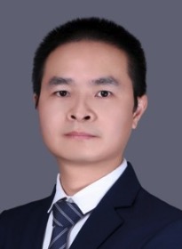 Hua Wang (王华)合作论文数School of Basic Medical Sciences, Peking University Health Science Center;International Cancer Institute, Peking University2
 Qingran Kong合作论文数2Lipu Song合作论文数1
 Siyuan Xia (夏思源)合作论文数Department of Human Cell Biology and Genetics, School of Medicine, Southern University of Science and Technology1Wei Deng (邓伟)合作论文数1Zhang Chenlu合作论文数1Wang Jiabao合作论文数1Yaqi Jin合作论文数1Nannan Zhao合作论文数1
 Xiawei Wei (魏霞蔚)合作论文数State Key Laboratory of Biotherapy, Sichuan University;National Clinical Research Center for Geriatrics, West China Hospital, Sichuan University1Maogong Dong合作论文数1
 Guoxing Yin合作论文数1
 Yong Wang合作论文数1
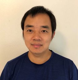 Jiameng Dan合作论文数Department of Epigenetics and Molecular Carcinogenesis, The University of Texas MD Anderson Cancer Center1Jingjing Yang合作论文数1
 Qian Zhang合作论文数Key Lab Aging & Canc Biol Zhejiang Prov, Hangzhou Normal Univ1
 Dan Wang合作论文数Tianjin Univ, Sch Sci, Dept Phys, Tianjin, Peoples R China1




