Supplementary Figure S6. Low-, medium-, and high dose modulation of heterodimer levels following a dose of venetoclax
Supplementary Figure S12. In vivo pharmacodynamic changes in Mcl-1, Bim, and Bad levels following cirtuvivint without or with venetoclax in MV4-11 and KG-1a tumors
Supplementary Figure S1. AMO-1 and MOLT-4 cells were treated with either navitoclax, S63845, or vehicle control
Supplementary Figure S11. Body weights of mice bearing either MV4-11 or KG-1a tumors
Abstract The National Cancer Institute’s Patient Derived Models Repository (NCI PDMR; https://pdmr.cancer.gov) has developed a national repository of Patient-Derived Models (PDMs) currently comprised of over 1000 patient-derived xenograft (PDX), 450 organoid (PDOrg), 500 tumor cell culture (PDC), and 425 cancer associated fibroblast (CAF) models. Over 450 PDXs have matched PDOrg and/or PDCs allowing for complimentary/parallel in vivo/in vitro studies. These PDMs are clinically annotated with molecular information available in a public database for the extramural community with additional molecular features including OncoKB annotated mutations, microsatellite instability (MSI), human leukocyte antigen (HLA) typing, and clinically relevant fusions. Researchers can use these clinical and molecular features or perform their own independent analyses using the public data to aid in their selection of preclinical models. Due to the large number of NCI PDMR models within histologies and research community interest, we have developed histology-based PDX TMAs to further facilitate the selection of models for cancer research. Each TMA panel includes up to 60 unique PDX models, with two 1.5mm cores/model plus murine control tissue. Quality control (QC) assessment of the TMA cores is performed by a pathologist. Each core is reviewed with an initial pass/fail threshold set to ≥10% human tumor/core area with ≥500 tumor cells. TMA slides pass QC if they meet these criteria and ≥75% of the models have at least one passing core. TMA blocks are QC’d at regularly set intervals to ensure all distributable slides meet these requirements. The first PDX TMA panel available for distribution this year (PANC I) contains 60 pancreatic cancer PDXs (predominantly pancreatic adenocarcinoma [PAAD]) derived from primary and metastatic lesions from treatment naïve through heavily pretreated patients. KRAS mutated models include 28 G12D, 15 G12V, 10 G12R, 3 G12C, 1 Q61H, and 3 KRAS wildtype. Other genes frequently mutated in pancreatic cancer are also found in this cohort including TP53, SMAD4, and CDKN2A. Also in development are four colorectal cancer TMAs panels: (1) a general set of colon adenocarcinomas (COAD) with features including early onset, non-European ancestry, and MSI-High; (2) KRAS mutated COAD; (3) Treatment naïve COAD and wildtype APC COAD; and (4) Rectal adenocarcinoma models. These TMAs can be used to stratify models by therapeutic target, develop predictive markers or classify differential signaling in disease subtypes, integrative analysis of genomic and protein expression, and discover or validate biomarkers of disease in an efficient and cost-effective way. Targeted model selection is of high importance to better understand the biology of these cancers and improve preclinical drug testing and screening design to translate novel therapeutics from bench to clinic. Citation Format: Cindy R. Timme, Lindsay Dutko, Sayak Ghatak, Ting-Chia Chang, Alice P. Chen, Li Chen, Biswajit Das, Tara Grinnage-Pulley, Shahanawaz Jiwani, Kaci Paulus, Chris A. Karlovich, Sergio Alcoser, Yvonne Evrard, Melinda G. Hollinghead, James H. Doroshow. Development of pancreatic and colon patient-derived xenograft (PDX) tumor microarrays (TMAs) from the NCI patient derived models repository [abstract]. In: Proceedings of the American Association for Cancer Research Annual Meeting 2026; Part 1 (Regular Abstracts); 2026 Apr 17-22; San Diego, CA. Philadelphia (PA): AACR; Cancer Res 2026;86(7 Suppl):Abstract nr 6061.
3031 Background: Trastuzumab deruxtecan (T-DXd) is a HER2-directed antibody conjugated to topoisomerase 1 inhibitor, deruxtecan, payload that is an effective treatment strategy across several tumor types and various HER2 expressions. The relative contributions of each of the underlying mechanisms driving the broad clinical activity require further elucidation for the ongoing rational development of this promising agent. To this end, our pilot study (NCT04294628) evaluates the pharmacodynamics (PD) of T-DXd in patients (pts) with solid tumors displaying a variety of HER2 expression levels. Methods: This multicenter pilot study enrolled pts with HER2-expressing advanced solid tumors as defined by HER2 immunohistochemistry (IHC) score of 1+ or greater or ERBB2 amplifications (amp) or mutations (mut). Baseline HER2 expression was evaluated by Ventana PATHWAY immunohistochemical (IHC) analysis. T-DXd was administered at 5.4 mg/kg intravenously once every 3 weeks, in 21-day cycles (C), with mandatory tumor biopsies collected at baseline, post-dose C1, and pre-dose C3. Blood samples for biomarker analyses were collected throughout the study. PD biomarkers for topoisomerase 1 (TOP1) target engagement, consequent DNA damage repair (DDR), and tumor immune microenvironment changes were analyzed. Overall response was also evaluated. Results: Sixty-one pts received T-DXd. Eligibility HER2 (eHER2) status was determined from pt records: 21 IHC 1+, 22 IHC 2+, 7 IHC 3+, 8 ERBB2 amp, 3 ERBB2 mut. Of the 41 pts with baseline biopsies centrally assessed for HER2 (bHER2) by IHC, 13 had bHER2 scores discordant with eHER2, including 8 patients who were eHER2 1-2+ but were bHER2 null. No new safety signals were observed. One pt had a complete response (cervical, bHER2 3+), 14 pts had confirmed partial responses (PR), including 3 who were bHER2 null (2 ovarian, 1 uterine), and 5 pts had unconfirmed PR (uPR). Of the 21 paired biopsies assessable for PD response, 15 demonstrated TOP1 target modulation and 14 of those 15 also demonstrated DDR induction. Substantial tumor infiltration and activation of CD8+ T cells at baseline and/or following T-DXd administration occurred in several patients and were particularly prevalent in those with response. In the one responding patient where lesion-specific analyses were possible, we observed significant diameter reduction (3.9 cm to 1.9 cm) and PD responses in a bHER2 null lesion. Conclusions: Clinical responses and target modulation were observed in pts irrespective of bHER2 expression. Lesion-specific analyses provide evidence of antitumor activity and target engagement even in a bHER2-null lesion. Genomic analyses to identify additional molecular determinants of response or resistance to T-DXd are ongoing. This project was funded in part by the National Cancer Institute, National Institutes of Health, under Contract No. 75N91019D00024. Clinical trial information: NCT04294628 .
While most gastrointestinal stromal tumors are driven by oncogenic mutations in KIT or PDGFRA, 10-15% exhibit functional loss of the succinate dehydrogenase (SDH) complex and genome-wide DNA hypermethylation. Excess methylation in SDH-deficient gastrointestinal stromal tumors disrupts genomic insulators, inducing aberrant expression of oncogenic ligands FGF3, FGF4, and activating an autocrine signaling loop mediated through FGFR1. We conducted a phase 2 trial of pan-fibroblast growth factor receptor inhibitor rogaratinib in patients with sarcoma and report here on the cohort of patients with advanced SDH-deficient GIST. The primary objective was to estimate objective response rate. Secondary objectives were to estimate progression-free survival (PFS) and assess safety and tolerability. Exploratory objectives were to evaluate serial measurements of FGF3 and FGF4 and fibroblast growth factor receptors in serial biopsies, to perform whole-exome sequencing in serial biopsies and to explore rogaratinib exposure with pharmacodynamic effects. Twenty-four patients received rogaratinib and ten experienced partial responses for an objective response rate of 41.7%. Median PFS was 31.0 months (95% confidence interval 20.2-not reached), and 1-year PFS was 77.4% (95% confidence interval 61.7-97.1). Toxicities were manageable and included hyperphosphatemia, fatigue and diarrhea. Elevations in phosphorous were seen across the cohort, consistent with target engagement of FGFR1. Whole-exome and next-generation sequencing revealed alterations in the SDH subunit coding genes (SDHx) as expected. This trial illustrates a successful demonstration of targeted cancer therapy predicated on an epigenetic mechanism of oncogene activation. Clinicaltrials.gov identifier: NCT04595747 .
Supplementary Figure S5. Low-, medium-, and high dose modulation of heterodimer levels following a dose of S63845
Supplementary Figure S9. Detection of Bak–Bax heterodimers and cCasp3 in xenograft models following a single dose of venetoclax
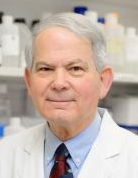 James H. Doroshow合作论文数Center for Cancer Research, National Cancer Institute39
 Yvonne A Evrard合作论文数Leidos Biomed Res Inc, Frederick Natl Lab Canc Res, Appl Dev Res Directorate, Lab Human Toxicol & Pharmacol, Frederick, MD USA31
 Melinda G. Hollingshead合作论文数Healthy Pet Mobile Vet31
 Biswajit Das合作论文数Department of Medicinal Chemistry, Ranbaxy Research Laboratories31
 Chris Karlovich合作论文数23
 P. Mickey Williams合作论文数20Dianne L. Newton合作论文数19Li Chen合作论文数19Alice Chen合作论文数18
 Ting-Chia Chang合作论文数Molecular Characterization Laboratory, Frederick National Laboratory for Cancer Research;Molecular Characterization Laboratory, Frederick National Laboratory for Cancer Research16Carter John A合作论文数13Lindsay Dutko合作论文数12Chelsea McGlynn合作论文数12Michelle Gottholm-Ahalt合作论文数12Michael Mullendore合作论文数12
 Sergio Y Alcoser合作论文数12Suzanne Borgel合作论文数12Gloryvee Rivera合作论文数11Tomas Vilimas合作论文数11Shannon Uzelac合作论文数11L. Chen合作论文数11Amanda Peach合作论文数10Nikitha Nair合作论文数10Tara Grinnage-Pulley合作论文数10Kelly Benauer合作论文数9Michelle Eugeni合作论文数9Justine Mills合作论文数9Dougherty Kelly M合作论文数9Alyssa Chapman合作论文数8Jesse Stottlemyer合作论文数8Emily Delaney合作论文数8Howard Stotler合作论文数8Marion Gibson合作论文数7Candace Mallow合作论文数7Cindy R. Timme合作论文数7
 Patidar Rajesh合作论文数National Cancer Institute, National Institutes of Health7
 Jerald Radich合作论文数Translational Science and Therapeutics Division, Fred Hutchinson Cancer Center;Molecular Oncology Lab, Fred Hutchinson Cancer Center;Division of Hematology and Oncology, School of Medicine, University of Washington7
 Lyndsay Harris合作论文数Division of Cancer Treatment & Diagnosis, National Cancer Institute7Kevin Plater合作论文数6
 Brandie Fullmer合作论文数6Chris Alan Karlovich合作论文数6Thomas Walsh合作论文数6McShane Lisa M合作论文数6Carrie Bonomi合作论文数6Thomas J. Forbes合作论文数6Tiffanie Miner合作论文数5Marianne Radzyminski合作论文数5Anna Lee Fong合作论文数5Corinne Camalier合作论文数5




