Immune marker staining, collagen features, and αSMA-associated stromal analyses in KC/AKC and KPC/AKPC tumors.
Clinical characteristics and mutation profiles of the patient-derived organoids used in this study.
Analyses of TGFβ ligand/receptor expression, reporter activity, inferred signaling interactions, and PSC differentiation in ATM-deficient tumor models.
PSC-induced chemotherapy resistance and in vitro and ex vivo assays assessing apoptosis, invasion, wound closure, and drug response after perturbation of tumor–CAF signaling.
Fibrosis and CAF marker analyses, MRI imaging, survival, proliferation, apoptosis, and flow cytometry from orthotopic treatment experiments.
ROS measurements, cytoskeletal marker expression, migration assays, and PSC differentiation analyses following genetic and pharmacologic perturbation of ROS and cytoskeletal pathways in ATM-deficient tumor models.
Number and proportion of cell types identified by single-nucleus multiomic analysis.
Single-cell analyses of BMP4 and CAF lineage signatures, CAF differentiation assays in PSC- (with BMP pathway perturbation) and CAF-based models, and subcutaneous assay with MRTX1133 and galunisertib.
Pancreatic cancer has a very poor prognosis due to its high resistance to chemotherapeutic drugs, late diagnosis, and early metastasis, making it the most aggressive and lethal cancer. Hence, there is a pivotal need to find new effective preventive and curative treatments. In this study, extracted and fractions obtained from date palm (Phoenix dactylifera L.) fruit using ethyl acetate and n-butanol as solvents. The prepared date fruit extracts and fractions were pharmacologically evaluated on three pancreatic cancer cell lines: PT45P1, PANC-1, and SUIT-2. Specific extracts demonstrated strong anti-proliferative activity. In particular Bu-IV was the most active n-butanol fraction tested at 500 μg/mL. Moreover, sub-fractions obtained from the ethyl acetate extract inhibited pancreatic cancer cell proliferation, especially fraction EA-III-2 tested at 50-150 μg/mL. These findings suggest that date fruit extracts contain nutraceuticals with potential therapeutic value in the treatment of pancreatic cancer. Future work will be focused on isolation and identification of the active compounds and explore their mechanisms of action.
Diagnostic performance obtained by four independent repetitions of creating a SVM classifier on the basis of the clinical parameters recorded for the 302 IPMN patients.
Background Pancreatic ductal adenocarcinoma (PDAC) comprises two clinically relevant subtypes. Currently, determining the tumor subtype relies on tissue biopsies. Unfortunately, these biopsies are spatially biased, highly invasive, difficult to obtain, and unsuitable for monitoring tumor dynamics. Methods We employed whole transcriptome sequencing (WTS) on circulating cell-free (cf) RNA in plasma samples from patients with well-characterized tumor subtypes. Additionally, quantitative protein mass spectrometry was utilized to identify minimally invasive markers for tumor subtypes. We validated our findings using independent liquid and tissue samples from large clinical trials and investigated treatment-induced subtype dynamics and responses. Results An exploratory analysis of 10 patients (four basal-like and six classical) was conducted using whole transcriptome sequencing (WTS). Following differential transcript abundance analysis and integration with expression data from tumor and non-tumor samples (N > 200), we identified 32 protein-coding subtype-specific cfRNA-defined transcripts. The subtype specificity of these transcripts was validated in two independent tissue cohorts comprising 195 and 250 cases, respectively. Three disease-relevant cfRNA-defined subtype markers ( DEGS1, KDELC1 , and RPL23AP7 ) consistently associated with basal-like tumors across all cohorts and were validated using machine learning. Further analysis of these markers using RT-ddPCR in over 160 patient sera and 24 samples from healthy donors revealed their predictive and prognostic value, as well as subtype specificity and therapy-induced dynamics. In both tumor and liquid biopsies, the overexpression of these markers was associated with poor overall and progression-free survival. Moreover, elevated tissue/liquid levels of the identified markers were linked to a poor response to systemic therapy and rapid disease recurrence in resected patients. Conclusion Our data provide support for the clinical significance of cfRNA markers in determining tumor subtypes and monitoring disease recurrence and therapy-induced subtype switches in pancreatic ductal adenocarcinoma (PDAC). Consequently, further validation studies in larger independent cohorts are warranted to confirm the robustness and generalizability of these findings.
Pancreatic ductal adenocarcinoma (PDAC) comprises two clinically relevant molecular subtypes that are currently determined using tissue biopsies, which are spatially biased and highly invasive. We used whole transcriptome sequencing of 10 plasma samples with tumor-informed subtypes, complemented by proteomic analysis for minimally invasive identification of PDAC subtype markers. Data were validated in independent large cohorts and correlated with treatment response and patient outcome. Differential transcript abundance analyses revealed 32 subtype-specific, protein-coding cell-free RNA (cfRNA) transcripts. The subtype specificity of these transcripts was validated in two independent tissue cohorts comprising 195 and 250 cases, respectively. Three disease-relevant cfRNA-defined subtype markers (DEGS1, KDELC1, and RPL23AP7) that consistently associated with basal-like tumors across all cohorts were identified. In both tumor and liquid biopsies, the overexpression of these markers correlated with poor survival. Moreover, elevated levels of the identified markers were linked to a poor response to systemic therapy and early relapse in resected patients. Our data indicate clinical applicability of cfRNA markers in determining tumor subtypes and monitoring disease recurrence.
Abstract Pancreatic ductal adenocarcinoma (PDAC)patients have a 5-year survival rate of less than 10%, partly due to 80% developing recurrence within 2 years after curative intent surgery. As surgical resectability criteria expand and techniques progress, better understanding of tumor biology regarding post-resection prognosis is needed to improve patient outcomes. Our study therefore investigated usefulness of liquid biopsy and proteomics analysis for stratification of post-resection risk groups. Patients undergoing standardized follow-up after resection of PDAC at the European Pancreas Center (Heidelberg, Germany) donated blood samples and clinical data after giving informed consent. Outcomes were extracted from the institutional database or Electronic Health Records and documented retrospectively. From all included patients (n=101) 7mL serum was drawn at different follow-up time points. Overall, 139 samples were collected. Protein expression values of 2953 proteins were determined via antibody microarrays. For analysis and statistics, GenePix 6.0, R and GraphPad Prism were used. First, a principal component analysis was performed on the complete data set. Then the data set was split in a training and test set and A subgroup of 43 patients experienced recurrence of PDAC within 24 months post resection (median: 11.39 months), classified as “early recurrence” (ER). Furthermore, 23 patients experienced recurrence between 26 and 144 months and were termed “late recurrence” (LR). 32 patients had a follow up > 26 months after resection and 6 months after sampling and remained recurrence-free; those were termed “long-term recurrence free survivors” (LT-RFS). Principal component analysis of protein microarray data revealed distinct clustering of LT-RFS together along the primary axis. Contrarily, all of the samples taken at time of recurrence were exclusively found in other quadrants, as were all samples but one of the ER subgroup preceeding diagnosis of recurrence. Samples of the LR subgroup appeared to shift from clustering with LT-RFS samples towards samples from ER and post-recurrence. Lasso regression yielded a panel of 10 proteins, with a specificity of 98.0%/90.9% and a sensitivity of 89.4%/65.0% (training/test set), for identification of samples from LT-RFS. The findings of our protein microarray analysis on PDAC recurrence monitoring suggest a distinct proteome of patients with RFS of >2a post resection. Furthermore, we found hints that the post-resection proteome undergoes a dynamic shift from recurrence-free phenotype towards a phenotype of recurrence. These data need validation in prospective cohorts to determine clinical relevance, however, they point towards opportunities offered by liquid biopsy for sophisticated disease monitoring post resection and for novel insights into mechanisms of disease in PDAC. A protein panel predictive of long-term RFS might enable personalized follow-up pathways and have a positive impact on frequency and invasiveness of post-resection diagnostic testing for PDAC patients. Citation Format: Teresa Colbatzky, Fawaz N. Al-Shaheri, Andrea S. Bauer, Nathalia Giese, Jörg D. Hoheisel, Ulrike Heger. Liquid biopsy shows distinctive proteome of long-term recurrence free survivors after PDAC resection [abstract]. In: Proceedings of the AACR Special Conference in Cancer Research: Pancreatic Cancer; 2023 Sep 27-30; Boston, Massachusetts. Philadelphia (PA): AACR; Cancer Res 2024;84(2 Suppl):Abstract nr A112.
Supplementary Materials and Methods, Supplementary Figure 1. Representation of the DNA sequence upstream of the miR-192 gene and location of differentially methylated CpG dimers. Supplementary Figure 2. Assessment of in vitro DNA methylation of the CpG island sequence upstream the miR-192 transcription start site (TSS) by agarose gel electrophoresis after digesting with restriction endonuclease AciI. Supplementary Figure 3. Heatmap showing differentially expressed microRNAs in PDAC compared to normal pancreas tissue. Supplementary Figure 4. Heatmap displaying differentially expressed microRNAs in CP compared to normal pancreas tissue. Supplementary Figure 5. Heatmap showing a three-way comparison of microRNA expression in PDAC, CP and healthy pancreas tissues. Supplementary Figure 6. Result of bisulfite sequencing of the CpGI located 2.3 kb upstream of miR-181c. Supplementary Figure 7. Analysis of the effect of 5-azacytidine treatment on miR-192 expression in cell lines BXPC-3 and PANC-1. Supplementary Figure 8. Receiver Operating Characteristic (ROC) curve displaying the ability of miR-192 expression to discriminate between CP and PDAC samples. Supplementary Figure 9. Correlation of miR-192 expression and tumor stage. Supplementary Table 1. List of analyzed patient samples with information about relevant clinical annotations and the types of analysis that were performed with each sample. Supplementary Table 2. List of all DNA primer sequences used in this study. Supplementary Table 3. Summary of the methylation status of the analyzed CpG islands found upstream of the TSSs of the set of 13 miRNAs that exhibited differential expression of a similar nature in both CP and PDAC and had CpGIs within 10 kb upstream their TSSs. Supplementary Table 4. List of predicted miR-192 targets.
 Andrea Bauer合作论文数57N Giese合作论文数30
 Thilo Hackert合作论文数Department of General, Visceral and Thoracic Surgery, Universitätsklinikum Hamburg-Eppendorf;Center for Surgical Sciences, Universitätsklinikum Hamburg-Eppendorf23
 Marcus Frohme合作论文数Molecular Biology and Functional Genomics, University of Applied Sciences Wildau20Nicole C. Hauser合作论文数19
 Kurt Fellenberg合作论文数18
 Markus Büchler合作论文数Department of Surgery, Heidelberg University18Mohamed Saiel Saeed Alhamdani合作论文数17Jacob Anette合作论文数14Lueong Smiths合作论文数14
 Christine Clayton合作论文数Center for Molecular Biology, Universitat Heidelberg;Department of Microbiology, University of Heidelberg13
 Strobel Oliver合作论文数13
 Markus Beier合作论文数12
 John P. Neoptolemos合作论文数Department of General, Visceral and Transplantation Surgery, Universität Heidelberg11
 Susanne Roth合作论文数10
 Fawaz Al-Shaheri合作论文数Department of Medical Laboratory Sciences, Jordan University of Science and Technology10
 Christoph Schroder合作论文数Sciomics GmbH10Diehl Frank合作论文数Sysmex Inostics GmbH10Wlad Kusnezow合作论文数10
 Yasser Riazalhosseini合作论文数Genetics Research Center, University of Social Welfare and Rehabilitation Sciences10
 Michael Boettcher合作论文数9Chao-Yang Zhang合作论文数9Jandaghi Pouria合作论文数Functional Genome Analysis, Deutsches Krebsforschungszentrum (DKFZ),9Ulf Hinz合作论文数9
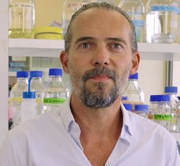 Nelson Dusetti合作论文数Cancrer Research Center of Marseille9
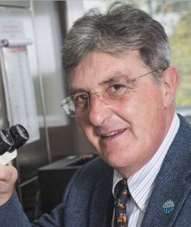 Aldo Scarpa合作论文数Pathological Anatomy Section, Department of Diagnostics and Public Health, Verona University9
 Martin Vingron合作论文数Department of Computational Molecular Biology, Max Planck Institute for Molecular Genetics9
 Marcel Scheideler合作论文数Institute for Genomics and Bioinformatics and Christian Doppler Laboratory for Genomics and Bioinformatics, Graz University of Technology9Boris Beckmann合作论文数9
 Eithne Costello合作论文数9Mark Hänle合作论文数8Klaus Kluck合作论文数8Dharini Srinivasan合作论文数8Rima Singh合作论文数8Thomas Engleitner合作论文数8
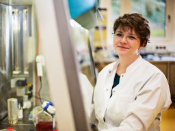 Anna-Lena Volckmar合作论文数Institut für Pathologie, Universitätsklinikum Heidelberg Heidelberg8
 Patrick C Hermann合作论文数Ulm Univ, Dept Internal Med 1, Ulm, Germany8
 Medhanie A. Mulaw合作论文数Clinical Cooperative Group “Leukemia, Helmholtz Zentrum Muenchen;Department of Medicine III, University of Munich-Grosshadern;Clinical Cooperative Group “Leukemia, University of Munich-Grosshadern8
 Ludwig Christina合作论文数8
 Konrad Steinestel合作论文数Institute of Pathology and Molecular Pathology, Bundeswehrkrankenhaus Ulm;Universität Ulm8
 Sparrer Konstantin M J合作论文数Max von Pettenkofer-Institute and Gene Center, Ludwig-Maximilians-University Munich8
 Evgeny A Moskalev合作论文数Friedrich Alexander Univ Erlangen Nurnberg, Univ Hosp Erlangen8
 Alexander Kleger合作论文数Universität Ulm, Univ. Ulm8Perkhofer Lukas合作论文数8Abaei Alireza合作论文数8
 Christopher Halbrook合作论文数Department of Cancer Biology, Mayo Clinic Florida;Department of Chemistry, Stony Brook University;Department of Cancer Biology, Stony Brook University8Albrecht Stenzinger合作论文数Institute of Pathology, University Hospital8
 Hans Lehrach合作论文数Max Planck Institute for Molecular Genetics8
 Roland Rad合作论文数School of Medicine and Health, Technical University of Munich;Institut für Molekulare Onkologie und Funktionelle Genomik, Klinikum Rechts der Isar, Technische Universität München8




