Clinical AI assumes that the influence of training data can persist indefinitely. This premise fails when patients withdraw consent, evidence evolves, or bias is identified. Machine unlearning aims to remove specific data influence without full retraining. We argue that unlearning readiness should be built into the infrastructure of high-risk healthcare AI across patient autonomy, clinical validity, and system governance, and we outline a governance pathway to keep updates auditable and clinically safe.
This pilot study characterizes the spectrum of atherosclerotic coronary artery pathologies in a porcine model of streptozotocin-induced diabetes, with a particular focus on the spontaneous progression of atherosclerotic plaques. Four pigs (designated P01 - P04) were subjected to an identical streptozotocin regimen to induce diabetes and studied using blood biochemistry, lipid profiling, coronary angiography, optical coherence tomography, and histological analysis of the coronary arteries to assess for the development of coronary atherosclerosis. All animals developed sustained hyperglycemia and dyslipidemia, with total cholesterol levels ranging from 14.9 to 36.7 mmol/L and low-density lipoprotein concentrations reaching up to 32 mmol/L. Notably, the study captured the full pathological continuum: from nearly no coronary abnormality in P01 and early atheromatous plaque formation in P02, to advanced atherosclerosis in P03, and finally, in P04, severe fibrofatty atheroma, and myocardial infarction. Unlike conventional animal myocardial infarction models that rely on interventional triggers, the infarction in P04 occurred spontaneously, demonstrating the spontaneous cascade of plaque development and acute plaque rupture with thrombosis. These findings demonstrate the feasibility of using streptozotocin-treated pigs to model key stages of diabetic coronary artery disease.
Medical vision-and-language models (MVLMs) have attracted substantial interest due to their capability to offer a natural language interface for interpreting complex medical data. Their applications are versatile and have the potential to improve diagnostic accuracy and decision-making for individual patients while also contributing to enhanced public health monitoring, disease surveillance, and policy-making through more efficient analysis of large data sets. MVLMS integrate natural language processing with medical images to enable a more comprehensive and contextual understanding of medical images alongside their corresponding textual information. Unlike general vision-and-language models trained on diverse, non-specialized datasets, MVLMs are purpose-built for the medical domain, automatically extracting and interpreting critical information from medical images and textual reports to support clinical decision-making. Popular clinical applications of MVLMs include automated medical report generation, medical visual question answering, medical multimodal segmentation, diagnosis and prognosis and medical image-text retrieval. Here, we provide a comprehensive overview of MVLMs and the various medical tasks to which they have been applied. We conduct a detailed analysis of various vision-and-language model architectures, focusing on their distinct strategies for cross-modal integration/exploitation of medical visual and textual features. We also examine the datasets used for these tasks and compare the performance of different models based on standardized evaluation metrics. Furthermore, we highlight potential challenges and summarize future research trends and directions. The full collection of papers and codes is available at: https://github.com/YtongXie/Medical-Vision-and-Language-Tasks-and-Methodologies-A-Survey.
Medical Large Multi-modal Models (LMMs) have demonstrated remarkable capabilities in medical data interpretation. However, these models frequently generate hallucinations contradicting source evidence, particularly due to inadequate localization reasoning. This work reveals a critical limitation in current medical LMMs: instead of analyzing relevant pathological regions, they often rely on linguistic patterns or attend to irrelevant image areas when responding to disease-related queries. To address this, we introduce HEAL-MedVQA (Hallucination Evaluation via Localization MedVQA), a comprehensive benchmark designed to evaluate LMMs' localization abilities and hallucination robustness. HEAL-MedVQA features (i) two innovative evaluation protocols to assess visual and textual shortcut learning, and (ii) a dataset of 67K VQA pairs, with doctor-annotated anatomical segmentation masks for pathological regions. To improve visual reasoning, we propose the Localize-before-Answer (LobA) framework, which trains LMMs to localize target regions of interest and self-prompt to emphasize segmented pathological areas, generating grounded and reliable answers. Experimental results demonstrate that our approach significantly outperforms state-of-the-art biomedical LMMs on the challenging HEAL-MedVQA benchmark, advancing robustness in medical VQA.
This pilot study characterizes the spectrum of atherosclerotic coronary artery pathologies in a porcine model of streptozotocin-induced diabetes, with a particular focus on the natural progression of atherosclerotic plaques. Four pigs (designated P01 - P04) were subjected to an identical streptozotocin regimen to induce diabetes and studied using blood biochemistry, lipid profiling, coronary angiography, optical coherence tomography, and histological analysis of the coronary arteries to assess for the development of coronary atherosclerosis. All animals developed sustained hyperglycemia and dyslipidemia, with total cholesterol levels ranging from 14.9 to 36.7 mmol/L and low-density lipoprotein concentrations reaching up to 32 mmol/L. Notably, the study captured the full pathological continuum: from nearly no coronary abnormality in P01 and early atheromatous plaque formation in P02, to advanced atherosclerosis in P03, and finally, in P04, severe fibrofatty atheroma, and myocardial infarction. Unlike conventional animal myocardial infarction models that rely on interventional triggers, the infarction in P04 occurred spontaneously, demonstrating the natural cascade of plaque development and acute plaque rupture with thrombosis. These findings demonstrate the feasibility of using streptozotocin-treated pigs to model key stages of diabetic coronary artery disease.
The ability to interpret and intervene model decisions is important for the adoption of computer-aided diagnosis methods in clinical workflows. Recent concept-based methods link the model predictions with interpretable concepts and modify their activation scores to interact with the model. However, these concepts are at the image level, which hinders the model from pinpointing the exact patches the concepts are activated. Alternatively, prototype-based methods learn representations from training image patches and compare these with test image patches, using the similarity scores for final class prediction. However, interpreting the underlying concepts of these patches can be challenging and often necessitates post-hoc guesswork. To address this issue, this paper introduces the novel Concept-based Similarity Reasoning network (CSR), which offers (i) patch-level prototype with intrinsic concept interpretation, and (ii) spatial interactivity. First, the proposed CSR provides localized explanation by grounding prototypes of each concept on image regions. Second, our model introduces novel spatial-level interaction, allowing doctors to engage directly with specific image areas, making it an intuitive and transparent tool for medical imaging. CSR improves upon prior state-of-the-art interpretable methods by up to 4.5% across three biomedical datasets. Our code is released at https://github.com/tadeephuy/InteractCSR.
Accurate medical image segmentation is challenging due to the high variance of out-of-distribution (OOD) data, which is costly for acquisition and annotation. However, existing methods often focus on the sub-scenarios of the OOD problem (e.g., Domain generalization) and approach this via a pretraining and fine-tuning domain adaptation paradigm, which does not explicitly utilize the intrinsic semantic relationship among those OOD tasks. To address this problem, we introduce a novel Attribute-Based Segmentation (CA-Seg) method to unify the OOD problems, where only the OOD object class semantic information is required to bridge the domain gap. CA-Seg contains two stages of learning: high-level semantic abstraction learning and low-level visual pattern learning. In the first stage of the training phase, we extract morphological knowledge of the tissue/organ of interest using an off-the-shelf Vision-Language Model (VLM), leveraging its rich language and image pattern association ability to describe class semantics into a set of human-understandable text attributes. Subsequently, the concepts serve as a condition to guide the low-level visual pattern learning by using the flow-matching or dice loss. During the adaptation phase, CA-Seg only requires the OOD class labels to rebind the attributes and the target object class to achieve good segmentation performance. CA-Seg addresses the limitations of data scarcity in the broad OOD problems and is computationally efficient in adapting to the OOD data, making it ideal in resource-constrained settings. We evaluated CA-Seg on 3 common OOD tasks in medical image segmentation, demonstrating a high cross-domain segmentation performance with limited data availability. Our code is available at https://github.com/iClaude1998/CA-seg.
Vision-language models have demonstrated remarkable success in general medical image analysis, yet their application in pediatric imaging remains significantly underexplored. These models show limited performance on pediatric datasets, primarily due to domain gaps stemming from anatomical differences, lower radiation doses, and pediatric-specific diseases. To this end, we present the first pediatric vision-language pre-training framework, dubbed PedCLIP, trained on a comprehensive pediatric imaging dataset comprising 404,670 X-rays of pediatric patients across diverse anatomical regions. To address anatomical diversity, we introduce a Mixture of Body part Experts design, with each expert sp ecializing in learning features from distinct anatomical regions. Experimental evaluation across eleven downstream tasks demonstrates that our model significantly outperforms current stateof-the-art vision-language models, achieving superior diagnostic accuracy in challenging pediatric conditions, including rare diseases such as pediatric inflammatory arthritis. Code is available: https://github.com/ tadeephuy/PedCLIP
Visual grounding (VG) is the capability to identify the specific regions in an image associated with a particular text description. In medical imaging, VG enhances interpretability by highlighting relevant pathological features corresponding to textual descriptions, improving model transparency and trustworthiness for wider adoption of deep learning models in clinical practice. Current models struggle to associate textual descriptions with disease regions due to inefficient attention mechanisms and a lack of fine-grained token representations. In this paper, we empirically demonstrate two key observations. First, current VLMs assign high norms to background tokens, diverting the model's attention from regions of disease. Second, the global tokens used for cross-modal learning are not representative of local disease tokens. This hampers identifying correlations between the text and disease tokens. To address this, we introduce simple, yet effective Disease-Aware Prompting (DAP) process, which uses the explainability map of a VLM to identify the appropriate image features. This simple strategy amplifies disease-relevant regions while suppressing background interference. Without any additional pixel-level annotations, DAP improves visual grounding accuracy by 20.74
A fundamental challenge in endoscopy is how to fabricate a small fiber-optic probe that can achieve comparable function to devices with large, complicated optics. To achieve high resolution over an extended depth of focus (DOF), the application of needle-like beams has been proposed. However, existing methods for miniaturized needle-beam designs fail to adequately correct astigmatism and other monochromatic aberrations, limiting the resolution of at least one axis. Here, we describe an approach to realize freeform beam-shaping endoscopic probes via two-photon polymerization three-dimensional (3D) printing. We present a design achieving <8 mu m lateral resolution with a DOF of similar to 800 mu m. The probe has a diameter of <260 mu m (without the torque coil and catheters) and is fabricated using a single printing step directly on the optical fiber. The probe was successfully utilized for intravascular imaging in living diabetic swine at multiple time points, as well as human atherosclerotic plaques ex vivo. To the best of our knowledge, this is the first report of a 3D-printed micro-optic for in vivo imaging of the coronary arteries. These results are a substantial step to enable the clinical adoption of both 3D-printed micro-optics and beam-tailoring devices.
BACKGROUND:Clinical work-up for suspected cardiac chest pain is resource intensive. Despite expectations, high-sensitivity cardiac troponin assays have not made decision making easier. The impact of recently validated rapid triage protocols including the 0-hour/1-hour hs-cTn protocols on care and outcomes may be limited by the heterogeneity in interpretation of troponin profiles by clinicians. We have developed machine learning (ML) models which digitally phenotype myocardial injury and infarction with a high predictive performance and provide accurate risk assessment among patients presenting to EDs with suspected cardiac symptoms. The use of these models may support clinical decision-making and allow the synthesis of an evidence base particularly in non-T1MI patients however prospective validation is required. OBJECTIVE:We propose that integrating validated real-time artificial intelligence (AI) methods into clinical care may better support clinical decision-making and establish the foundation for a self-learning health system. DESIGN:This prospective, multicenter, open-label, cluster-randomized clinical trial within blinded endpoint adjudication across 12 hospitals (n = 20,000) will randomize sites to the clinical decision-support tool or continue current standard of care. The clinical decision support tool will utilize ML models to provide objective patient-specific diagnostic probabilities (ie, likelihood for Type 1 myocardial infarction [MI] versus Type 2 MI/Acute Myocardial Injury versus Chronic Myocardial Injury etc.) and prognostic assessments. The primary outcome is the composite of cardiovascular mortality, new or recurrent MI and unplanned hospital re-admission at 12 months post index presentation. SUMMARY:Supporting clinicians with a decision support tool that utilizes AI has the potential to provide better diagnostic and prognostic assessment thereby improving clinical efficiency and establish a self-learning health system continually improving risk assessment, quality and safety. TRIAL REGISTRATION:ANZCTR, Registration Number: ACTRN12620001319965, https://www.anzctr.org.au/.
We conducted the first comprehensive association analysis of a coronary artery disease (CAD) cohort within the recently released UK Biobank (UKB) whole genome sequencing dataset. We employed fine mapping tool PolyFun and pinpoint rs10757274 as the most likely causal SNV within the 9p21.3 CAD risk locus. Notably, we show that machine-learning (ML) approaches, REGENIE and VariantSpark, exhibited greater sensitivity compared to traditional single-SNV logistic regression, uncovering rs28451064 a known risk locus in 21q22.11. Our findings underscore the utility of leveraging advanced computational techniques and cloud-based resources for mega-biobank analyses. Aligning with the paradigm shift of bringing compute to data, we demonstrate a 44% cost reduction and 94% speedup through compute architecture optimisation on UK Biobank’s Research Analysis Platform using our RAPpoet approach. We discuss three considerations for researchers implementing novel workflows for datasets hosted on cloud-platforms, to pave the way for harnessing mega-biobank-sized data through scalable, cost-effective cloud computing solutions.
The scarcity of annotated Magnetic Resonance Imaging (MRI) tumor data presents a major obstacle to accurate and automated tumor segmentation. While existing data synthesis methods offer promising solutions, they often suffer from key limitations: manual modeling is labor intensive and requires expert knowledge. Deep generative models may be used to augment data and annotation, but they typically demand large amounts of training pairs in the first place, which is impractical in data limited clinical settings. In this work, we propose Tumor Fabrication (TF), a novel two-stage framework for unpaired 3D brain tumor synthesis. The framework comprises a coarse tumor synthesis process followed by a refinement process powered by a generative model. TF is fully automated and leverages only healthy image scans along with a limited amount of real annotated data to synthesize large volumes of paired synthetic data for enriching downstream supervised segmentation training. We demonstrate that our synthetic image-label pairs used as data enrichment can significantly improve performance on downstream tumor segmentation tasks in low-data regimes, offering a scalable and reliable solution for medical image enrichment and addressing critical challenges in data scarcity for clinical AI applications.
Counterfactual explanations (CFE) for deep image classifiers aim to reveal how minimal input changes lead to different model decisions, providing critical insights for model interpretation and improvement. However, existing CFE methods often rely on additional image encoders and generative models to create plausible images, neglecting the classifier's own feature space and decision boundaries. As such, they do not explain the intrinsic feature space and decision boundaries learned by the classifier. To address this limitation, we propose Mirror-CFE, a novel method that generates faithful counterfactual explanations by operating directly in the classifier's feature space, treating decision boundaries as mirrors that “reflect” feature representations in the mirror. Mirror-CFE learns a mapping function from feature space to image space while preserving distance relationships, enabling smooth transitions between source images and their counterfactuals. Through extensive experiments on four image datasets, we demonstrate that Mirror-CFE achieves superior performance in validity while maintaining input resemblance compared to state-of-the-art explanation methods. Finally, mirror-CFE provides interpretable visualization of the classifier's decision process by generating step-wise transitions that reveal how features evolve as classification confidence changes.
BACKGROUND:With widespread adoption of high-sensitivity troponin assays, more individuals with myocardial injury are now identified, with type 1 myocardial infarction (T1MI) being less common despite having the most well-established evidence base to inform care. This study assesses the temporal time course of cardiovascular events among various forms of myocardial injury. METHOD:Consecutive hospital encounters were identified. Using the first episode of care during the sampling period, myocardial injury classifications (i.e., T1MI, acute injury/type 2 myocardial infarction [T2MI], chronic injury, and no injury) were established via two machine learning algorithms. The temporal time course of increased hazard for mortality, recurrent myocardial infarction, heart failure, and arrhythmia over 3 years were explored. RESULTS:There were 176,787 index episodes; 6.9% were classified as T1MI, 6.0% as acute injury/T2MI, and 26.7% as chronic injury. Although each classification was associated with an early increased risk of all-cause mortality compared with no injury (incidence rate ratio [IRR]<30 days: T1MI: 19.97 [95% confidence interval 12.50-32.69]; acute injury/T2MI: 26.51 [16.80-42.97]; chronic injury: 15.37 [10.22-23.95]), the instantaneous relative hazard for recurrent myocardial infarction was highest in those with initial T1MI (IRR<30 days: T1MI: 28.81 [22.75-36.76]; acute injury/T2MI: 10.23 [7.60-13.77]; chronic injury:5.54 [4.34-7.41]). In contrast, the instantaneous hazard for heart failure in those with initial acute injury/T2MI and chronic injury remained increased over long-term follow up unlike in T1MI (IRR1 3 yrs: T1MI: 5.52 [4.99-6.09]; acute injury/T2MI: 10.36 [9.51-11.30]; chronic injury:7.40 [6.90-7.94]). CONCLUSIONS:The substantial and persistent rate of late cardiac events highlights the need to establish an evidence base for the therapeutic management of "non-T1MI" diagnostic classifications and suggests opportunity to improve late outcomes using existing and emerging therapies.
Medical Large Multi-modal Models (LMMs) have demonstrated remarkable capabilities in medical data interpretation. However, these models frequently generate hallucinations contradicting source evidence, particularly due to inadequate localization reasoning. This work reveals a critical limitation in current medical LMMs: instead of analyzing relevant pathological regions, they often rely on linguistic patterns or attend to irrelevant image areas when responding to disease-related queries. To address this, we introduce HEAL-MedVQA (Hallucination Evaluation via Localization MedVQA), a comprehensive benchmark designed to evaluate LMMs' localization abilities and hallucination robustness. HEAL-MedVQA features (i) two innovative evaluation protocols to assess visual and textual shortcut learning, and (ii) a dataset of 67K VQA pairs, with doctor-annotated anatomical segmentation masks for pathological regions. To improve visual reasoning, we propose the Localize-before-Answer (LobA) framework, which trains LMMs to localize target regions of interest and self-prompt to emphasize segmented pathological areas, generating grounded and reliable answers. Experimental results demonstrate that our approach significantly outperforms state-of-the-art biomedical LMMs on the challenging HEAL-MedVQA benchmark, advancing robustness in medical VQA.
Echocardiography is the diagnostic workhorse in cardiology, but interpretation can be inconsistent. Machine learning (ML) and artificial intelligence (AI) are increasingly being used in echocardiography, effecting significant improvements in accuracy and reproducibility. This chapter reviews state-of-the-art ML applications including segmentation, diagnostic classification, and outcome prediction.
The integration of vision-language models such as CLIP and Concept Bottleneck Models (CBMs) offers a promising approach to explaining deep neural network (DNN) decisions using concepts understandable by humans, addressing the black-box concern of DNNs. While CLIP provides both explainability and zero-shot classification capability, its pre-training on generic image and text data may limit its classification accuracy and applicability to medical image diagnostic tasks, creating a transfer learning problem. To maintain explainability and address transfer learning needs, CBM methods commonly design post-processing modules after the bottleneck module. However, this way has been ineffective. This paper takes an unconventional approach by re-examining the CBM framework through the lens of its geometrical representation as a simple linear classification system. The analysis uncovers that post-CBM fine-tuning modules merely rescale and shift the classification outcome of the system, failing to fully leverage the system’s learning potential.We introduce an adaptive module strategically positioned between CLIP and CBM to bridge the gap between source and downstream domains. This simple yet effective approach enhances classification performance while preserving the explainability afforded by the framework. Our work offers a comprehensive solution that encompasses the entire process, from concept discovery to model training, providing a holistic recipe for leveraging the strengths of GPT, CLIP, and CBM. Code is available at: https://github.com/AIML-MED/AdaCBM .
 Zhibin Liao合作论文数School of Computer and Mathematical Sciences, Faculty of Sciences, Engineering and Technology, University of Adelaide;Australian Institute for Machine Learning, University of Adelaide35
 Jagat P Narula合作论文数Icahn School of Medicine, Mount Sinai18
 Gustavo Carneiro合作论文数Centre for Vision, Speech and Signal Processing, School of Computer Science and Electronic Engineering, University of Surrey17
 Anton Van Den Hengel合作论文数School of Computer Science, The University of Adelaide14
 Yutong Xie合作论文数V3ALab, University of Adelaide;Australian Institute for Machine Learning, Faculty of Sciences, Engineering and Technology, University of Adelaide12
 Leonard Hofstra合作论文数University Maastricht Cardiovascular Research Institute12
 Farouc Jaffer合作论文数Cardiology Division, Department of Medicine, Massachusetts General Hospital;Cardiovascular Research Center, Massachusetts General Hospital;Harvard Medical School11
 Peter Psaltis合作论文数Royal Adelaide Hospital;South Australian Health and Medical Research Institute;The Queen Elizabeth Hospital;Faculty of Health and Medical Sciences, Adelaide Medical School, University of Adelaide10
 Minh-Son To合作论文数9
 Stephen Nicholls合作论文数Monash University;Monash Victorian Heart Institute;Victorian Heart Hospital9
 Christina A. Bursill合作论文数University of Sydney Heart Research Institute9Chris P. Reutelingsperger合作论文数9Eric A. Osborn合作论文数9Rajvinder Singh合作论文数Gastroenterology & Hepatology, Lyell McEwin Hospital8Artiom Petrov合作论文数8
 Guansong Pang合作论文数School of Computing and Information Systems, Singapore Management University7
 Giovanni Ughi合作论文数Department of Radiology, T.H. Chan School of Medicine, University of Massachusetts;Gentuity, LLC;Spryte Medical7Slomka Piotr J合作论文数7
 Guillermo J. Tearney合作论文数Harvard Medical School, Harvard University;School of Dental Medicine, Harvard University7
 Robert A. Mclaughlin合作论文数School of Biomedicine, Faculty of Health and Medical Sciences, The University of Adelaide7
 Minh H. Phan合作论文数School of Electrical, Computer and Telecommunications Engineering, University of Wollongong7
 Marc Dweck合作论文数Centre for Cardiovascular Science, University of Edinburgh6
 Mohsen Dorraki合作论文数Electr. Eng. Dept., Amirkabir Univ. of Technol.;c;Electr. Eng. Dept., Amirkabir Univ. of Technol.6
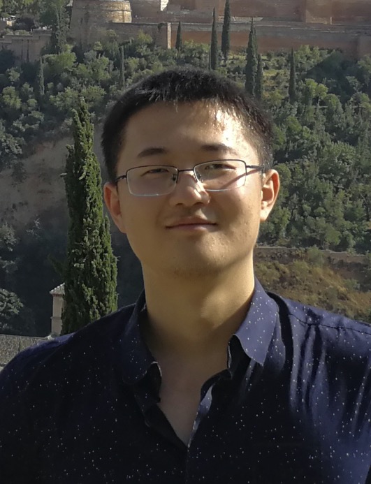 Jianpeng Zhang合作论文数Zhejiang University;Alibaba DAMO Academy6
 Simon Thiele合作论文数Printoptix GmbH6
 Harald Giessen合作论文数4th Physics Institute, University of Stuttgart6
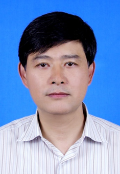 Jiawen Li (李家文)合作论文数Micro Nano Engineering Laboratory, Department of Precision Machinery and Precision Instrumentation, University of Science and Technology of China6
 Yong Xia (夏勇)合作论文数Northwestern Polytechnical University6
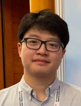 Yuanhong Chen合作论文数SonyAI6Dagfinn Lovhaug合作论文数6
 Gabriel Maicas合作论文数University of Adelaide5
 Yu Tian合作论文数University of Pennsylvania5
 Vannan Mani A合作论文数5
 Derek Chew合作论文数College of Medicine and Public Health, Flinders University;Monash Heart and Victorian Heart Institute5Rodney W Kirk合作论文数5
 Navneet Narula合作论文数5
 Chunhua Shen (沈春华)合作论文数College of Computer Science and Technology, Zhejiang University5
 Qi Chen合作论文数Australian Institute for Machine Learning, The University of Adelaide;Vision, Ask, Answer, Act Laboratory, The University of Adelaide5
 Yuyuan Liu (刘育源)合作论文数Department of Engineering Science, University of Oxford5Partho P. Sengupta合作论文数5
 Kewen Liao合作论文数School of Information Technology, Faculty of Science Engineering and Built Environment, Deakin University5J Narula合作论文数4
 Philip Adamson合作论文数4Leo Hofstra合作论文数4Susanne W.M. van den Borne合作论文数4
 Pavel Ruchka合作论文数Inst Tech Opt, Univ Stuttgart4
 John P. Greenwood合作论文数Baker Heart and Diabetes Institute;University of Melbourne;Monash University;University of Leeds;The Alfred Hospital4Nikant Sabharwal合作论文数4Jasper Prijs合作论文数4




