Supplementary figures S1-5, materials and methods. 1) Supplementary figures and associated legends: - Figure S1. ERK silencing decreases breast cancer cell migration and invasion but does not influence cell proliferation. - Figure S2. ERK2 does not interact with actin or keratin 8 and ERK2 phosphorylation is supported by vimentin but not actin. - Figure S3. Fluorescence recovery of wild-type Slug-GFP after photobleaching (FRAP). - Figure S4. Mass spectrometric identification of ERK1/2-dependent phosphorylation sites on Slug and validation using a newly generated Slug phospho-serine-87 antibody. - Figure S5. The phosphorylation status of Slug does not influence repression of E-Cadherin, Slug nuclear localisation or stability. 2) Supplementary materials and methods 3) Supplementary references
Reactivation of specific PP2A complexes is required for PME-1 depletion-mediated synthetic lethality. Supplementary figure related to Figure 3.
PME-1 depletion sensitizes GBM cells to multikinase inhibitor staurosporine. Supplementary figure related to Figure 1.
Clinically relevant kinase inhibitors display synthetic lethality in PME-1 depleted glioma cells. Supplementary figure related to Figure 2.
Abstract Epithelial–mesenchymal transition (EMT) in cells is a developmental process adopted during tumorigenesis that promotes metastatic capacity. In this study, we advance understanding of EMT control in cancer cells with the description of a novel vimentin–ERK axis that regulates the transcriptional activity of Slug (SNAI2). Vimentin, ERK, and Slug exhibited overlapping subcellular localization in clinical specimens of triple-negative breast carcinoma. RNAi-mediated ablation of these gene products inhibited cancer cell migration and cell invasion through a laminin-rich matrix. Biochemical analyses demonstrated direct interaction of vimentin and ERK, which promoted ERK activation and enhanced vimentin transcription. Consistent with its role as an intermediate filament, vimentin acted as a scaffold to recruit Slug to ERK and promote Slug phosphorylation at serine-87. Site-directed mutagenesis established a requirement for ERK-mediated Slug phosphorylation in EMT initiation. Together, these findings identified a pivotal step in controlling the ability of Slug to organize hallmarks of EMT. Cancer Res; 75(11); 2349–62. ©2015 AACR.
PME-1 and HDAC4 expression can predict glioma patient response to kinase inhibitors. Supplementary figure related to Figure 6.
List of PME-1 regulated PP2A B-subunits and their known interactor proteins.
Bioimaging has now entered the era of big data with faster-than-ever development of complex microscopy technologies leading to increasingly complex datasets. This enormous increase in data size and informational complexity within those datasets has brought with it several difficulties in terms of common and harmonized data handling, analysis, and management practices, which are currently hampering the full potential of image data being realized. Here, we outline a wide range of efforts and solutions currently being developed by the microscopy community to address these challenges on the path towards FAIR bioimaging data. We also highlight how different actors in the microscopy ecosystem are working together, creating synergies that develop new approaches, and how research infrastructures, such as Euro-BioImaging, are fostering these interactions to shape the field.
siRNA target sequences used in the study.
HDAC4 inhibition sensitize GBM cells to kinase inhibitors. Supplementary figure related to Figure 5.
Structure-activity relationship (SAR) analysis of STS derivatives to identify active synthetic lethality in PME-1 depleted cells.
We wanted to create a highly-qualified network of expertise, know-how, and cutting-edge technologies, and innovation platforms, by bringing together the most qualified imaging centers in Europe.We run the gamut from noninvasive imaging to invasive validation and from invasive discovery to noninvasive translation. We talk to each other, collaborate, understand each other's needs … This creates an atmosphere of openness and mutual consideration.Euro-BioImaging provides an avenue for scientists at early career stages to gain access to imaging technologies and expertise that will truly enhance their research. Euro-BioImaging ERIC (European Research Infrastructure Consortium) is a European, publicly funded, nonprofit research infrastructure (https://www.eurobioimaging.eu/). Our mission is to provide open access to biological and biomedical imaging technologies, training, and image data services to all researchers, regardless of research interest or affiliation, in both academia and industry. To achieve this, we bring together 137 of the most prestigious imaging facilities (status December 2021), employing 500+ imaging specialists, which make their services, technologies, and expertise available to our users. These facilities, grouped together in entities called Nodes, are based across 16 member states in Europe and the European Molecular BioIogy Laboratory (EMBL) and offer around 50 different imaging technologies, both for biological and biomedical imaging. Each facility has a highly unique profile, not only in imaging technology, but also in surrounding infrastructure and multidisciplinary research environment. All have demonstrated their scientific and technical excellence and commitment to user service in a stringent evaluation by the Scientific Advisory Board of Euro-BioImaging. In this Backstory, we explain the motivation behind forming an interdisciplinary research infrastructure like Euro-BioImaging, challenges that we faced along implementation, and explain how such an infrastructure can create opportunities to bring together communities in support of excellent research (Figure 1). Coordination of such a large, multisited infrastructure requires close collaboration of the international Hub with all the different sites. The Euro-BioImaging Hub consists of a Statutory Seat in Finland (Turku), a community-specific Bio-Hub for biological imaging at EMBL (Heidelberg), and a community-specific Med-Hub for biomedical imaging in Italy (Torino). When world-class research instruments are placed in centers operating with open access principle and highly skilled staff in place, the efficiency and productivity of instrument use, as well as the quality and reproducibility of the obtained data will all improve significantly. Instead of being mere local service providers, imaging core facilities residing in internationally recognized research centers are becoming pioneering and strategically-led powerhouses of science and provide key resources of collaborative and open research operating at the frontiers of science. Precisely for this reason we wanted to launch a Europe-wide research infrastructure dedicated to imaging. First, we wanted to create a highly-qualified network of expertise, know-how, and cutting-edge technologies, and innovation platforms, by bringing together the most qualified imaging centers in Europe. Second, we wanted to democratize access to the imaging resources – technologies, expertise, advanced training, and image data services - that are available at our Nodes. In that way, every researcher, both from academia and industry, can apply for Euro-BioImaging services whenever they have a project requiring imaging technologies and expertise, but do not have the equipment or the skills to perform the experiments at their home institute. By providing open access to imaging technologies, training, and data services, Euro-BioImaging enables European scientists across different disciplines to carry out cutting-edge research, allowing them to address key societal challenges including health and aging, agricultural and marine research, climate action, and environment and to boost all branches of economy related to life sciences, health, diagnostics, pharma, and biotechnology. With a world-class infrastructure based on open access, we foster excellent science and aim to drive recognition of the importance of imaging in the Life Sciences on an international level. The preparatory phase of Euro-BioImaging began in 2009. A major factor for success – and a major challenge at the start – was to build an international imaging community of core facility scientists representing 26 different national imaging communities, who by now know and trust each other and understand the benefits of working collaboratively. Euro-BioImaging played a vital role in bringing together these imaging scientists and in building coordinated, national imaging communities around Europe, such as the one in Sweden: "In parallel to the start of the Euro-BioImaging preparatory phase, the Swedish Bioimaging community was established, and thanks to the funding that we got from the Swedish Research Council in 2010, we implemented a coordinated network for biological and medical imaging. The network had around 150 members representing 11 Swedish universities and virtually all bioimaging facilities (7) that were nationally accessible in 2010," says Julia Fernandez-Rodriguez, Head of the Center for Cellular Imaging Core Facility of the University of Gothenburg, part of the Swedish National Microscopy Infrastructure (NMI), Euro-BioImaging's Swedish Node. "In 2014, the Swedish Research Council launched a specific open call for biological imaging infrastructures in Sweden to be included on the national roadmap (strategic areas of research investment). One important aspect that helped us to get Imaging on the national roadmap was the fact that our community was already coordinated nationally (Swedish BioImaging), with a clear organization for collaboration and user access, and was cooperating internationally with Euro-BioImaging." "Then came the challenge of refining our community even further. Between 2014 and 2015, the Swedish Research Council evaluated all the facilities that applied to form our national microscopy infrastructure. This phase was extremely competitive! We had to show how we were going to run the infrastructure, the operational plan, governance, implement user access, etc. In addition, we had to show that each facility was unique in terms of competence and instrumentation," says Julia. "Many different imaging facilities from different Swedish Universities applied to this call. Luckily, the evaluation was carried out by an independent international advisory panel with clear research infrastructure criteria. This was crucial to avoid conflicts within our national community. It would have been too challenging to make these hard decisions internally," reflects Julia. Today, five facilities – distributed in Stockholm, Uppsala, Gothenburg, and Umeå – are part of the National Microscopy Infrastructure. But the challenging and hard work of community building is paying off. "Today, being coordinated nationally and organized internationally as part of Euro-BioImaging, helps us to gain new funding and more exciting research collaborations. As an example, in 2020, NordForsk launched the Nordic Research Infrastructure Hubs initiative, and granted a consortium of national imaging infrastructures, four of which are also Euro-BioImaging Nodes, the Bridging Nordic Microscopy Infrastructures (BNMI) grant (220,000 Euros). With this application, we aim to create a strong and well-interlinked network between the different microscopy infrastructures in the Nordic countries: Denmark, Finland, Iceland, Norway, and Sweden. The funding is already contributing to strengthen international competitiveness and facilitating the development of world-leading Nordic microscopy environments, by organizing scientific and technical Symposia, workshops and knowledge-exchange seminars, shadowing programs for facility staff, and short-term scientific mobility grants for researchers and increase the training and innovation activities among the participant Nordic countries. Being connected internationally via Euro-BioImaging is extremely important when applying for this type of grant," concludes Julia Rodriguez-Fernandez. Our technology portfolio is extensive, with Euro-BioImaging Nodes offering around 50 different biological and biomedical imaging technologies (Figure 2). Within our research infrastructure, we bring together biological and biomedical imaging communities who are determined to work together, sometimes on a European-level, sometimes nationally, sometimes even within a Node. Nodes that offer both biological and biomedical imaging technologies are called "Mixed" Nodes - and Marc van Zandvoort, Maastricht University, is coordinator of the Advanced Microscopy and Multimodal Imaging (AMMI) Node – Maastricht, one of the first "Mixed" Nodes to join Euro-BioImaging. "As a multimodal Node, we connect state-of-the-art (light and electron) microscopy with high-end, innovative molecular and noninvasive imaging technologies, such as MassSpec Imaging and PET," explains Marc. "This is really interesting from a scientific standpoint because we can really go from bench to bedside and back again. The AMMI Node – Maastricht is organized to work together across various imaging modalities. We run the gamut from noninvasive imaging to invasive validation and from invasive discovery to noninvasive translation. We talk to each other, collaborate, and understand each other's needs … This creates an atmosphere of openness and mutual consideration. Of course, multimodal projects can be demanding – it is a challenge to train users from a variety of different backgrounds, and data management and analysis in these projects are particularly complex. We work with various resolutions, scales, and various types of information - and they all have to be linked. Despite the challenge, using multiple modalities provides a true added-value. In fact, applying different techniques to the same samples, tissues, or regions of interest both in vitro and intravitally, provides complementary information and allows a deeper understanding of the processes underlying, for example, the onset, development, and response to treatments of diseases," says Marc. "Ironically, the real challenge for us is to find users who want to do these complex, interdisciplinary projects. If the user comes for example from optical microscopy, it is really hard to convince them to leave their comfort zone and use a new technique that they know nothing about," regrets Marc. "That is why we are so heavily involved in translational and interdisciplinary teaching programs, including a newly accredited Master in Imaging within the University of Maastricht (with an accent on multimodal techniques) and various PhD and Postdoc courses, like those on "Advanced Optical Microscopy" and "Artificial Intelligence." But the biggest success story so far involves a newly-awarded PhD project, within the Maastricht Imaging Valley. "We have one PhD student who will work on a comorbidity model – involving oncology and atherosclerosis. First of all, this PhD project bridges four different Maastricht schools (CARIM, GROW, NUTRIM, and MHeNS), specialized in different disease topics, and is supported by the Faculty of Health, Medicine, and Life sciences. Consequently, the student will have four tutors – one specialized in cardiovascular research, one specialized in neurosciences, one specialized in oncology, and one in optical imaging. Secondly, what makes this project so interesting and challenging is that it involves imaging at all scales - from super resolution microscopy to noninvasive techniques (like MRI, PET) – and even involves human behavior. Two of the Node leaders from the AMMI Node are involved in this project." Imaging is a crucial part of research projects in many different disciplines and along the whole research trajectory from basic to translational research. Therefore, Euro-BioImaging can bring together researchers from different backgrounds and at different levels of imaging expertise in a single project. Today's scientists work in a complex world where multidisciplinary/cross-disciplinary studies based on new technologies are often required. Increasing cooperation between institutions and disciplines is expected and necessary. Euro-BioImaging is embedded in the landscape of European research infrastructures (RIs) (https://lifescience-ri.eu/home.html) and maintains strong links with other RIs in the life sciences and beyond. Through EU-funded projects, Euro-BioImaging provides open-access biomedical and biological imaging technologies while collaborating with other European Research Infrastructures across disciplines to widen the array of services available at scientists' fingertips. Euro-BioImaging user, Roberta Ranieri, provides a compelling example of this interdisciplinarity. Roberta Ranieri was a PhD student at the University of Perugia, Italy, in the lab of Professor Martelli, where she was able to utilize her unique scientific background in cancer biology investigation and drug development methods to study a specific form of acute myeloid leukemia (AML). She was applying her previous laboratory experience, skills, and know-how to screen thousands of drugs and compounds in a search for new effective treatments. This complex, interdisciplinary research was aided by the Horizon 2020-funded CORBEL project that connected Professor Martelli and Roberta with two European Life Science Research Infrastructures, namely EU-OPENSCREEN and Euro-BioImaging. Her project was one of almost 40 user projects that were selected and granted access, funded by CORBEL, to at least two RIs of their choice. In the CORBEL project, Euro-BioImaging was the most requested research infrastructure, underlining the importance of imaging technologies to cross-disciplinary research in the life sciences. "The CORBEL experience, within a larger ERC-funded project awarded to Professor Martelli, of the University of Perugia, will hopefully be a step toward novel therapeutic options to treat AML, which are desperately needed. The collaborative spirit of CORBEL and its interdisciplinary approach clearly made our research more significant and will have a positive impact on human health," explains Roberta Ranieri. Between 2022 and 2025, Euro-BioImaging will collaborate with the other European life science research infrastructures in the framework of the Horizon Europe-funded emergency project, ISIDORe, to enable access to imaging and other much needed services for researchers urgently working on identifying and understanding COVID-19 variants as well as increasing Europe's preparedness for tackling infectious diseases (Figure 3). With the advances in imaging technology, more and more new technologies are available to users, making training in the correct use of the technologies and the connected sample preparation and data analysis crucial. The Euro-BioImaging Nodes offer a wide range of training opportunities that are targeted at users, students, and facility staff. Training courses at our Nodes cover a wide range of topics from basic introductory courses to advanced technologies in both biological and biomedical imaging. These courses, mostly combining theory and hands-on learning, are taught in English, and are open for anyone to apply to. A list of upcoming training courses is available on our website (https://www.eurobioimaging.eu/content/training). During the pandemic, many of the Euro-BioImaging Nodes developed innovative approaches to allow continued training to take place remotely. Training on the theoretical aspects of microscopy and those for image data analysis could be quite easily transferred to a remote setting, although this presents a challenge for the majority of courses that include strong practical components. Several Nodes now offer training that includes remote control of imaging instruments via remote desktop solutions, allowing the students to gain experience in controlling all parameters of the instrument, whereas the Node staff on site provides support with sample handling. At the same time, Nodes are running virtual courses with multi-camera setups to allow the students to see the details of practical steps, for example of sample preparation, from multiple angles and close up. Several of the Euro-BioImaging Nodes participate in MSc degree programmes. In addition, companies on our Euro-BioImaging Industry Board (https://www.eurobioimaging-industryboard.com/) offer internships to MSc students to gain insight into highly qualified job profiles in industry to help them make an informed choice about their future career. In addition, Euro-BioImaging projects are a great way for students to gather data and build their skill sets, as in the case of Euro-BioImaging user and study abroad student Marcos Gonzalez Lopez. In 2020, Marcos González López was a Master's student in "Cell and Gene therapies" from Spain, who became a Euro-BioImaging user at the CELLIM facility, part of our Advanced Light Microscopy and Medical Imaging Node Brno CZ, while pursuing his Master's degree research. Passionate about stem cells and genetic engineering, Marcos was looking forward to his second year of studies with more hands-on learning, and therefore he started a cooperation with Jan Křivánek's lab in the Department of Embryology and Histology at Masaryk University in Brno to pursue his Master's thesis. A scholarship from the Czech Ministry of Education, Youth, and Sports (MŠMT) helped to convince Marcos to take advantage of this opportunity to study abroad – despite the COVID-19 situation. The closeness, proximity of the university with the CELLIM core facility made the study-abroad opportunity even more attractive. Marcos could apply for access to the facility as a Euro-BioImaging user, and with support from the experts at the Node, learn how to use different imaging techniques, such as confocal microscopy, and gain experience on image data processing and further analysis. "Being able to work in this facility alongside other experts has been an extremely rich experience for me – even against the backdrop of COVID-19," says Marcos Gonzalez Lopez. "The state-of-the-art equipment available at the CELLIM imaging facility is crucial to my work – and the competent and friendly staff have been particularly supportive of his project." Stories like this are compelling examples of how Euro-BioImaging provides an avenue for scientists at early career stages to gain access to imaging technologies and expertise that will truly enhance their research – and perhaps define their future career path. Today, Marcos is a PhD student in the Biomedical Sciences Program at Masaryk University, specializing in Cell and Tissue Morphology. The Euro-BioImaging Hub team organizes a number of topic-focused Expert Groups which are open to all staff from our Nodes. "Our Expert Groups serve as important platforms to bring together the community around topics of particular interest, such as quality and data management, remote access, and communication," says Alessandra Viale, Scientific Project Manager at Euro-BioImaging Med-Hub. "The work of the expert groups paves the way toward higher quality of offered services by sharing best practices and lays the groundwork for a more homogeneous landscape for users accessing different Nodes." The expert groups with their regular meetings are also a great way for facility staff all across Europe to get to know each other and build connections that support building of expertise and interdisciplinary collaborations. "In addition to our topic-focused expert groups, we also have 3 technology expert groups – for medical imaging, light microscopy and electron microscopy." says Johanna Bischof, Scientific Project Manager at Euro-BioImaging Bio-Hub. "The technology expert groups offer a great avenue for the experts at the Euro-BioImaging Nodes across Europe to come together and exchange about their areas of expertise and keep up to date on the latest technology developments." In addition, these groups provide a good opportunity to exchange and learn from industry partners, as Claudia Pfander, Euro-BioImaging Industry Board Coordinator, explains, "Imaging companies actively contribute by supporting and providing advanced training on technology to Node staff or by sharing knowledge and best practices in one of the Expert Groups. For example, they participate in our Remote Access group, aiming to support facilities to offer imaging services remotely, which has been crucial during the pandemic, but also offers opportunities for a more democratic and ecological access to imaging in the future. Active engagement with the community helps companies to better identify areas where their customers need more support or even new developments and products to address their research questions." As we look to the future, at Euro-BioImaging we always aim to expand our user base by cooperating closely with universities and research institutions across Europe, industry, European Life Science Research Infrastructures, and the Euro-BioImaging family. One of the challenges is to sustainably fund user access to our infrastructure and truly democratize imaging, i.e., make the cutting-edge technologies and know-how available to a user regardless of their background. In response to this challenge, Euro-BioImaging participated in several Horizon Europe calls in 2021. ISIDORe, part of the HERA project for pandemic preparedness funded by Horizon Europe, was awarded in July 2021, and with this project, Euro-BioImaging will be able to provide funding for scientists undertaking infectious disease research at a Euro-BioImaging Node. In 2020–2021, the first years of operations for Euro-BioImaging, despite the global pandemic and lockdowns in Europe we saw some flavor of the breadth and capabilities of the Nodes and saw how imaging technologies have been innovatively used to answer timely research questions, including COVID, cancer, cardiovascular research, neurology, and also marine and plant biology. Still, we are quite convinced that the really significant advances of Euro-Bioimaging will be seen in the coming years. In future, we hope our infrastructure will continue to expand to benefit our users, helping them make discoveries that provide better understanding of the basis of our world as well as scientific solutions to its grand challenges.
The vast majority of cervical and 75% of oropharyngeal carcinomas are triggered by infection with a type of high-risk oncogenic human papillomavirus (HPV). It is well-known that E6 and E7 oncoproteins are critical for viral-induced cancer, and hence, they represent valuable targets for therapeutic intervention in HPV-mediated cancers. Our earlier research on the cembranoid, anisomelic acid (AA) showed that, AA has the potential to induce apoptosis in HPV cells by the depletion of E6 and E7 oncoproteins. The present study describes the structure-activity relationship and the evaluation of synthetic AA like compounds, i.e simplified cembranoid-like structures, as HPV inhibitors against some papilloma cell lines. Both from experimental and computational results, we observed that these compounds induced apoptosis by the same E6/E7-based mechanism as AA, but at earlier time points, thus being far more effective than AA. Further, the data indicated that only part of the structure of AA is required for the molecular action. Based on these results, we identified some novel and potential compounds for specific treatment of HPV-associated carcinomas.
With increased co-firing of coal and different biomasses in power plants, there is a growing need to study and characterize the produced, often heterogeneous ashes and to assess their utilization as secondary raw materials. In this work, we compare different methods for characterizing the ash compositions. These methods include scanning electron microscopy-energy dispersive X-ray analysis (SEM-EDS), X-ray fluorescence (XRF) and inductively coupled plasma emission spectroscopy (ICP-OES). The content of carbonaceous residues in the ashes was measured with CHNS analyzer. The methods and the pretreatment required for each of the analysis were studied in detail for three ashes: (1) coal, (2) peat and wood, (3) peat and wood residue with dolomite addition. The limitations of each method will be discussed. This work gives guidelines for selecting a rapid and reliable method for analyzing the suitability of the ash for a particular application.
The transcriptional network and protein regulators that govern T helper 17 (Th17) cell differentiation have been studied extensively using advanced genomic approaches. For a better understanding of these biological processes, we have moved a step forward, from gene- to protein-level characterization of Th17 cells. Mass spectrometry-based label-free quantitative (LFQ) proteomics analysis were made of in vitro differentiated murine Th17 and induced regulatory T (iTreg) cells. More than 4,000 proteins, covering almost all subcellular compartments, were detected. Quantitative comparison of the protein expression profiles resulted in the identification of proteins specifically expressed in the Th17 and iTreg cells. Importantly, our combined analysis of proteome and gene expression data revealed protein expression changes that were not associated with changes at the transcriptional level. Our dataset provides a valuable resource, with new insights into the proteomic characteristics of Th17 and iTreg cells, which may prove useful in developing treatment of autoimmune diseases and developing tumor immunotherapy.
Abstract Introduction: The bromodomain and extraterminal (BET) family of proteins are chromatin readers that recognize and bind to specific acetylated histones and promote the transcription of several important cell identity genes. BET bromodomain inhibitors have shown promising antitumor activity in a variety of pre-clinical cancer models, as BET inhibition abrogates the transcription of several key oncogenes in a cell type-specific manner. It is known that inhibition of BET proteins effectively inhibits the proliferation of estrogen receptor positive (ER+) breast cancer cells, at least in part through repression of ER and MYC signaling. However, many additional cancer-associated genes are likely to underlie the growth inhibitory effects of BET inhibitors in breast cancer. The purpose of this study was to determine the anticancer activity of the novel BET bromodomain inhibitor ODM-207 in pre-clinical ER+ breast cancer models, and further, to look for cancer-associated signaling pathways suppressed by BET inhibitors. Methodology and results: ODM-207 is a novel, highly selective BET bromodomain inhibitor structurally distinct from JQ1 and its benzodiazepine-related derivatives. In this study, we show that ODM-207 effectively inhibits the proliferation of ER+ breast cancer cell lines when measured by cell viability assays as well as suppresses the growth of patient-derived xenograft tumors. Furthermore, we wanted to investigate the anticancer signaling pathways regulated by ODM-207 as well as the prototypical BET inhibitor JQ1 in breast cancer cells. For this purpose, we performed RNA sequencing on two ER+ breast cancer cell lines after 24h treatment with the aforementioned BET inhibitors. We found that both BET inhibitors targeted several genes and pathways important for breast cancer progression. For example, the targets included CDK4 and CDK6, two cell cycle kinases fundamental for the development and treatment of ER+ breast cancer. The RNA sequencing results were further validated in vitro, and were utilized as a basis for combination therapy assessment. Conclusions: Our results indicate that the novel BET bromodomain inhibitor ODM-207, which is currently in Phase I clinical trials for treating solid tumors, causes significant growth inhibition and cell cycle arrest in pre-clinical models of ER+ breast cancer, and regulates multiple crucial signaling pathways involved in breast cancer cell cycle and survival. Citation Format: Julia Lindqvist, Mari Björkman, Reetta Riikonen, Daniel Nicorici, Elina Mattila, Chandrasekhar Abbineni, Mahaboobi Jaleel, John Eriksson, Pekka Kallio, Anu-Maarit Moilanen. Therapeutic targeting of estrogen receptor positive breast cancer with the BET bromodomain inhibitor ODM-207 [abstract]. In: Proceedings of the American Association for Cancer Research Annual Meeting 2018; 2018 Apr 14-18; Chicago, IL. Philadelphia (PA): AACR; Cancer Res 2018;78(13 Suppl):Abstract nr 3970.
Background: The bromodomain and extraterminal (BET) family of proteins are chromatin readers that promote the transcription of several important cell identity genes. BET inhibitors have shown promising antitumor activity in a variety of pre-clinical cancer models, as BET inhibition abrogates the transcription of several key oncogenes in a cell type-specific manner. Hence, the purpose of this study was to determine the anticancer activity of the novel BET inhibitor ODM-207 in ER+ breast cancer models and to look for cancer-associated signaling pathways suppressed by BET inhibitors. Methods: ER+ breast cancer cell lines were studied for sensitivity to ODM-207 and the in vivo efficacy was assessed using the ER+ Ma3366 patient-derived xenograft model. For gene expression analyses, breast cancer cells were treated with ODM-207 or reference BET inhibitor JQ1 and differentially expressed genes were analysed by RNA-sequencing. The ability of ODM-207 to regulate anticancer signaling pathways was validated by western blotting. Synergistic drug interactions were profiled using five-concentration dose response matrices. Results: ODM-207 is a novel BET inhibitor structurally distinct from JQ1 and its benzodiazepine-related derivatives. In this study, we show that ODM-207 effectively inhibits the proliferation of ER+ breast cancer cell lines as well as suppresses the growth of patient-derived xenograft tumors. Furthermore, ODM-207 and the JQ1 targeted several pathways important for cancer progression such as the DNA damage and repair pathways. Conclusions: Our results indicate that ODM-207, which is currently in Phase I clinical trials for treating solid tumors, causes significant growth inhibition in pre-clinical models of ER+ breast cancer, and regulates signaling pathways involved in breast cancer cell survival. Legal entity responsible for the study: Orion Corporation, Orion Pharma. Funding: Orion Corporation, Orion Pharma. Disclosure: A. Moilanen, M. Björkman, R. Riikonen, D. Nicorici, E. Mattila, P. Kallio: Employee: Orion Corporation, Orion Pharma. J. Lindqvist: Employee: Orion Corporation, Orion Pharma, Åbo Akademi University. C. Abbineni, M. Jaleel: Employee: Aurigene Discovery Technologies Limited. J. Eriksson: Employee: Åbo Akademi.
Abstract Quantitative 3D imaging of live single tumor cells enables direct insights into the intricacies of cellular machinery while offering new ways of assessing intercellular variability. Since its invention, X-ray computed tomography has been indispensable in the clinic for cancer diagnostic and prognostic purposes due to its quantitative manner of imaging owing to the isotropic 3D spatial resolution. Although its principles have recently begun to be implemented in the optical spectral range for single cell imaging, the applications have mainly been directed towards imaging fixed cells in absorption mode for studying an important hallmark of cancer - nuclear architecture. We present an approach that utilizes the concept of computed tomography for quantitative functional 3D imaging of live single cells. The method offers truly isotropic 3D spatial resolution and enables imaging of natural suspension cells, such as non-solid tumor or immune system cells, as opposed to cells attached to substrates, as is common to the majority of other imaging approaches. We report on technical characteristics of the method as well as experimental findings of a nuclear and mitochondrial dynamics study in human myelogenous leukemia and mouse macrophage cells as a validation. The absolute quantification capability of the method makes it a powerful tool in the field of oncology by enabling direct studies of cellular and nuclear architecture dynamics in the context of tumorigenesis and progression. Citation Format: Laimonas Kelbauskas, Rishabh M. Shetty, Bin Cao, Kuo-Chen Wang, Dean Smith, Hong Wang, Shih-Hui Chao, Brian Ashcroft, Margaret Kritzer, Honor Glenn, Erik Niemela, Roger H. Johnson, John Eriksson, Deirdre R. Meldrum. Computed tomography for quantitative imaging of live cancer cells with isotropic 3D spatial resolution. [abstract]. In: Proceedings of the AACR Special Conference on Engineering and Physical Sciences in Oncology; 2016 Jun 25-28; Boston, MA. Philadelphia (PA): AACR; Cancer Res 2017;77(2 Suppl):Abstract nr B15.
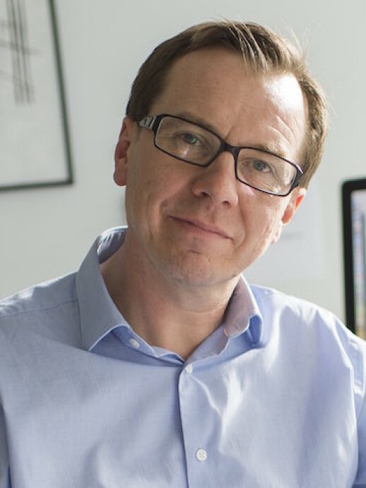 Jukka Westermarck合作论文数Institute of Biomedicine, University of Turku12
 Hannu Haapasalo合作论文数Department of Pathology, Tampere University11
 Pirjo Laakkonen合作论文数11Emilia Peuhu合作论文数University of Turku, Åbo Akademi University;Department of Biosciences, Åbo Akademi University11Mikael Jumppanen合作论文数University of Turku10
 Ahmed Shafiq U合作论文数Institute of Cancer Sciences, University of Glasgow10
 Oxana V. Denisova合作论文数Institute for Molecular Medicine Finland, University of Helsinki10
 Amanpreet Kaur合作论文数University of Turku, Åbo Akademi University;Department of Pathology, University of Turku;University of Turku, Åbo Akademi University;Åbo Akademi University, Åbo Akademi University10
 Xi Qiao合作论文数10Olayinka Raheem合作论文数10
 Anthony Chalmers合作论文数School of Cancer Sciences, University of Glasgow;Institute of Cancer Sciences, University of Glasgow10
 Fang Cheng (程芳)合作论文数School of Pharmaceutical Sciences (Shenzhen), Sun Yat-sen University5de Thonel Aurélie合作论文数4
 Cecilia Sahlgren合作论文数Department of Biomedical Engineering, Eindhoven University of Technology;Department of Natural and Health Sciences, Faculty of Science and Engineering, Åbo Akademi University;Turku Centre for Biotechnology4James B Lorens合作论文数3De Franceschi Nicola合作论文数Department of Biology, University of Padova;Centre for Biotechnology, University of Turku;University of Padova, University of Turku3Kaukonen Riina合作论文数3Mai Anja合作论文数3Torvaldson Elin合作论文数3Saari Markku合作论文数3Virtakoivu Reetta合作论文数3
 Ylermi Soini合作论文数Department of Pathology, University of Oulu and Oulu University Hospital3
 Veli-Matti Kosma合作论文数Institute of Clinical Medicine, School of Medicine, Faculty of Health Sciences, University of Eastern Finland;Cancer Center of Eastern Finland, University of Eastern Finland3
 Garry L Corthals合作论文数Turku Ctr Biotechnol, Univ Turku3
 Arto Mannermaa合作论文数Institute of Clinical Medicine, School of Medicine, Faculty of Health Sciences, University of Eastern Finland3
 Ghaffar Muharram合作论文数3
 Diana M. Toivola合作论文数Department of Medicine;VA Medical Center;Department of Medicine, VA Medical Center3Christine Gilles合作论文数University of Liege Laboratory of Tumor and Developmental Biology, CHU-Sart-Tilman Liege3
 Susumu Y. Imanishi合作论文数Graduate School of Environmental and Human Sciences, Meijo University3
 Elina Mattila合作论文数VTT Technical Research Centre of Finland3
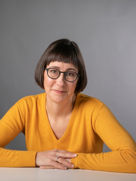 Johanna Ivaska合作论文数Department of Life Technologies, University of Turku;Cell adhesion and Cancer Group, Turku Biosience Centre, University of Turku3Antje Keppler合作论文数2m bjorkman合作论文数2Kari Nousiainen合作论文数2
 Johanna Bischof合作论文数Tufts Univ, Allen Discovery Ctr, Medford, MA 02155 USA2
 Erik Niemela合作论文数2
 Robert Goldman合作论文数Department of Cell and Developmental Biology, Feinberg School of Medicine, Northwestern University2
 Pekka Kallio合作论文数2
 Carmen Garrido合作论文数2Ann-Sofi Härmälä-Braskén合作论文数2
 Reetta Riikonen合作论文数2
 Daniel Nicorici合作论文数Tampere International;Center for Signal Processing;Tampere University of Technology;Center for Signal Processing, Tampere University of Technology2Tore Lindholm合作论文数2Vivek M. Rangnekar合作论文数Department of Radiation Medicine Combs Research Building, University of Kentucky2Marko J. Kallio合作论文数2
 Per Venge合作论文数Diagnostics Development2Chandrasekhar Abbineni合作论文数2h swahn合作论文数1
 Ola Ohlsson合作论文数1




