PURPOSE:A multi-institutional planning study was performed to evaluate the frequency that current guidelines established by Radiation Therapy Oncology Group (RTOG) protocols and other literature for lung stereotactic body radiotherapy (SBRT) treatments are followed.METHODS:A total of 300 patients receiving lung SBRT treatments in four different institutions were retrospectively reviewed. The treatments were delivered using Linac based SBRT (160 patients) or image guided robotic radiosurgery (140). Most tumors were located peripherally (250/300). Median fractional doses and ranges were 18 Gy (8-20 Gy), 12 Gy (6-15 Gy), and 10 Gy (5-12 Gy) for three, four, and five fraction treatments, respectively. The following planning criteria derived from RTOG trials and the literature were used to evaluate the treatment plans: planning target volumes, PTVV 100 ≥ 95% and PTVV 95 ≥ 99%; conformality indices, CI100% < 1.2 and CI50% range of 2.9-5.9 dependent on PTV; total lung-ITV: V20Gy < 10%, V12.5Gy < 15%, and V5Gy < 37%; contralateral lung V5Gy < 26%; and maximum doses for spinal cord, esophagus, trachea/bronchus, and heart and great vessels. Populations were grouped by number of fractions, and dosimetric criteria satisfaction rates (CSRs) were reported.RESULTS:Five fraction regimens were the most common lung SBRT fractionation (46%). The median PTV was 27.2 cm(3) (range: 3.8-419.5 cm(3)). For all plans: mean PTVV 100 was 94.5% (±5.6%, planning CSR: 69.8%), mean PTVV 95 was 98.1% (±4.1%, CSR: 69.5%), mean CI100% was 1.14 (±0.21, CSR: 79.1%, and 16.5% within minor deviation), and mean CI50% was 5.63 (±2.8, CSR: 33.0%, and 28.0% within minor deviation). When comparing plans based on location, peripherally located tumors displayed higher PTVV 100 and PTVV 95 CSR (71.5% and 71.9%, respectively) than centrally located tumors (61.2% and 57.1%, respectively). Overall, the planning criteria were met for all the critical structure such as lung, heart, spinal cord, esophagus, and trachea/bronchus for at least 85% of the patients.CONCLUSIONS:Among the various parameters that were used to evaluate the SBRT plans, the CI100% and CI50% were the most challenging criteria to meet. Although the CSRs of organs at risk were higher among all cases, their proximity to the PTV was a significant factor.
Purpose: To assess the variability in treatment planning of stereotactic body radiotherapy (SBRT) of lung lesions, a multi‐institutional planning comparison study was performed. Methods: A total of 150 patients receiving lung SBRT treatments in five different institutions were reviewed. Treatment plans were generated using Varian/Eclipse™ (82), CMS/Xio™ (53) and Cyberknife/Multiplan™ (15) systems. Most tumors were located peripherally (126/150) and in right lung (82/150). Various dose fractionations were used: 3x18Gy (5 cases), 3x20Gy (13), 4x12Gy (18), 4x12.5Gy (9), 5x10Gy (65), 5x12Gy (38), and 5x8or9Gy (2). The following planning criteria derived from RTOG trials and literature were used to evaluate the 4 or 5 fraction SBRT plans (133 cases): PTV‐V100>=95°/o, PTVV95>=99%, conformality index (CI‐100%) <1.2, CI‐50% isodose <4 for PTV>50cc or <5 for PTV<50cc, total lung‐ITV: V20Gy<10%, V12.5Gy<15%, V5Gy<37%, contralateral lung V5Gy<26%, cord: maximum<25 Gy, D0.25cc<22.5Gy, esophagus: max<30Gy, D5cc<27.5Gy, trachea/bronchus maximum<40Gy, heart & great vessels maximum<45Gy. The mean, standard deviation (±) and dosimetric criteria pass rates (PR) were reported. Results: The median PTV volume was 27.0cc (range:3.8/188.9cc). The mean PTV‐V100 was 96.0% (±3.6, PR:87.1%). The mean PTV‐V95 was 99.4% (±2.1, PR:90.2%) and the mean CI‐100% was 1.16 (±0.18, PR:74.0%). The mean CI‐50% was 4.14 (±0.67, PR:46.4%) for PTV>50cc (28 cases) and 7.36 (±3.36, PR:29.4%) for PTV<50cc (102 cases). The planning criteria were met for all the critical structure such as lung, heart, cord, esophagus, and trachea for at least 90% of the patients. The mean of all OAR criteria PR was 97.7% for peripheral and 88.6% for central lesions. Conclusion: Five fraction regimens were the most common lung SBRT fractionation (70%). Among the various parameters that were used to evaluate the SBRT plans, the CI‐100% and CI‐50% were the most challenging criteria to meet. Although the OAR PRs were higher among all cases, their proximity to the PTV was a significant factor.
PURPOSEVolumetric modulated arc therapy (VMAT) treatment planning studies have been reported to provide good target coverage and organs at risk (OARs) sparing in total marrow irradiation (TMI). A comprehensive dosimetric study simulating the clinical situation as close as possible is a norm in radiotherapy before a technique can be used to treat a patient. Without such a study, it would be difficult to make a reliable and safe clinical transition especially with a technique as complicated as VMAT-TMI. To this end, the dosimetric feasibility of VMAT-TMI technique in terms of treatment planning, delivery efficiency, and the most importantly three dimensional dose distribution accuracy was investigated in this study. The VMAT-TMI dose distribution inside a humanlike Rando phantom was measured and compared to the dose calculated using RapidArc especially in the field junctions and the inhomogeneous tissues including the lungs, which is the dose-limiting organ in TMI.METHODSThree subplans with a total of nine arcs were used to treat the planning target volume (PTV), which was determined as all the bones plus the 3 mm margin. Thermoluminescent detectors (TLDs) were placed at 39 positions throughout the phantom. The measured TLD doses were compared to the calculated plan doses. Planar dose for each arc was verified using mapcheck.RESULTSTLD readings demonstrated accurate dose delivery, with a median dose difference of 0.5% (range: -4.3% and 6.6%) from the calculated dose in the junctions and in the inhomogeneous medium including the lungs.CONCLUSIONSThe results from this study suggest that RapidArc VMAT technique is dosimetrically accurate, safe, and efficient in delivering TMI within clinically acceptable time frame.
Purpose: To investigate the dosimetric feasibility of VMAT‐TMI technique in terms of treatment planning and delivery efficiency and accuracy for a Rando phantom using Eclipse RapidArc technology. Methods: Total body irradiation (TBI) has been an integral part of pre‐conditioning regimens for the treatment of hematologic malignancies. Normal tissue toxicity is a common complication of TBI inclusive regimens especially at doses higher than 12 Gy due to inhomogeneous and excessive dose to organs at risk (OARs). Fixed gantry linear accelerator (linac) and Tomotherapy intensity modulated total bone marrow (IM‐TMI) techniques have been reported to reduce doses to OARs. However, the treatment time of more than one hour with both techniques may limit their clinical application. Volumetric modulated arc therapy (VMAT) may be able to increase the treatment delivery efficiency while increasing the target conformality and reducing the radiation dose to OARs. In order to treat all the bones in the Rando phantom, three sub‐plans with a total of nine arcs were used. A total of 100 thermoluminescent detectors (TLDs) were placed at 39 positions throughout the phantom. The measured TLD doses were compared to the calculated plan doses. Planar dose for each arc was verified using MapCheck and MapPhan. Results: Excellent target coverage with the prescription dose and dose reduction to the OARs were achieved similar to the fixed gantry linac IM‐TMI technique. The number of monitor units was decreased by 50%, while the delivery efficiency increased by more than 60%. TLD readings demonstrated accurate dose delivery, with a median dose difference of 0.5% from the calculated dose. Planar dose distributions of all arcs satisfied the 3% and 3 mm gamma criteria with an average pass rate of 97.8%. Conclusions: Results from this study suggest that RapidArc VMAT‐TMI technique is clinically feasible.This study is supported by Varian Medical Systems
BACKGROUND:Patients with end-stage renal disease are at increased risk for sudden cardiac death, although the utility of implantable cardioverter-defibrillators (ICDs) in these patients is unknown.OBJECTIVES:The purpose of this study was to evaluate whether end-stage renal disease is an independent risk factor for appropriate ICD therapy for ventricular tachycardia (VT) or ventricular fibrillation (VF) and to compare the long-term survival of ICD recipients with and without end-stage renal disease.METHODS:A retrospective cohort study was performed on ICD recipients at a single center. The primary endpoint was first appropriate ICD therapy for VT/VF. The secondary endpoint was survival.RESULTS:The study included 585 patients, 19 (3.2%) of whom had end-stage renal disease prior to device implantation. Average follow-up time was 2.2 +/- 2.4 years, during which time 156 patients (26.7%) received appropriate ICD therapy. End-stage renal disease was strongly associated with appropriate ICD therapy (hazard ratio 2.30, 95% confidence interval 1.17-4.54) and remained a significant predictor following adjustment for implant indication, ejection fraction, diabetes, hypertension, and beta-blocker use. Survival was significantly shorter in the end-stage renal disease patients, with a median survival time of 3.2 +/- 0.6 (SEM) years in the dialysis cohort and 7.4 +/- 0.5 (SEM) years in those without end-stage renal disease (log rank P = .009). The majority of deaths in the end-stage renal disease cohort were due to non-device-related infection.CONCLUSION:In this cohort, end-stage renal disease was the single greatest predictor of ICD therapies for VT/VF. The survival rate was significantly shorter than that of ICD recipients without end-stage renal disease, suggesting that comorbidities in end-stage renal disease patients meeting current implant indications may reduce the survival benefit of ICD placement in this population.
PC-SPES is a potent eight-herb formulation sold directly to consumers; it has promising efficacy in the treatment of prostate cancer (CaP). The product induces a castrate status in most, if not all, men, resulting in a 50% or greater prostate-specific antigen reduction in the great majority of men with androgen-dependent CaP and in more than one half of the men with androgen-independent CaP. The duration of response is not yet clear. The efficacy of PC-SPES appears to exceed that of androgen ablation alone, but is not necessarily separate from an estrogenic effect. Common side effects include gynecomastia, nipple tenderness, loss of libido, and impotency; uncommon side effects include a 4% incidence of thromboembolic phenomena, but also two reports of bleeding diatheses. The mechanisms of action may involve downregulation of the androgen receptor, induction of apoptosis by way of inhibition of the bcl-2 gene, and increased expression of p53. Two marker compounds in PC-SPES are baicalin and oridonin, both of which exhibit antiproliferative effects in CaP cell lines. Thousands of men are currently obtaining this nonprescription medicine, and physicians should ask patients specifically about its use. PC-SPES is of great interest in men with androgen-independent CaP, an area in which future research should be primarily directed.
Objectives. To examine the nuclear chromatin characteristics of epithelial cells, looking for an SPHB-mediated effect on nuclear DNA structure and organization. Saw palmetto herbal blend (SPHB) causes contraction of prostate epithelial cells and suppression of tissue dihydrotestosterone levels in men with symptomatic benign prostatic hyperplasia, but a fundamental mechanism remains unknown.Methods. A 6-month randomized trial, comparing prostatic tissue of men treated with SPHB (n = 20) or placebo (n = 20), was performed. At baseline, the two groups were similar in age (65 versus 64 years), symptoms (international Prostate Symptom Score 18 versus 17), uroflow (maximal urinary flow rate 10 versus 11 mL/s), prostate volume (59 versus 58 cm(3)), prostate-specific antigen (4.2 versus 2.7 ng/mL), and percentage of epithelium (17% versus 16%). Prostatic tissue was obtained by sextant biopsy before and after treatment. Five-micron sections were Feulgen stained and quantitatively analyzed using the AutoCyte QUIC-DNA imaging system. Images were captured from 200 randomly selected epithelial cell nuclei, and 60 nuclear morphometric descriptors (NMDs) (eg, size, shape, DNA content, and textural features) were determined for each nucleus. Logistic regression analysis was used to assess the differences in the variances of the NMDs between the treated and untreated prostate epithelial cells.Results. At baseline, the SPHB and placebo groups had similar NMD values. After 6 months of placebo, no significant change from baseline was found in the NMDs. However, after 6 months of SPHB, 25 of the 60 NMDs were significantly different compared with baseline, and a multivariate model for predicting treatment effect using 4 of the 25 was created (P <0.001). The multivariate model had an area under the receiver operating characteristic curve of 94% and an accuracy of 85%.Conclusions. Six months of SPHB treatment appears to alter the DNA chromatin structure and organization in prostate epithelial cells. Thus, a possible molecular basis for tissue changes and therapeutic effect of the compound is suggested. (C) 2002, Elsevier Science Inc.
Immunological ReviewsVolume 72, Issue 1 p. 119-141 Molecular Interactions in T-Cell-Mediated Cytotoxicity Benjamin Bonavida, Benjamin Bonavida Department of Microbiology and Immunology, UCLA School of Medicine, Los Angeles, U.S.A.Search for more papers by this authorThomas Bradley, Thomas Bradley Department of Microbiology and Immunology, UCLA School of Medicine, Los Angeles, U.S.A.Search for more papers by this authorJohn Fan, John Fan Department of Microbiology and Immunology, UCLA School of Medicine, Los Angeles, U.S.A.Search for more papers by this authorJohn Hiserodt, John Hiserodt Department of Microbiology and Immunology, UCLA School of Medicine, Los Angeles, U.S.A.Search for more papers by this authorRita Effros, Rita Effros Department of Microbiology and Immunology, UCLA School of Medicine, Los Angeles, U.S.A.Search for more papers by this authorHannah Wexler, Hannah Wexler Department of Microbiology and Immunology, UCLA School of Medicine, Los Angeles, U.S.A.Search for more papers by this author Benjamin Bonavida, Benjamin Bonavida Department of Microbiology and Immunology, UCLA School of Medicine, Los Angeles, U.S.A.Search for more papers by this authorThomas Bradley, Thomas Bradley Department of Microbiology and Immunology, UCLA School of Medicine, Los Angeles, U.S.A.Search for more papers by this authorJohn Fan, John Fan Department of Microbiology and Immunology, UCLA School of Medicine, Los Angeles, U.S.A.Search for more papers by this authorJohn Hiserodt, John Hiserodt Department of Microbiology and Immunology, UCLA School of Medicine, Los Angeles, U.S.A.Search for more papers by this authorRita Effros, Rita Effros Department of Microbiology and Immunology, UCLA School of Medicine, Los Angeles, U.S.A.Search for more papers by this authorHannah Wexler, Hannah Wexler Department of Microbiology and Immunology, UCLA School of Medicine, Los Angeles, U.S.A.Search for more papers by this author First published: June 1983 https://doi.org/10.1111/j.1600-065X.1983.tb01075.xCitations: 20 Supported in part by the National Cancer Institute grants CA12800, CA-09120, and by the Cancer Research Coordinating Committee, Berkeley AboutPDF ToolsRequest permissionExport citationAdd to favoritesTrack citation ShareShare Give accessShare full text accessShare full-text accessPlease review our Terms and Conditions of Use and check box below to share full-text version of article.I have read and accept the Wiley Online Library Terms and Conditions of UseShareable LinkUse the link below to share a full-text version of this article with your friends and colleagues. Learn more.Copy URL Share a linkShare onFacebookTwitterLinked InRedditWechat Citing Literature Volume72, Issue1June 1983Pages 119-141 RelatedInformation
In the past few years the first steps have been taken towards an understanding of the molecular basis of T-cell-mediated cytotoxicity. Particular attention has been paid to the part played in cytoxicity by idiotype-bearing antigen-receptors and other molecules on the T-cell surface. This article reviews these studies and focuses on the information obtained from studies on the inhibition of cytotoxic T cells with antisera or monoclonal antibodies directed against molecules on their surface.
The preparation of a xenogeneic rat anti-mouse CTL serum, RAT∗, which potently inhibits CTL function in the absence of complement, has recently been described. In the present study, this serum is further characterized. The membrane antigens recognized by RAT∗ were shown by FACS analysis to be detected most intensely on CTL, but are also present in lesser amounts on splenic T cells, thymocytes, and B cells. The T-cell proliferative response induced by Con A, PHA, TCGF, and modified self were all inhibited by RAT∗, but no effect was seen on LPS-induced B-cell proliferation. In addition, the in vitro induction of secondary CTL was not observed in the RAT∗-treated cultures. Finally, the effect of RAT∗ pretreatment is shown to be irreversible. It is proposed that RAT∗ serum reacts with receptors on T cells which are intimately involved in the “activation” step during the induction or expression phases of the response.
 Benjamin E. Bonavida合作论文数Department of Microbiology, Immunology, and Molecular Genetics, David Geffen School of Medicine, University of California , Los Angeles5
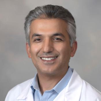 Murat Surucu合作论文数Department of Radiation Oncology, School of Medicine, Stanford University4
 Rita Effros合作论文数David Geffen School of Medicine, University of California, Los Angeles4
 John Hiserodt合作论文数4
 Bulent Aydogan合作论文数Department of Radiation and Cellular Oncology, University of Chicago4Hannah M. Wexler合作论文数David Geffen School of Medicine, UCLA3
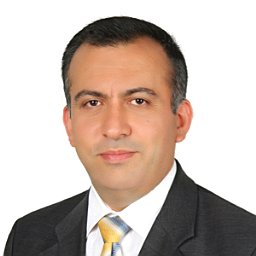 Mete Yeginer合作论文数Faculty of Medicine, Hacettepe University2Harkenrider Matthew M合作论文数Department of Radiation Oncology, Loyola University Medical Center2
 John Roeske合作论文数Medical Center, Loyola University Chicago;Division of Medical Physics, Stritch School of Medicine, Loyola University Chicago2
 Leonard S. Marks合作论文数David Geffen School of Medicine, University of California, Los Angeles2Gulbin O. Kavak合作论文数2F. Vali合作论文数2Jonathan Hsu合作论文数1R KAHLE合作论文数1Mc Miller合作论文数1Varro E. Tyler合作论文数1Sophie Chen合作论文数1JC HISERODT合作论文数1S Kessler合作论文数1Jay Tiongson合作论文数1A. Woerner合作论文数1
 Rod S Passman合作论文数Department of Medicine, Feinberg School of Medicine, Northwestern University1Jason C Robin合作论文数Department of Medical Education, Feinberg School of Medicine, Northwestern University1
 Christina Ciaccio合作论文数Department of Pediatrics, The University of Chicago;Department of Pediatrics-Allergy & Immunology, The University of Chicago1
 Alan W. Partin合作论文数Department of Urology, Johns Hopkins Medicine1Weinberg Kenneth M合作论文数1
 Peter Nelson合作论文数Stuart and Molly Sloan Precision Oncology Institute, Fred Hutch Cancer Center;Human Biology Division, Fred Hutch Cancer Center;University of Washington School of Medicine1
 Arie Belldegrun合作论文数Ronald Reagan UCLA Medical Center;Department of Urology, University of California, Los Angeles1
 Jonathan Epstein合作论文数Johns Hopkins University School of Medicine;Johns Hopkins Hospital1
 Alan H. Kadish合作论文数Northwestern University1
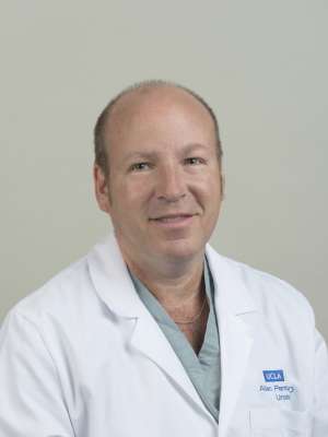 Allan J. Pantuck合作论文数Department of Urology, University of California, Los Angeles;Jonsson Comprehensive Cancer Center, University of California, Los Angeles;Center for Integrative Medicine, University of California, Los Angeles1
 David Heber合作论文数Herbalife Nutrition Institute;Center for Human Nutrition, Department of Medicine, David Geffen School of Medicine, University of California, Los Angeles;Division of Clinical Nutrition, Department of Medicine, David Geffen School of Medicine, University of California, Los Angeles1Robert S. Dipaola合作论文数1Floyd E. Leaders合作论文数1Thomas Bradley合作论文数1Wes D. Bales合作论文数1Michael Quadrini合作论文数1
 James A Radosevich合作论文数1Franklin C. Lowe合作论文数1Madhavi Reddy合作论文数1
 Robert W. Veltri合作论文数Brady Urological Institute, Johns Hopkins University School of Medicine1
 Mercedes Carnethon合作论文数Department of Preventive Medicine, Feinberg School of Medicine, Northwestern University1
 Jeffrey Goldberger合作论文数Division of Cardiovascular Medicine, Department of Medicine, Miller School of Medicine, University of Miami1Irwin Scher合作论文数1Patrick Choi合作论文数1
 Matthew Koshy合作论文数1ML Macairan合作论文数1




