Disrupted sleep is commonly reported during hematopoietic stem cell transplant. In this study, we use actigraphy to measure sleep parameters, and qualitative measures of quality of life, depression, and sleep in pediatric and young adult transplant recipients to describe their time course through transplant. Eight patients had evaluable actigraphy data, and 10 patients completed the surveys. The median age of the 6 male and 7 female participants was 13.94 years old. Sleep duration and ef ficiency measured by actigraphy were suboptimal prior to transplant, then declined to a nadir between Day +7 to +14. Self -reported sleep quality, depression, and quality of life were worst at Day +14 to +30 but improved by Day +100. Findings support efforts to improve sleep, which may improve recovery, mental health and quality of life.
One important goal of circadian medicine is to apply time- of- day dosing to improve the efficacy of chemotherapy. However, limited knowledge of how the circadian clock regulates DNA repair presents a challenge to mechanism- based clinical application. We studied time- series genome- wide nucleotide excision repair in liver and kidney of wild type and three different clock mutant genotypes (Cry1-/-Cry2-/-, Per1-/-Per2-/-, and Bmal1-/-). Rhythmic repair on the nontranscribed strand was lost in all three clock mutants. Conversely, rhythmic repair of hundreds of genes on the transcribed strand (TSs) persisted in the livers of Cry1-/-Cry2-/- and Per1-/-Per2-/- mice. We identified a tissue- specific, promoter element- driven repair mode on TSs of collagen and angio-genesis genes in the absence of clock activators or repressors. Furthermore, repair on TSs of thousands of genes was altered when the circadian clock is disrupted. These data contribute to a better understanding of the regulatory role of the circadian clock on nucleotide excision repair in mammals and may be invaluable toward the design of time- aware platinum- based interventions in cancer.
Collegiate athletes must satisfy the academic obligations common to all undergraduates, but they have the additional structural and social stressors of extensive practice time, competition schedules, and frequent travel away from their home campus. Clearly such stressors can have negative impacts on both their academic and athletic performances as well as on their health. These concerns are made more acute by recent proposals and decisions to reorganize major collegiate athletic conferences. These rearrangements will require more multi-day travel that interferes with the academic work and personal schedules of athletes. Of particular concern is additional east-west travel that results in circadian rhythm disruptions commonly called jet lag that contribute to the loss of amount as well as quality of sleep. Circadian misalignment and sleep deprivation and/or sleep disturbances have profound effects on physical and mental health and performance. We, as concerned scientists and physicians with relevant expertise, developed this white paper to raise awareness of these challenges to the wellbeing of our student-athletes and their co-travelers. We also offer practical steps to mitigate the negative consequences of collegiate travel schedules. We discuss the importance of bedtime protocols, the availability of early afternoon naps, and adherence to scheduled lighting exposure protocols before, during, and after travel, with support from wearables and apps. We call upon departments of athletics to engage with sleep and circadian experts to advise and help design tailored implementation of these mitigating practices that could contribute to the current and long-term health and wellbeing of their students and their staff members.
A growing body of research has identified circadian-rhythm disruption as a risk factor for metabolic health. However, the underlying biological basis remains complex, and complete molecular mechanisms are unknown. There is emerging evidence from animal and human research to suggest that the expression of core circadian genes, such as circadian locomotor output cycles kaput gene ( CLOCK ), brain and muscle ARNT-Like 1 gene ( BMAL1 ), period ( PER ), and cyptochrome ( CRY ), and the consequent expression of hundreds of circadian output genes are integral to the regulation of cellular metabolism. These circadian mechanisms represent potential pathophysiological pathways linking circadian disruption to adverse metabolic health outcomes, including obesity, metabolic syndrome, and type 2 diabetes. Here, we aim to summarize select evidence from in vivo animal models and compare these results with epidemiologic research findings to advance understanding of existing foundational evidence and potential mechanistic links between circadian disruption and altered clock gene expression contributions to metabolic health-related pathologies. Findings have important implications for the treatment, prevention, and control of metabolic pathologies underlying leading causes of death and disability, including diabetes, cardiovascular disease, and cancer.
Studies in shift workers and model organisms link circadian disruption to breast cancer. However, molecular circadian rhythms in noncancerous and cancerous human breast tissues and their clinical relevance are largely unknown. We reconstructed rhythms informatically, integrating locally collected, time-stamped biopsies with public datasets. For noncancerous breast tissue, inflammatory, epithelial–mesenchymal transition (EMT), and estrogen responsiveness pathways show circadian modulation. Among tumors, clock correlation analysis demonstrates subtype-specific changes in circadian organization. Luminal A organoids and informatic ordering of luminal A samples exhibit continued, albeit dampened and reprogrammed rhythms. However, CYCLOPS magnitude, a measure of global rhythm strength, varied widely among luminal A samples. Cycling of EMT pathway genes was markedly increased in high-magnitude luminal A tumors. Surprisingly, patients with high-magnitude tumors had reduced 5-y survival. Correspondingly, 3D luminal A cultures show reduced invasion following molecular clock disruption. This study links subtype-specific circadian disruption in breast cancer to EMT, metastatic potential, and prognosis.
Heterozygous de novo or inherited gain-of-function mutations in the MTOR gene cause Smith-Kingsmore syndrome (SKS). SKS is a rare autosomal dominant condition, and individuals with SKS display macrocephaly/megalencephaly, developmental delay, intellectual disability, and seizures. A few dozen individuals are reported in the literature. Here, we report a cohort of 28 individuals with SKS that represent nine MTOR pathogenic variants. We conducted a detailed natural history study and found pathophysiological deficits among individuals with SKS in addition to the common neurodevelopmental symptoms. These symptoms include sleep-wake disturbance, hyperphagia, and hyperactivity, indicative of homeostatic imbalance. To characterize these variants, we developed cell models and characterized their functional consequences. We showed that these SKS variants display a range of mechanistic target of rapamycin (mTOR) activities and respond to the mTOR inhibitor, rapamycin, differently. For example, the R1480_C1483del variant we identified here and the previously known C1483F are more active than wild-type controls and less responsive to rapamycin. Further, we showed that SKS mutations dampened circadian rhythms and low-dose rapamycin improved the rhythm amplitude, suggesting that optimal mTOR activity is required for normal circadian function. As SKS is caused by gain-of-function mutations in MTOR, rapamycin was used to treat several patients. While higher doses of rapamycin caused delayed sleep-wake phase disorder in a subset of patients, optimized lower doses improved sleep. Our study expands the clinical and molecular spectrum of SKS and supports further studies for mechanism-guided treatment options to improve sleep-wake behavior and overall health.
STUDY OBJECTIVES:Genetics impacts sleep, yet, the molecular mechanisms underlying sleep regulation remain elusive. In this study, we built machine learning models to predict sleep genes based on their similarity to genes that are known to regulate sleep.METHODS:We trained a prediction model on thousands of published datasets, representing circadian, immune, sleep deprivation, and many other processes, using a manually curated list of 109 sleep genes.RESULTS:Our predictions fit with prior knowledge of sleep regulation and identified key genes and pathways to pursue in follow-up studies. As an example, we focused on the NF-κB pathway and showed that chronic activation of NF-κB in a genetic mouse model impacted the sleep-wake patterns.CONCLUSION:Our study highlights the power of machine learning in integrating prior knowledge and genome-wide data to study genetic regulation of complex behaviors such as sleep.
INTRODUCTION:Chronotherapy is the timing of medication according to biological rhythms of the host to optimize drug efficacy and minimize toxicity. Efficacy and myelosuppression of azathioprine/6-mercaptopurine (AZA/6-MP) are correlated with the metabolite 6-thioguanine, while the metabolite 6-methylmercaptopurine correlates with hepatotoxicity.METHODS:This was a single-center, 10-week prospective crossover trial involving 26 participants with inactive inflammatory bowel disease (IBD) on a stable dose and time of AZA or 6-MP therapy. Participants were switched to the opposite delivery time (morning or evening) for 10 weeks, and metabolite measurements were at both time points.RESULTS:In the morning vs evening dosing, 6-thioguanine levels were 225.7 ± 155.1 vs 175.0 ± 106.9 ( P < 0.01), and 6-methylmercaptopurine levels were 825.1 ± 1,023.3 vs 2,395.3 ± 2,880.3 ( P < 0.01), with 69% (18 out of 26) of participants had better metabolite profiles in the morning. Participants with optimal dosing in the morning had an earlier chronotype by corrected midpoint of sleep.DISCUSSION:In the first study on a potential role of chronotherapy in IBD, we found (i) morning dosing of AZA or 6-MP resulted in more optimal metabolite profiles and (ii) host chronotype could help identify one-third of patients who would benefit from evening dosing. Circadian regulation of metabolic enzymes of AZA/6-MP activity in the liver is the likely cause of these differences. This pilot study confirms the need to incorporate chronotherapy in future multicenter clinical trials on IBD disease.
. To the editor: In inpatients unable to sustain themselves enterally, standard i.v. total parenteral nutrition (TPN) is often administered over a 20-to 24-hour feeding period. However, feeding time is a dominant entraining cue for peripheral mammalian clocks and, importantly, a major driver of metabolism (1). It is well established that eating over long periods such as these contributes to metabolic dysregulation and poor sleep. Moreover, the first report of long-term TPN, given at home to adults with chronic bowel disease, was administered overnight for 12–18 hours (2). Time-restricted feeding is actively being studied in adult outpatients with obesity and those with healthy weight, with evaluation of weight loss or metabolic syndrome manifestations. The benefits of limiting the time of day and daily feeding duration in the inpatient setting in pediatric patients experiencing an acute but expected recoverable intestinal insult are unknown but likely important to study. We conducted a single-center, randomized, nonblinded, controlled pilot study from May 2020 to September 2022 to establish the safety and feasibility of daytime-restricted nutrition in hematopoietic stem cell transplant (HSCT) recipients and to evaluate the effect of nutritional supplementation timing on post-HSCT outcomes (ClinicalTrials.gov, NCT04549038). This study was approved by the Cincinnati Children’s Hospital Medical Center Institutional Review Board and performed in accordance with the principles of the Declaration of Helsinki. Patients or […]
Intermittent hypoxia (IH) is a major clinical feature of obstructive sleep apnea (OSA). The mechanisms that become dysregulated after periods of exposure to IH are unclear, particularly in the early stages of disease. The circadian clock governs a wide array of biological functions and is intimately associated with stabilization of hypoxia-inducible factors (HIFs) under hypoxic conditions. In patients, IH occurs during the sleep phase of the 24-hour sleep–wake cycle, potentially affecting their circadian rhythms. Alterations in the circadian clock have the potential to accelerate pathological processes, including other comorbid conditions that can be associated with chronic, untreated OSA. We hypothesized that changes in the circadian clock would manifest differently in those organs and systems known to be impacted by OSA. Using an IH model to represent OSA, we evaluated circadian rhythmicity and mean 24-hour expression of the transcriptome in 6 different mouse tissues, including the liver, lung, kidney, muscle, heart, and cerebellum, after a 7-day exposure to IH. We found that transcriptomic changes within cardiopulmonary tissues were more affected by IH than other tissues. Also, IH exposure resulted in an overall increase in core body temperature. Our findings demonstrate a relationship between early exposure to IH and changes in specific physiological outcomes. This study provides insight into the early pathophysiological mechanisms associated with IH.
The molecular circadian clock is regulated by a transcriptional translational feedback loop. However, the post-translational control mechanisms are less understood. The NRON complex is a large ribonucleoprotein complex, consisting of a lncRNA and several proteins. Components of the complex play a distinct role in regulating protein phosphorylation, synthesis, stability, and translocation in cellular processes. This includes the NFAT and the circadian clock pathway. PSMD11 is a component of the NRON complex and a lid component of the 26S proteasome. Among the PSMD family members, PSMD11 has a more specific role in circadian clock function. Here, we used cell and biochemical approaches and characterized the role of PSMD11 in regulating the stability and nuclear translocation of circadian clock proteins. We used size exclusion chromatography to enrich the NRON complex in the cytosolic and nuclear fractions. More specifically, PSMD11 knockdown affected the abundance of PER2 and CRY2 proteins and the nuclear translocation of CRY1. This changed the relative abundance of CRY1 and CRY2 in the nucleus. Thus, this work defines the role of PSMD11 in the NRON complex regulating the nuclear translocation of circadian repressors, thereby enabling cellular circadian oscillations.
Disruption of the circadian clock is linked to cancer development and progression. Establishing this connection has proven beneficial for understanding cancer pathogenesis, determining prognosis, and uncovering novel therapeutic targets. However, barriers to characterizing the circadian clock in human pancreas and human pancreatic cancer–one of the deadliest malignancies–have hindered an appreciation of its role in this cancer. Here, we employed normalized coefficient of variation (nCV) and clock correlation analysis in human population-level data to determine the functioning of the circadian clock in pancreas cancer and adjacent normal tissue. We found a substantially attenuated clock in the pancreatic cancer tissue. Then we exploited our existing mouse pancreatic transcriptome data to perform an analysis of the human normal and pancreas cancer samples using a machine learning method, cyclic ordering by periodic structure (CYCLOPS). Through CYCLOPS ordering, we confirmed the nCV and clock correlation findings of an intact circadian clock in normal pancreas with robust cycling of several core clock genes. However, in pancreas cancer, there was a loss of rhythmicity of many core clock genes with an inability to effectively order the cancer samples, providing substantive evidence of a dysregulated clock. The implications of clock disruption were further assessed with a Bmal1 knockout pancreas cancer model, which revealed that an arrhythmic clock caused accelerated cancer growth and worse survival, accompanied by chemoresistance and enrichment of key cancer-related pathways. These findings provide strong evidence for clock disruption in human pancreas cancer and demonstrate a link between circadian disruption and pancreas cancer progression.
Healthy individuals exhibit blood pressure variation over a 24-hour period with higher blood pressure during wakefulness and lower blood pressure during sleep. Loss or disruption of the blood pressure circadian rhythm has been linked to adverse health outcomes, for example, cardiovascular disease, dementia, and chronic kidney disease. However, the current diagnostic and therapeutic approaches lack sufficient attention to the circadian rhythmicity of blood pressure. Sleep patterns, hormone release, eating habits, digestion, body temperature, renal and cardiovascular function, and other important host functions as well as gut microbiota exhibit circadian rhythms, and influence circadian rhythms of blood pressure. Potential benefits of nonpharmacologic interventions such as meal timing, and pharmacologic chronotherapeutic interventions, such as the bedtime administration of antihypertensive medications, have recently been suggested in some studies. However, the mechanisms underlying circadian rhythm-mediated blood pressure regulation and the efficacy of chronotherapy in hypertension remain unclear. This review summarizes the results of the National Heart, Lung, and Blood Institute workshop convened on October 27 to 29, 2021 to assess knowledge gaps and research opportunities in the study of circadian rhythm of blood pressure and chronotherapy for hypertension.
Studies in shift workers and model organisms link circadian disruption to breast cancer. However, molecular rhythms in non-cancerous and cancerous human breast tissues are largely unknown. We reconstructed rhythms informatically, integrating locally collected, time-stamped biopsies with public datasets. For non-cancerous tissue, the inferred order of core-circadian genes matches established physiology. Inflammatory, epithelial-mesenchymal transition (EMT), and estrogen responsiveness pathways show circadian modulation. Among tumors, clock correlation analysis demonstrates subtype-specific changes in circadian organization. Luminal A organoids and informatic ordering of Luminal A samples exhibit continued, albeit disrupted rhythms. However, CYCLOPS magnitude, a measure of global rhythm strength, varied widely among Luminal A samples. Cycling of EMT pathway genes was markedly increased in high-magnitude Luminal A tumors. Patients with high-magnitude tumors had reduced 5-year survival. Correspondingly, 3D Luminal A cultures show reduced invasion following molecular clock disruption. This study links subtype-specific circadian disruption in breast cancer to EMT, metastatic potential, and prognosis.
Background: Adolescent sleep is an underappreciated risk factor for hypertension. Blood pressure is regulated by circadian processes, which are affected by bedtime and sleep duration. Aim of review: We review the literature associating low sleep duration and late bedtimes with high blood pressure and consider potential interventions to improve adolescent sleep and its effects on blood pressure. Key scientific concepts of review: Both intrinsic factors, such as race and sex, and extrinsic factors, such as school start time and sleep hygiene, impact adolescent sleep patterns. Inadequate and delayed sleep is associated with an increased risk of hypertension in adolescents, likely compounded by other behaviors associated with poor sleep, and multiple approaches are needed to address this growing problem.
Objective Seizures are known to occur with diurnal and other rhythms. To gain insight into the neurophysiology of periodicity of seizures, we tested the hypothesis that intracranial high-frequency oscillations (HFOs) show diurnal rhythms and sleep-wake cycle variation. We further hypothesized that HFOs have different rhythms within and outside the seizure-onset zone (SOZ). Methods In drug-resistant epilepsy patients undergoing stereotactic-EEG (SEEG) monitoring to localize SOZ, we analyzed the number of 50-200 Hz HFOs/channel/minute (HFO density) through a 24-hour period. The distribution of HFO density during the 24-hour period as a function of the clock time was analyzed with cosinor model, and for non-uniformity with the sleep-wake cycle. Results HFO density showed a significant diurnal rhythm overall and both within and outside SOZ. This diurnal rhythm of HFO density showed significantly lower amplitude and longer acrophase within SOZ compared to outside SOZ. The peaks of difference in HFO density within and outside SOZ preceded the seizures by approximately 4 hours. The difference in HFO density within and outside SOZ also showed a non-uniform distribution as a function of sleep-wake cycle, with peaks at first hour after arousal and ±2 hours around sleep onset. Conclusions Our study shows that the diurnal rhythm of intracranial HFOs is more robust outside the SOZ. This suggests cortical tissue within SOZ generates HFOs relatively more uniformly throughout the day with attenuation of expected diurnal rhythm. The difference in HFO density within and outside SOZ also showed non-uniform distribution according to clock times and the sleep-wake cycle, which can be a potential biomarker for preferential times of pathological cortical excitability. A temporal correlation with seizure occurrence further substantiates this hypothesis.
SignificanceWe successfully identified theduperallele as a null mutation of Cryptochrome 1 in Syrian hamsters. Here, we have shown the use of fast homozygosity mapping as an effective approach to identify causal mutations in mammals, despite lacking chromosomal genome information. In the course of this work, we improved the draft Syrian hamster genome and generated datasets necessary to exploit Syrian hamsters as a modern genetic research model. The unique physiological features of Syrian hamsters make them a desirable model to investigate human diseases, including circadian disorders, cancer, heart function, metabolism, and infectious diseases (e.g., severe acute respiratory syndrome coronavirus 2).
 Gang Wu合作论文数Univ Penn, Dept Syst Pharmacol & Translat Therapeut, Philadelphia, PA 19104 USA38
 Lauren J. Francey合作论文数The Children's Hospital of Philadelphia, The Children's Hospital of Philadelphia33Ron C Anafi合作论文数Department of Medicine, Perelman School of Medicine, University of Pennsylvania26
 Steve A. Kay合作论文数The Keck School of Medicine, University of Southern California;The Scripps Research Institute25Julie E. Baggs合作论文数22
 David F Smith合作论文数Dept Otolaryngol Head & Neck Surg, Univ Cincinnati20
 Marc D Ruben合作论文数Center for Chronobiology, Cincinnati Children’s Hospital Medical Center19michael dean hughes合作论文数16
 Satchidananda Panda合作论文数Regulatory Biology Laboratory, Salk Institute for Biological Studies15
 Andrew I. Su合作论文数Department of Integrative Structural and Computational Biology, The Scripps Research Institute14Trey K. Sato合作论文数14
 Christopher Bradfield合作论文数McArdle Laboratory for Cancer Research, Department of Oncology, University of Wisconsin-Madison14
 Garret A. FitzGerald合作论文数Institute for Translational Medicine and Therapeutics, Department of Systems Pharmacology and Translational Therapeutics, Perelman School of Medicine, University of Pennsylvania14
 Andrew Chuanyin Liu合作论文数Department of Cell Biology, The Scripps Research Institute14
 Anand Venkataraman合作论文数ZS14John R. Walker合作论文数13
 Joseph S. Takahashi合作论文数Department of Neuroscience, Howard Hughes Medical Institute, University of Texas Southwestern11Nicholas F. Lahens合作论文数11S Panda合作论文数10Olarerin-George Anthony O合作论文数Perelman School of Medicine, the University of Pennsylvania;Perelman School of Medicine, University of Pennsylvania;University of Pennsylvania, University of Pennsylvania8Dye Thomas J合作论文数8Robert E. Schmidt合作论文数7
 Chidambaram Ramanathan合作论文数Coll Hlth Sci, Univ Memphis7Yin Yeng Lee合作论文数Department of Pharmacology and Systems Physiology, University of Cincinnati College of Medicine, Cincinnati, OH 45229, USA7
 Russell N. Van Gelder合作论文数Department of Ophthalmology, University of Washington, Seattle7Lee Yool合作论文数Department of Biological Sciences and 21st Frontier Program in Neuroscience, Seoul National University7
 Amita Sehgal合作论文数Sehgal Lab, Perelman School of Medicine, University of Pennsylvania;Department of Neuroscience, University of Pennsylvania School of Medicine7Gu Y Z合作论文数6
 Hayer Katharina E合作论文数Department of Systems Pharmacology and Translational Therapeutics, University of Pennsylvania Perelman School of Medicine6
 Hiroki Ueda (上田泰己)合作论文数Laboratory for Systems Biology, Center for Developmental Biology, RIKEN;Functional Genomics Subunit, Center for Developmental Biology, RIKEN;Tokushima University;Osaka University;Kyoto University;National Institute of Genetics6Yang Shen合作论文数Shanghai Veterinary Research Institute, Chinese Academy of Agricultural Science6
 Chi Van Dang合作论文数School of Medicine, Johns Hopkins University6
 Sumit Chanda合作论文数Department of Immunology and Microbiology, Scripps Research6
 Kevin R. Hayes合作论文数Laboratory for Cancer Research;University of Wisconsin Medical School;Laboratory for Cancer Research, University of Wisconsin Medical School6
 Brian J. Altman合作论文数Univ Penn, Philadelphia, PA 19104 USA6Jeremy S. Caldwell合作论文数6Radu Daniel Rudic合作论文数Georgia Health Sciences University, Georgia Health Sciences University6Loren J Miraglia合作论文数5D. F. Smith合作论文数5P McNamara合作论文数5Kavakli Ibrahim Halil合作论文数School of Medicine, University of North Carolina5
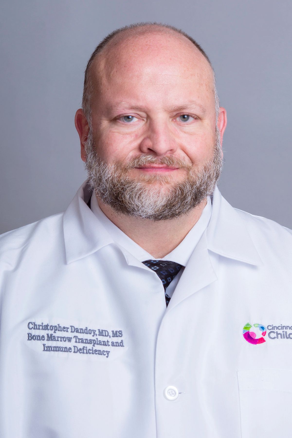 Christopher E. Dandoy合作论文数Cincinnati Children's Hospital Medical Center;University of Cincinnati Cancer Center;University of Cincinnati College of Medicine5
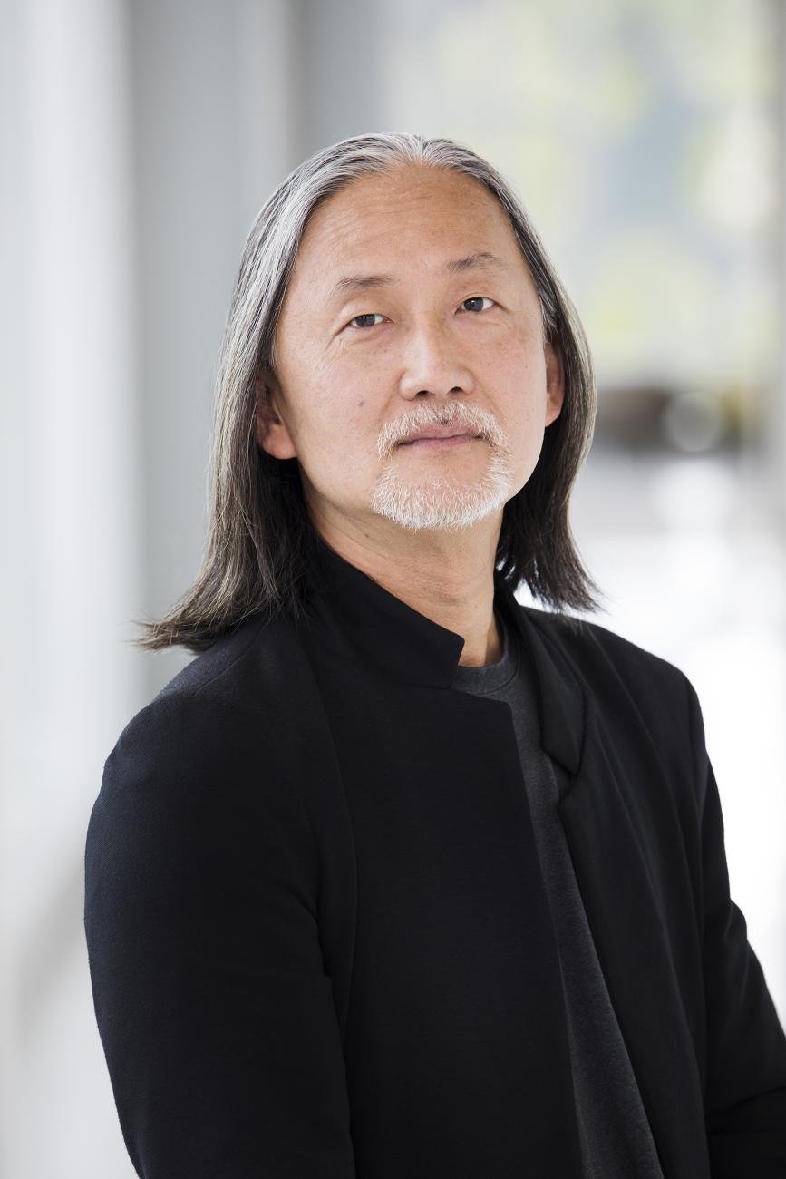 Junhyong Kim合作论文数The Kim Lab, Department of Biology, University of Pennsylvania;DataHelix, LLC5Growe Jacqueline合作论文数5
 Aziz Sancar合作论文数Department of Biochemistry and Biophysics, School of Medicine, University of North Carolina;Lineberger Comprehensive Cancer Center, School of Medicine, University of North Carolina5
 Karl Kornacker合作论文数Division of Sensory Biophysics, The Ohio State University5
 Hilmar Lapp合作论文数Novartis Research Foundation Genomics Institute5
 Erik Herzog合作论文数Department of Biology, College of Arts & Sciences, Washington University in St. Louis5
 Mathew T. Pletcher合作论文数Bayer School of Natural and Environmental Sciences, Duquesne University5




