Purpose:Uterine fibroids commonly impact women of reproductive age, with the submucosal subtype known to adversely affect fertility. Recently, non-cavity distorting intramural fibroids have been found to impair the uterine environment leading many reproductive endocrinologists to surgically remove these fibroids prior to infertility treatment. However, the frequency with which this intervention occurs in actual practice remains in question. Patients and Methods:A database of 407 US-based reproductive endocrinology and infertility (REI) clinics was synthesized from the Centers for Disease Control & Prevention National ART Surveillance System and the Society for Assisted Reproductive Technology's Clinic List. Medical directors at each site were invited to complete a REDCap-administered cross-sectional survey on management of non-cavity distorting intramural fibroids in patients pursuing infertility treatment. Results:A total of 112 (28%) REI physicians completed the survey, representing a variety of clinic settings, locations, and volumes. The majority of respondents (72%) consider surgical management of non-cavity distorting intramural fibroids based on factors such as size, with myomas 4.1-6 cm or larger more likely to be considered for removal. Approximately 42% of REI physician respondents surgically remove non-cavity distorting intramural fibroids themselves, while 58% refer to other gynecologic surgeons. Conclusion:Many physicians consider the removal of non-cavity distorting intramural fibroids prior to infertility treatment. Additional research is needed to better understand the indications for surgical management of various fibroid subtypes and the true scope of removal in current practice.
OBJECTIVE:To demonstrate the step-by-step technique for performing a transabdominal oocyte retrieval with a vaginal transducer in patients where transvaginal access is not an option. DESIGN:Video article. SUBJECTS:The first patient is a 38-year-old gravida 0 woman with prior midline laparotomy for myomectomy and endometriosis resection. In vitro fertilization was recommended after unsuccessful natural conception attempts. Her baseline antimüllerian hormone was 17 pmol/L (2.38 ng/mL). The second patient is a 26-year-old gravida 0 woman with history of multiple abdominal and pelvic surgeries because of congenital vaginal and anal atresia. She presented with locally advanced cancer of the neovagina. Her baseline antimüllerian hormone was 24 pmol/L (3.36 ng/mL). The patients included gave consent for publication and posting of the video online including social media, the journal website, scientific literature websites (such as PubMed, ScienceDirect, and Scopus, etc.) and other applicable sites. EXPOSURE:Both patients underwent controlled ovarian stimulation using a gonadotropin-releasing hormone antagonist protocol. Oocyte retrievals were performed transabdominally using a 7 MHz vaginal transducer fitted with a disposable needle guide and a 17 gauge, 25-cm single lumen aspiration needle. Follicular aspiration was performed with a pedal-activated vacuum pump with suction pressures ranging between 105-140 mmHg. MAIN OUTCOME MEASURES:Oocyte yield after transabdominal oocyte retrieval. RESULTS:The first patient's in vitro fertilization cycle yielded 12 oocytes, of which 8 fertilized with conventional insemination; four blastocysts were cryopreserved. A frozen embryo transfer is planned imminently. Twenty-one oocytes were retrieved in the second patient, of which 12 mature oocytes were cryopreserved for future use. She is currently undergoing radiation therapy. CONCLUSION:Transabdominal oocyte retrieval using a vaginal transducer is a safe, effective, and feasible method of oocyte retrieval in select patients where ovaries are not accessible transvaginally. This technique may be more easily adopted where abdominal transducer needle guides and adapters are not readily accessible, offering a practical alternative for oocyte retrieval in patients with complex pelvic anatomy or pathology.
The fallopian tube serves as a sperm reservoir, and it is the site where the oocytes become fertilized. Here, we describe development of an organ-on-a-chip microfluidic model of the fallopian tube (FT Chip) lined by primary human epithelial cells and stromal fibroblasts derived from the FT ampulla. Abundant tissue folds lined by hormone-responsive, epithelial cells resembling those seen in vivo formed on-chip, but not in epithelial organoids cultured in gel cultures. Comparative time-resolved analysis of human sperm versus oocyte-sized microparticles introduced into the epithelial channel in the presence of estradiol revealed that sperm movement was significantly reduced, while the oocyte-sized particles increased, relative to movements in acellular chips. When the non-hormonal contraceptive TDI-11861 was administered to the chip, dose-dependent inhibition of human sperm motility was detected. Thus, this FT Chip may offer a human preclinical tool to study FT physiology and assess the efficacy and mechanism of action of contraceptives. ### Competing Interest Statement D.E.I. holds equity in Emulate Inc., chairs its scientific advisory board, and is a member of its board of directors. Gates Foundation, [INV-056272]
Previous studies have examined the predictors of PFAS concentrations among pregnant women and children. However, no study has explored the predictors of preconception PFAS concentrations among couples in the United States. This study included 572 females and 279 males (249 couples) who attended a U.S. fertility clinic between 2005 and 2019. Questionnaire information on demographics, reproductive history, and lifestyles and serum samples quantified for PFAS concentrations were collected at study enrollment. We examined the PFAS distribution and correlation within couples. We used Ridge regressions to predict the serum concentration of each PFAS in females and males using data of (1) socio-demographic and reproductive history, (2) diet, (3) behavioral factors, and (4) all factors included in (1) to (3) after accounting for temporal exposure trends. We used general linear models for univariate association of each factor with the PFAS concentration. We found moderate to high correlations for PFAS concentrations within couples. Among all examined factors, diet explained more of the variation in PFAS concentrations (1-48%), while behavioral factors explained the least (0-4%). Individuals reporting White race, with a higher body mass index, and nulliparous women had higher PFAS concentrations than others. Fish and shellfish consumption was positively associated with PFAS concentrations among both females and males, while intake of beans (females), peas (male), kale (females), and tortilla (both) was inversely associated with PFAS concentrations. Our findings provide important data for identifying sources of couples' PFAS exposure and informing interventions to reduce PFAS exposure in the preconception period.
Prenatal per and polyfluoroalkyl substances (PFAS) exposure is associated with adverse birth outcomes. There is an absence of evidence on the relationship between maternal and paternal preconception PFAS exposure and birth outcomes. This study included 312 mothers and 145 fathers with a singleton live birth from a preconception cohort of subfertile couples seeking fertility treatment at a U.S. clinic. PFAS were quantified in serum samples collected before conception. Gestational age (GA) and birthweight (BW) were abstracted from delivery records. We also assessed low birthweight (BW < 2500 g) and preterm birth (GA < 37 completed weeks). We utilized multivariable linear regression, logistic regression, and quantile-based g computation to examine maternal or paternal serum concentrations of individual PFAS and mixture with birth outcomes. Maternal serum concentrations of perfluorooctanesulfonate (PFOS), perfluorohexanesulfonate (PFHxS), and the total PFAS mixture were inversely associated with birthweight. Maternal PFOS concentration was associated with a higher risk of low birthweight. Conversely, paternal PFOS and PFHxS concentrations were imprecisely associated with higher birthweight. No associations were found for gestational age or preterm birth. The findings have important implications for preconception care. Future research with larger sample sizes would assist in validating these findings.
Per- and polyfluoroalkyl substances (PFAS) exposure was associated with changes in thyroid function in pregnant mothers and the general population. Limited such evidence exists in other susceptible populations such as females with fertility problems. This cross-sectional study included 287 females seeking medically assisted reproduction at a fertility clinic in Massachusetts, United States, between 2005 and 2019. Six long-alkyl chain PFAS, thyroid hormones, and autoimmune antibodies were quantified in baseline serum samples. We used generalized linear models and quantile g-computation to evaluate associations of individual PFAS and their total mixture with thyroid biomarkers. Most females were White individuals (82.7%), had graduate degrees (57.8%), and nearly half had unexplained subfertility (45.9%). Serum concentrations of all examined PFAS and their mixture were significantly associated with 2.6%-5.6% lower total triiodothyronine (TT3) concentrations. Serum concentrations of perfluorononanoate (PFNA), perfluorodecanoate (PFDA), and perfluoroundecanoate (PFUnDA), and of the total mixture were associated with higher ratios of free thyroxine (FT4) to free triiodothyronine (FT3). No associations were found for PFAS and TSH or autoimmune antibodies. Our findings support the thyroid-disrupting effect of long alkyl-chain PFAS among a vulnerable population of subfertile females.
Previous studies reported that exposures to per- and polyfluoroalkyl substances (PFAS), largely in higher exposed populations, were associated with elevated risk of polycystic ovary syndrome (PCOS). However, studies evaluating PCOS risk in populations with lower background exposures to PFAS are limited. This study aimed to examine the associations between serum PFAS concentrations and PCOS risk among women attending a U.S. academic fertility clinic during 2005-2019. A total of 502 females who sought fertility evaluation and assisted reproduction treatments were included. Nine PFAS were quantified in non-fasting serum samples collected at study entry. Diagnosis of PCOS was based on the Rotterdam criteria. We used logistic regression to examine the odds ratio (OR) of PCOS in relation to individual PFAS concentrations (continuous and by tertiles) and quantile g-computation (QGC) and Bayesian Kernel Machine Regression (BKMR) to examine the joint associations of PFAS mixture with PCOS. Most participants were White and had a graduate degree or higher. Per doubling of serum perfluorooctane sulfonate (PFOS) and perfluorohexane sulfonate (PFHxS) concentrations were associated with higher odds of PCOS [OR (95%CI): 1.70 (1.06, 2.81) and 1.45 (1.02, 2.08) for PFOS and PFHxS respectively]. There was a dose-response relationship of PFOS with PCOS risk (p of trend by PFOS tertiles = 0.07). Both QGC and BKMR identified PFOS as the most important contributor among the mixture to PCOS risk. No clear joint effects were found for other PFAS or PFAS mixtures on PCOS risk. Our findings are consistent with existing evidence in populations with higher background PFAS concentrations and highlight the adverse effects of PFAS exposure on reproductive health. Findings can inform public health measures and clinical care to protect populations vulnerable to PCOS, in part, due to environmental exposures.
Objective: To objectively grade all video publications in Fertility and Sterility during the year 2021 and compile a list of the top 10 surgical videos. Design: A descriptive presentation of the 10 highest-scoring video publications from Fertility and Sterility in the year 2021. Setting: Not applicable. Patient/Animals: Not applicable. Interventions: J.F., Z.K., J.P.P., and S.R.L. acted as independent reviewers of all video publications. A standardized scoring method was used to score all videos. Main Outcome Measures: Up to 5 points were awarded for each of the following categories: scientific merit or clinical relevance of the topic; clarity of the video; use of an innovative surgical technique; and video editing or the use of marking tools on the video to highlight important features or surgical landmarks. This allowed a maximum score of 20 for each video. The number of YouTube views and likes was used as a tiebreaker if >= 2 videos scored similarly. The interclass coefficient from a 2-way random effects model was calculated to assess the agreement among the 4 independent reviewers. Result(s): A total of 36 videos were published in Fertility and Sterility during the year 2021. After averaging scores from all 4 reviewers, a top-10 list was created. The overall interclass correlation coefficient for the 4 reviews was 0.89 (95% confidence interval, 0.89-0.94). Conclusion(s): An overall substantial agreement was noted among the 4 reviewers. A total of 10 videos reigned supreme from a list of very competitive publications that had already undergone the peer review process. The subject matter of these videos ranged from complex surgical procedures, including uterine transplantation, to common procedures, such as GYN ultrasound. (c) 2023 by American Society for Reproductive Medicine.) El resumen este disponible en Espanol al final del articulo.
In 2012, the prequel to the “Alien” movie franchise, “Prometheus,” was released. In the now-infamous C-section scene, archeologist Elizabeth Shaw, played by Noomi Rapace, uses a MedPod 720i to extract a rapidly growing squid-like creature from her abdomen after unknowingly being used in an experiment. The scene beautifully depicted the interface of image-guided surgery, artificial intelligence (AI), and robotic surgery at its pinnacle. For every surgeon who saw this scene, a glimpse of what the future of surgery holds was on proud display. Image-guided surgery is not novel. We have been using transabdominal ultrasounds to guide dilation and curettage, hysteroscopic lysis of adhesions, and uterine septum repairs. Oocyte retrievals have matured from a laparoscopic-directed procedure to a transvaginal ultrasound-guided procedure that is safer and more efficient. Even embryo transfers have progressed from a blind procedure to one that is now guided by transabdominal ultrasound. Laparoscopic approaches to fibroid treatment are now using laparoscopic ultrasounds to aid in localization of fibroids intraoperatively that would otherwise be missed because of the lack of haptic feedback. All surgeons rely on preoperative imaging to help prepare them for surgery. For example, many surgeons who specialize in fibroid surgery rely on ultrasonography, sonohysterography, or magnetic resonance imaging to help them decide on the best surgical approach, gauge potential blood loss, and determine the anticipated number of fibroids that will be removed. Better imaging quality and radiologic protocols are making magnetic resonance imaging an integral part of diagnostic testing for patients with suspected endometriosis. Now, with easier and cheaper access to 3-dimensional (3D) printers, images can be converted to 3D models to help the surgeon practice their approach in the simulation laboratory before doing the surgery (1Pugliese L. Marconi S. Negrello E. Mauri V. Peri A. Gallo V. et al.The clinical use of 3D printing in surgery.Updates Surg. 2018; 70: 381-388Crossref PubMed Scopus (100) Google Scholar). In addition, more companies have invested in augmented reality, in which surgeons can use virtual reality headsets and, using preoperative images, create a simulated environment in which they can practice their movements and approach their surgery. Mercorio et al. (2Mercorio A. Zizolfi B. Barbuto S. Danzi R. Di Spiezio Sardo A. Moawad G. et al.3D imaging reconstruction and laparoscopic robotic surgery: a winning combination for a 5 complex case of multiple myomectomy.Fertil Steril. 2023; 120: 202-204Abstract Full Text Full Text PDF Scopus (1) Google Scholar) in Italy have beautifully shown us in their video a developing technology that allows 3D images of the fibroid uterus, generated from preoperative imaging, with each fibroid color coded, to be overlayed over the actual operative image during robotic surgery. The 3D images guide the surgeon to each fibroid, and because they are removed from the actual uterus, they are no longer visible in the 3D image. The belief is that this can facilitate efficient removal of all the fibroids and not rely on haptic feedback, which is limited with traditional laparoscopy and nonexistent in robotic surgery. Currently, the limit of this specific technology is that it still requires a second person to manipulate the 3D image overlay. Many companies are working on enhanced graphics and better real-time analytics to provide in-depth insights as the surgeon progresses through the surgery. Further refining this approach is the introduction of machine learning, deep learning, and computer vision to help create semiautonomous actions that can guide a surgeon because they operate on complex pathology. Training deep learning models requires an enormous number of images to help convolutional neural networks decipher the data (3Gumbs A.A. Frigerio I. Spolverato G. Croner R. Illanes A. Chouillard E. et al.Artificial intelligence surgery: how do we get to autonomous actions in surgery?.Sensors (Basel). 2021; 21: 1-18Crossref Scopus (30) Google Scholar). These data are now being stored in many institutions because surgeons capture videos of their surgeries and these are saved in the medical record system. These videos must be de-identified, annotated and eventually shared with the AI research community if image-based surgery is to advance. What can this information do? Imagine that you are doing surgery on a patient with stage IV endometriosis. As you start to enter the retroperitoneum on the left side, an image overlay starts to delineate the anatomic structures for you—the ureter is highlighted, the internal iliac, the uterine artery, and the obturator nerve. Warnings go off as you approach too closely to the adhered rectum in the cul-de-sac. When doing robotic surgery, the robot will attenuate your movements when needed and give you analytics and advice on how to approach certain pathology. The possibilities are endless. Robotic autonomy has always been a driving factor in surgical robot development. Even with the rapid development of AI, it is unlikely that we will ever have a MedPod 720i in our surgical suite, performing surgeries or oocyte retrievals and embryo transfers, in the near future. However, embracing the technology highlighted in videos such as this is how the field will progress toward the inevitable intertwining of image-guided surgery, AI, and minimally invasive surgery. Three-dimensional imaging reconstruction and laparoscopic robotic surgery: a winning combination for a complex case of multiple myomectomyFertility and SterilityVol. 120Issue 1PreviewTo demonstrate the intraoperative use of three-dimensional (3D) imaging reconstruction for a complex case of multiple myomectomy assigned to robot-assisted laparoscopic surgery. Full-Text PDF
BACKGROUND: Few studies have directly compared different surgical procedures for uterine fibroids with respect to long-term health-related quality of life outcomes and symptom improvement.OBJECTIVE: We examined differences in change from baseline to 1-, 2-, and 3-year follow-up in health-related quality of life and symptom severity among patients who underwent abdominal myomectomy, laparoscopic or robotic myomectomy, abdominal hysterectomy, laparoscopic or robotic hysterectomy, or uterine artery embolization.STUDY DESIGN: The COMPARE-UF registry is a multiinstitutional prospective observational cohort study of women undergoing treatment for uterine fibroids. A subset of 1384 women aged 31 to 45 years who underwent either abdominal myomectomy (n=237), laparoscopic myomectomy (n=272), abdominal hysterectomy (n=177), laparoscopic hysterectomy (n=522), or uterine artery embolization (n=176) were included in this analysis. We obtained demographics, fibroid history, and symptoms by questionnaires at enrollment and at 1, 2, and 3 years posttreatment. We used the UFS-QoL (Uterine Fibroid Symptom and Quality of Life) questionnaire to ascertain symptom severity and health-related quality of life scores among participants. To account for potential baseline differences across treatment groups, a propensity score model was used to derive overlap weights and compare total health-related quality of life and symptom severity scores after enrollment with a repeated measures model. For this health-related quality of life tool, a specific minimal clinically important difference has not been determined, but on the basis of previous research, a difference of 10 points was considered as a reasonable estimate. Use of this difference was agreed upon by the Steering Committee at the time when the analysis was planned.RESULTS: At baseline, women undergoing hysterectomy and uterine artery embolization reported the lowest health-related quality of life scores and highest symptom severity scores compared with those undergoing abdominal myomectomy or laparoscopic myomectomy (P<.001). Those undergoing hysterectomy and uterine artery embolization reported the longest duration of fibroid symptoms with a mean of 6.3 years (standard deviation, 6.7; P<.001). The most common fibroid symptoms were menorrhagia (75.3%), bulk symptoms (74.2%), and bloating (73.2%). More than half (54.9%) of participants reported anemia, and 9.4% women reported a history of blood transfusion. Across all modalities, total health-related quality of life and symptom severity score markedly improved from baseline to 1-year with the largest improvement in the laparoscopic hysterectomy group (Uterine Fibroids Symptom and Quality of Life: delta= [+] 49.2; symptom severity: delta= [-] 51.3). Those undergoing abdominal myomectomy, laparoscopic myomectomy, and uterine artery embolization also demonstrated significant improvement in health-related quality of life (delta= [+]43.9, [+]32.9, [+] 40.7, respectively) and symptom severity (delta= [-]41.4, [-] 31.5, [-] 38.5, respectively) at 1 year, and the improvement persisted from baseline for uterine-sparing procedures during second (Uterine Fibroids Symptom and Quality of Life: delta= [+]40.7, [+]37.4, [+]39.3 SS: delta= [-] 38.5, [-] 32.0, [-] 37.7 and third year (Uterine Fibroids Symptom and Quality of Life: delta= [+] 40.9, [+]39.9, [+]41.1 and SS: delta= [-] 33.9, [-]36.5, [-] 33.0, respectively), posttreatment intervals, however with a trend toward decline in degree of improvement from years 1 and 2. Differences from baseline were greatest for hysterectomy; however, this may reflect the relative importance of bleeding in the Uterine Fibroids Symptom and Quality of Life, rather than clinically meaningful symptom recurrence among women undergoing uterus-sparing treatments.CONCLUSION: All treatment modalities were associated with significant improvements in health-related quality of life and symptom severity reduction 1-year posttreatment. However, abdominal myomectomy, laparoscopic myomectomy and uterine artery embolization indicated a gradual decline in symptom improvement and health-related quality of life by third year after the procedure.
Current consensus regarding diagnostic evaluation for deep endometriosis (DE) is lacking. Our objective is to investigate the practice patterns of gynecologic surgeons using pelvic imaging to evaluate and treat endometriosis, including the role of MRI. This survey study was sent to members of the Society of Reproductive Surgeons (SRS) after IRB approval. Data collected included physician demographics, practice patterns related to endometriosis, and pelvic imaging modalities used (including MRI) in perioperative planning. Responses were collected using multiple-choice items and modified Likert-type scales. Descriptive statistics were used to analyze quantitative data. The results are presented as preliminary data as data collection is currently ongoing. At the time of writing,20 participants have completed the online survey. 85% had training backgrounds in Reproductive Endocrinology & Infertility, 10% in general OB/GYN, and 5% in Minimally Invasive Gynecologic Surgery (MIGS). 75% and 25% were from academic practice settings and private practice settings, respectively. All respondents evaluate patients with endometriosis, and 50% (n=10) identify 1-5 cases per month of stage 3-4 endometriosis and DE, with 30% (n=6) and 10% (n=2) identifying >6 cases of each, respectively. Half of respondents (n=10) surgically manage the cases of DE they identify. The other half of respondents indicated they do not surgically manage DE, with (80% n=8) referring to MIGS for management. The majority (95%, n=19) order diagnostic imaging, with 65% (n=13) using ultrasound and 35% (n=7) using pelvic MRI as their primary imaging modality. Ninety percent (n=18) have MRI capability at their institutions, with only 50% (n=10) having MRI protocols specific for endometriosis. Fifty percent selected either "agree" or "strongly agree" that MRI imaging has helped with perioperative planning for patients suspected of DE. Among an initial survey of reproductive surgeons, although all respondents encounter DE, many refer to MIGS for surgical management. While almost all respondents order imaging to evaluate endometriosis, relatively fewer providers use MRI as their primary imaging modality and there is not broad use of endometriosis-specific protocols for DE.
BACKGROUND AND AIM: This study aimed to examine the associations between serum per and polyfluoroalkyl substances (PFAS) concentrations and their mixture with thyroid biomarkers among females presenting for fertility investigation. METHOD: Our study included 287 females seeking medically assisted reproduction at a fertility center in Massachusetts, United States (U.S.). Non-fasting blood samples were collected at recruitment and quantified for serum concentrations of six long-alkyl chain PFAS compounds, thyroid hormones [thyroid stimulating hormones (TSH), free triiodothyronine (FT3), free thyroxine (FT4), total triiodothyronine (TT3), total thyroxine (TT4)], and two autoimmune antibodies [peroxidase antibody (TPOAb) and thyroglobulin antibody (TgAb)]. The ratio of FT4 to FT3 was calculated. We used multivariable linear regressions and quantile-based g-computation to evaluate associations of individual PFAS and their total mixture with thyroid biomarkers, adjusting for confounders. RESULTS: The mean (SD) age of the study participants was 34.5 (4.1) years. Most were White females (82.7%) and had graduate degrees (57.8%), and nearly half had unexplained infertility (45.9%). We found that higher serum concentrations of four PFAS compounds, namely perfluorooctanoate (PFOA), perfluorononanoate (PFNA), perfluorodecanoate (PFDA), perfluoroundecanoate (PFUnDA), and the total PFAS mixture were associated with lower TT3 levels. Specifically, each quartile increase in serum concentrations of the total PFAS mixture was associated with a 4.45% (95% CI: -7.15%, -1.67%) decrease in TT3. Further, we found higher serum concentrations of PFNA, PFDA, PFUnDA, and the total PFAS mixture to be associated with a higher FT4/FT3 ratio. No associations were found for TT4, TSH, or autoimmune antibodies. CONCLUSIONS: In this study of females attending a fertility clinic, we found that select PFAS compounds and the total PFAS mixture were associated with lower TT3 levels and higher FT4/FT3 ratios. Our findings suggest that PFAS induces thyroid disruption among a vulnerable population of subfertile females, especially among those with unexplained infertility.
Multiple viewpoints are often expressed regarding clinical care in reproductive surgery when it comes to pelvic pathology. Given this diversity, we invited 3 different gynecologic surgical groups to provide their perspectives regarding the management and treatment in 4 relatively common clinical scenarios. In this open format, we reflect on the old parable, Six Blind Men and An Elephant, which has its origins in the Rigveda, an ancient Indian collection of hymns from 1500 to 1000 BCE (1DeCourcy Ireland J. The Udāna: inspired utterances of the Buddha. Kandy. Buddhist Publication Society, Sri Lanka1997Google Scholar). In a later Buddhist translation, Buddha’s disciples asked for guidance regarding “those who indulged in constant dispute.” Buddha answered, “Once upon a time there was a Raja (king) who asked his followers to show an elephant to six men who were born blind.” To one, he presented the trunk of the elephant, to another its tusk, ears, side, leg, and tail, saying to each one that was the elephant. When finally each man was asked to describe the elephant, each adamantly replied, with the first blind man arguing that: “An elephant is like a giant snake”; the second, after feeling the elephant’s pointed tusk said: “You are wrong, this creature is as sharp and deadly as a spear.”; the third, touching one of the elephant’s ears said: “What we have here is a fan.”; The fourth, touching one of the elephant’s legs said: “What we have here is an animal like a tree.” The fifth, feeling the elephant’s side said: “I believe an elephant is like a wall.” Although the sixth blind men scoffed after tugging on the elephant’s coarse tail: “Why, this is nothing more than a piece of old rope.” The Raja asked the blind men, “How can each of you be so certain you are right?” As the 6 blind men considered, but remained silent, the Raja replied, “Since each man touched only one part, if you put the parts together, you will see the truth,” and the blind men agreed and said: “To learn the truth (facts), we must put all the parts together.” In the last 20 years, the landscape of reproductive endocrine infertility (REI) has undergone an enormous transition. Assisted reproductive technologies (ARTs) have become the mainstay of many practices, and there are those within the specialty that question the role of reproductive surgery given the success rates of ART (2Feinberg E.C. Levens E.D. DeCherney A.H. Infertility surgery is dead: only the obituary remains?.Fertil Steril. 2008; 89: 232-236Abstract Full Text Full Text PDF PubMed Scopus (53) Google Scholar). Conversely, there are many others in the field who argue that reproductive surgery continues to be innovative and remains an important component of treating the infertile couple and women of childbearing age, especially those with fibroids, symptomatic endometriosis, hydrosalpinges, ovarian cysts, intrauterine adhesions, and müllerian anomalies (3Raff M. DeCherney A. Reproductive surgery and in vitro fertilization: the future reevaluated.Fertil Steril. 2019; 112: 197-202Abstract Full Text Full Text PDF PubMed Scopus (4) Google Scholar). Furthermore, REI surgeons have been the pioneers of microsurgery and minimally invasive surgery, founding the Society of Reproductive Surgeons in 1984, and serving as a forum for those members of the American Society of Reproductive Medicine with special interest and competency in reproductive surgery. However, as the specialty has progressed, which gynecologic subspecialty is best suited to manage women with reproductive pathology requiring fertility-preserving surgery has been raised. This shift can be attributed to a variety of factors, including the advancement in ART and robot-assisted surgery, a dearth of dedicated REI surgeons, and a continued need to manage reproductive pathologies. Over the last 15–20 years, along with the development of a gynecologic fellowship and certification in minimally invasive surgery, there has been an increase in minimally invasive gynecologic surgeons (MIGS) and gynecologic oncologists (GOs) performing “fertility-sparing surgery,” which was historically performed only by the REI surgeons. Additionally, in the setting of complex or advanced pathology, many benign reproductive pathologies may be referred to the gynecologic oncologist for management. We find the REI-MIGS-GO approach toward “fertility-sparing surgery” like the blind men and elephant parable. Each is a distinct discipline looking at the same subject. Similar to touching different parts of the elephant, MIGS, GO, and REI have experience, and perspectives that are unique and different. Although there are clear similarities between the surgical disciplines, we often are at odds, trying to convince others of the superior aspects of a particular diagnostic and treatment approach. We forget the ultimate goal for many women of reproductive age, which requires consideration of the potential impact on fertility and using techniques to minimize any reduction in fecundity. We hear the Raja in our heads speak to each of us: “Since each of us comes with a different perspective, if we put our heads (parts) together, we will together see the way (truth).” As providers coming from diverse paths with training focused on different outcomes and pathophysiology, the nuances on clinical management regarding “fertility-sparing surgery” may be unique between subspecialists. In this month’s issue of Fertility & Sterility, Fertile Discussion, we review how each gynecologic surgical specialty may view frequently encountered clinical scenarios from a pre, intra, and postoperative management perspective and the roles that each play in women's future fertility needs.
In October 2021, the ASRM MAC2021 was introduced as a new, interactive, and integrated system for the classification of mullerian anomalies. We sought to gather user experience and feedback to serve as the basis for feedback and development of an updated version of this platform. A 4-question survey was added as a pop-up to the home page of the website. The survey questions asked the user to 1) rate their overall experience with using the ASRM MAC 2021 tool 2) determine whether they were simply capable of or confident in applying their learning to clinical practice 3) rate how engaging the interactions were and 4) provide open-ended feedback for the purpose of modification and enhancement of the MAC2021. A modified Likert scale (1- very dissatisfied, 2-dissatisfied, 3-neither dissatisfied nor satisfied, 4-satisfied,5-very satisfied) was implemented. Google analytics was used to monitor website traffic and usage. From January 2023 to April 2023, responses were solicited. One-hundred-twenty-eight anonymous responses were received regarding overall experience using the MAC2021 tool with over 85% (n=110) of users rating the tool 4 or 5 with a total weighted average of 4.5 for the overall experience. Fifty-six percent (n=19) of users were able to apply their new found knowledge to clinical practice, and 47.1% (n=6) were confident in applying what they learned. Regarding engagement of the platform, of the 26 responses, 88.5% gave a 4 or a 5 with an overall rating of 4.4. Open-ended feedback suggested more images as well as a comparison with normal anatomy; implementing an index or a table of contents to simplify accessing previously navigated pages; instructional surgery/procedure videos; searchability feature; enhanced visualization of the anomalies; and a mobile-friendly application. With respect to foot traffic, 50,634 total page views were recorded since implementation to December 31st, 2022. The top five countries from which users accessed this platform were the United States, India, Brazil, Mexico, and Russia, with more than half of the views originating from a mobile device (52.3%, n=26,532). The survey responses show that the ASRM MAC2021 tool has been well received and is a powerful tool that helps health care providers with the diagnosis and management of Mullerian anomalies. The constant feedback from users will help improve in real time the platform, its usability, and ensure continued satisfaction and universality of this tool as research on Mullerian anomalies and technology evolves. Given most users accessed this platform from a mobile device, there is a clear need for a mobile application.
The disposition of unused embryos is a pressing problem. Despite asking couples to indicate their disposition preference in the event of multiple hypothetical scenarios, decisions often change during fertility treatment and following prolonged embryo cryostorage. We sought to identify factors that may be associated with changes in embryo disposition decisions. This cross-sectional study analyzed consent forms signed through the EngagedMD platform from patients undergoing IVF using autologous gametes that includes embryo disposition decisions within the past year. Consents evaluated patient's directives for disposal in the event of dissolution of relationship, death of one or both partners, and termination of care with the option to donate to research, to other couples (Embryo Donation [ED]), or give to surviving spouse. Comparisons of consents were also made between initial and subsequent cycles and associations were evaluated based on demographic variables including age, ethnicity, insurance coverage, and interval of time between executed consents (<6 or >6 months). One hundred and four charts were reviewed. The mean age of females and their partner were 34.3 +/-4.9 and 37.6 +/-5.4 years; 61.2% were self-described as Caucasian, 14.1% Black and 11.8% Asian. 59.1% had no living children and 40.9% had at least one child; 61.7% and 30.9% were cash and insurance paying, respectively. Most commonly, in the event of dissolution of relationship and termination of care, 46.8% and 41.8% elected to thaw and discard any remaining embryos, respectively. In the event of death of one partner, 76.5% would give to surviving partner; while 42.7% would donate to other couples in the event of death of both. Of those who underwent a 2nd cycle (fresh or frozen ET), where our program requires reconsenting and includes the same embryo directives (n=52),38.5% changed from their initial embryo disposition, 8 (40%) had one modification, 6 (30%) had two, 6 (30%) had ≥3 changes. ED was the choice for embryo disposition in 78.9% the event of relationship dissolution, death of one or both partners, and termination of care while only one (5.2%) opted to donate to research. Changes in decisions were not associated with patient or partner age, living child(ren), IVF coverage, ethnicity, and just as likely to change within or after 6 months of reaffirming their consent. Two couples (1.9%) specifically stated their change in decision was based on the recent Dobbs vs Jackson Supreme Court decision. Changes in embryo disposition occur frequently between IVF cycles, though no specific factor appears to be associated with this change. This may suggest inadequate counselling or potentially changes in patients' perceptions of their embryos' identity and moral status. Revisiting these decisions from cycle to cycle may present an opportunity to clarify patient concerns and may proactively avert a long-term issue including abandonment.
Tubal factor is the primary reason for subfertility. If a woman diagnosed with proximal tubal blockage is found to have open tubes in a subsequent examination, a variety of etiologies can explain the discordant results, including previous tubal spasm, improper introduction of dye, and displacement of intraluminal tubal debris ( 1 Kodaman P.H. Arici A. Seli E. Evidence-based diagnosis and management of tubal factor infertility. Curr Opin Obstet Gynecol. 2004; 16: 221-229 Crossref PubMed Scopus (84) Google Scholar ). Elevated pressure of contrast injection ( 1 Kodaman P.H. Arici A. Seli E. Evidence-based diagnosis and management of tubal factor infertility. Curr Opin Obstet Gynecol. 2004; 16: 221-229 Crossref PubMed Scopus (84) Google Scholar ) and, empirically, extensive manipulation of the uterine cervix could potentially induce tubal spasm. Because many studies on the interstitial part of the fallopian tube have dealt with its gross anatomy only ( 2 Sweeney III, W.J. The interstitial portion of the uterine tube—its gross anatomy, course, and length. Obstet Gynecol. 1962; 17: 431-432 Google Scholar ) and there has been controversy about the presence of a sphincteric mechanism ( 3 Ueno J. A study on the so called 'sphincteral apparatus' of the fallopian tube. Jpn J Obstet Gynecol. 1933; 86: 569 Google Scholar ), modern high-resolution episcopic microscopy (HREM) was used to evaluate the structure of the interstitial fallopian tube in detail. Young transgender men undergoing gender reassignment surgery volunteered their normal uteri for the HREM analysis.
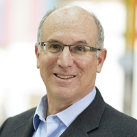 Russ Hauser合作论文数Department of Environmental Health, Harvard T.H. Chan School of Public Health, Harvard University;Department of Epidemiology, Harvard T.H. Chan School of Public Health, Harvard University;Center for the Environment, Harvard University34
 Paige L. Williams合作论文数Harvard T.H. Chan School of Public Health, Harvard University23
 Irene Souter合作论文数20
 Jorge E. Chavarro合作论文数Department of Epidemiology, Harvard T.H. Chan School of Public Health;Department of Nutrition, Harvard T.H. Chan School of Public Health;Medicine-Brigham and Women's Hospital, Harvard Medical School19
 Steven R. Lindheim合作论文数Department of Obstetrics and Gynecology, Boonshoft School of Medicine, Wright State University;Division of Reproductive Endocrinology and Infertility, Wright State University17
 Antonia M. Calafat合作论文数Division of Laboratory Sciences, National Center for Environmental Health, Centers for Disease Control and Prevention16Dimitriadis Irene合作论文数15
 Audrey Jane Gaskins合作论文数Division of Epidemiology, Statistics, and Prevention Research, Eunice Kennedy Shriver National Institute of Child Health and Human Development15
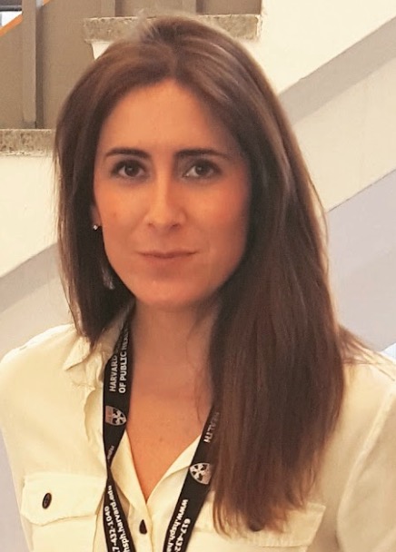 Lidia Mínguez-Alarcón合作论文数Harvard Medical School, Brigham and Women's Hospital13
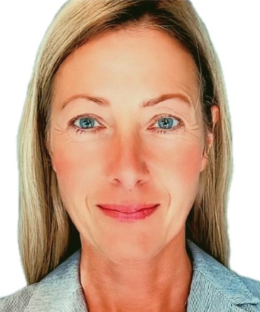 Carmen Messerlian合作论文数The Vie Institute for Generational Health;The Institute for Quantitative Social Sciences, Harvard University;Vie Science, Inc.;Ivy Thrive, LLC;Massachusetts General Hospital;Department of Environmental Health, Harvard T.H. Chan School of Public Health, Harvard University;Department of Epidemiology, Harvard T.H. Chan School of Public Health, Harvard University12
 John Meeker合作论文数School of Public Health, University of Michigan10Jennifer B. Ford合作论文数9Yu-Han Chiu合作论文数8
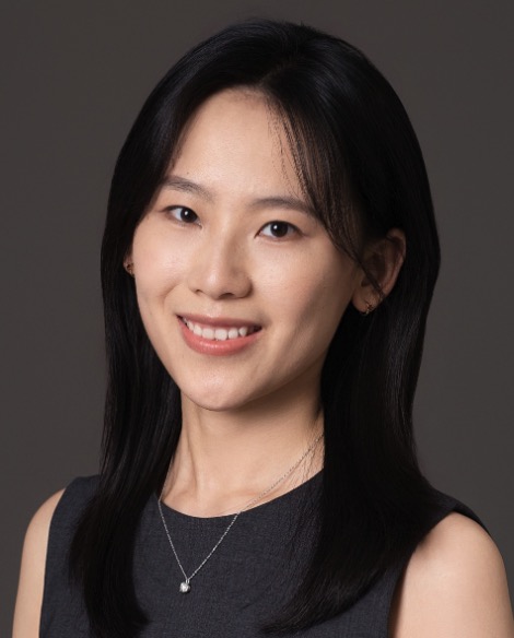 Yu Zhang (张羽)合作论文数College of Environmental Sciences and Engineering, Peking University8
 Vicente Mustieles合作论文数University of Granada8
 Feiby L Nassan合作论文数Department of Environmental Health, Harvard TH Chan School of Public Health;Biogen Inc;Department of Environmental Health, Harvard TH Chan School of Public Health8
 Batsis Maria合作论文数Massachusetts General Hospital Fertility Center, Harvard Medical School;Section on Endocrinology and Genetics, Program on Developmental Endocrinology and Genetics &Eunice Kennedy Shriver National Institute of Child Health and Human Development, National Institutes of Health;Eunice Kennedy Shriver National Institute of Child Health and Human Development, National Institutes of Health8Keller Myra合作论文数8
 Aaron Styer合作论文数8Ayanna Coburn-Sanderson合作论文数7Nicole Torres合作论文数7
 Prudhvi Thirumalaraju合作论文数Brigham & Womens Hosp, Harvard Med Sch7
 Manoj Kumar Kanakasabapathy合作论文数Brigham & Womens Hosp, Harvard Med Sch7
 Hadi Shafiee合作论文数Brigham and Women's Hospital, Harvard Medical School7
 Yang Sun合作论文数7Afeiche Myriam C合作论文数Department of Biostatistics, and cDepartment of Epidemiology, Harvard School of Public Health;d Division of Preventive Medicine and Public Health, University of Murcia School of Medicine;University Department of Growth and Reproduction, University of Copenhagen;School of Medicine, University of Rochester;Department of Preventive Medicine, Mount Sinai School of Medicine;and hChanning Division of Network Medicine, Department of Medicine, Brigham and Women's Hospital and Harvard Medical School, Boston, MA.7Martin Leah J合作论文数School of Public Health, University of Alberta7
 Charles L. Bormann合作论文数Department of Obstetrics Gynecology & Repro. Bio, Harvard Medical School;Massachusetts General Hospital;Brigham and Women's Hospital7Cigdem Tanrikut合作论文数7
 Yixin Wang (汪一心)合作论文数School of Public Health, Shanghai Jiao Tong University6Evelyn Minis合作论文数6Zainab Bibi合作论文数6Victoria W. Fitz合作论文数6Kaitlyn James合作论文数6Karissa C Hammer合作论文数6
 Samantha M. Pfeifer合作论文数Ronald O. Perelman and Claudia Cohen Center for Reproductive Medicine, Weill Cornell Medicine, Cornell University6
 Tamarra James-Todd合作论文数6
 Pietro Bortoletto合作论文数Feinberg School of Medicine, Northwestern University6
 Bala Bhagavath合作论文数6Thomas L. Toth合作论文数6Olivia First合作论文数5Flyckt Rebecca L合作论文数5
 Andrea Bellavia合作论文数Harvard T.H. Chan School of Public Health, Harvard University5Karmon Anatte E合作论文数5Diane L. Wright合作论文数5Shelley Ehrlich合作论文数5
 Stefan Ehrlich合作论文数4




