Uveal melanoma (UM), the most common intraocular cancer in adults, metastasizes to the liver in 90% of cases. Integrating DNA sequencing of 144 metastases with decade-long patient outcomes, we found that liver metastases are more enriched for BAP1-mutant, monosomy 3 clones, compared to extrahepatic metastases. BAP1 loss in liver metastases independently predicts poorer post-metastatic survival. Patterns of dissemination are set early in primary tumors, enabling molecular profiling to forecast metastatic spread.
Sequence of primers used for mouse genotyping and cell validation, and sequence of guide RNAs targeting mouse Bap1.
ABSTRACT Background The TI‐RADS guides the initial management of thyroid nodules, but the malignancy risk across different categories of cytologically indeterminate thyroid nodules (CITNs), particularly AUS and FN, remains unclear. Methods We reviewed thyroid FNA specimens diagnosed as AUS or FN with TI‐RADS scores of 3, 4, or 5 from 2017 to 2025. Clinical data, molecular results, and surgical outcomes were analyzed. Results A total of 919 CITNs were identified: 304 with TI‐RADS 3, 454 with TI‐RADS 4, and 161 with TI‐RADS 5. In the absence of molecular alterations, the risk of malignancy (ROM) was low for AUS and FN in both TI‐RADS 3 and 4 categories (< 10%). In contrast, TI‐RADS 5 nodules diagnosed as AUS or FN had the highest ROM (56%). ROM similarly increased with the TI‐RADS score. For AUS, ROM was 14% (TI‐RADS 3), 31% (TI‐RADS 4), and 60% (TI‐RADS 5). For FN, ROM increased from 26% to 44% to 57% across TI‐RADS 3, 4, and 5, respectively. Notably, even AUS nodules without detectable mutations had a high ROM of 44% in TI‐RADS 5, compared to 10% and 15% in TI‐RADS 3 and 4, respectively. Conclusion Our findings demonstrate distinctive ROMs among AUS and FN stratified by TI‐RADS category and underscore the variable utility of molecular testing. TI‐RADS 5 CITNs carry a significantly higher ROM even in the absence of molecular alterations, suggesting the importance of combining TI‐RADS and TBSRTC.
BACKGROUND:Thyroid nodules, found in up to 68% of adults, can be risk-stratified by high-resolution ultrasound. This study evaluates the concordance between "suspicious" Thyroid Imaging Reporting and Data System (TI-RADS) 3-5 nodules and cytologic and molecular findings from fine-needle aspiration. METHODS:A total of 1199 suspicious thyroid nodules stratified as TI-RADS 3 (n = 384), TI-RADS 4 (n = 569), and TI-RADS 5 (n = 246) were studied. Cytopathologic and molecular results were correlated with clinical data from electronic medical records. RESULTS:Cytology, histology, and molecular testing revealed a lower risk of malignancy in TI-RADS 3 and 4 nodules compared to TI-RADS 5. Among TI-RADS 3 nodules, 75% (287 of 384) were cytologically benign, with only one case diagnosed as papillary thyroid carcinoma (PTC), and 8 of 48 resected nodules (17%) confirmed as carcinoma. In TI-RADS 4, 10% (56 of 569) were suspicious for or diagnosed as PTC by cytology, with 85 of 130 (65%) resected nodules confirmed as carcinoma. In TI-RADS 5, 44% (109 of 246) were cytologically suspicious for or diagnosed as PTC, and 129 of 154 (84%) resected nodules were malignant. High-risk mutations were more frequent in TI-RADS 5 than in TI-RADS 3 and 4. Overall malignancy rates were 2.0% (eight of 384) for TI-RADS 3, 14% (77 of 569) for TI-RADS 4, and 52% (129 of 246) for TI-RADS 5 nodules. CONCLUSION:TI-RADS 3, 4, and 5 nodules demonstrate distinct cytologic, molecular, and histologic features. TI-RADS 3 and 4 nodules are associated with lower malignancy risks, whereas TI-RADS 5 nodules exhibit a high risk of malignancy and are associated with higher mortality.
Objective Diagnoses rendered using the frozen section (FS) technique during surgical procedures are used to guide intraoperative decisions. Therefore, diagnostic FS errors have the potential to affect patient safety and quality of care. Diagnostic FS errors arise due to both technical and interpretative factors and present a challenge to surgical pathology laboratories to recognize, document, and manage in a timely fashion. Thus, there is a need to monitor discrepancies between FS and permanent diagnoses and effectively communicate with the clinical teams when an error is discovered to ensure an opportunity for timely interventions, if clinically indicated.Methods Our FS practice is complex, with many contributing variables, such as a partially generalized FS pathology practice model among pathology faculty and/or surgeons with specific subspecialty expertise and different physical locations of FS facilities. We implemented a comprehensive frozen section quality assurance (FSQA) program using custom software solutions aimed at improving patient safety by monitoring recognition, increasing documentation, and facilitating communication in cases where there is a discordance between intraoperative and permanent diagnoses.Results Our FSQA program allows for categorizing frozen section discrepancies according to the source of error, such as interpretive vs technical errors, to understand how errors arise and to develop appropriate mitigation strategies for reducing errors.Conclusions Overall, our intervention to improve FSQA has engaged pathology faculty in a uniform and systematic manner, and our data show that our new FSQA program led to a markedly shortened time interval of FSQA, allowing for timely management and resolution of errors.
Conjunctival melanoma is a rare, life- and sight-threatening ocular malignancy sharing molecular features with cutaneous and mucosal melanoma. Despite current clinical approaches, high recurrence rates and frequent metastases pose significant challenges in management. Immune checkpoint inhibitors such as ipilimumab and nivolumab have revolutionized cutaneous melanoma treatment, but their efficacy in conjunctival melanoma remains largely unexplored. Herein, the authors present the case of metastatic palpebral conjunctival melanoma in a 59-year-old male successfully treated with a first-line combination of ipilimumab and nivolumab without adjuvant therapies or local surgeries. Local disease resolution was achieved after only 7 months of treatment, and the immune checkpoint inhibitor regimen was well-tolerated with limited systemic adverse effects and no ocular side effects.
Ocular manifestations in chronic lymphocytic leukemia (CLL) have been reported in 30% to 40% of patients and may be a result of direct tissue infiltration, concomitant blood dyscrasias, or a result of therapeutic intervention. Leukemia cutis, defined as infiltration of the epidermis or dermis by neoplastic lymphocytes, is rare. Herein, we present a case report of a patient with leukemia who presented with periorbital edema and ecchymosis. This is the first known case to date of periorbital CLL successfully treated with low-dose radiation therapy (4 Gy in 2 fractions). Clinicians should be aware of the possibility of ocular involvement from CLL, given the importance of prompt diagnosis and treatment.
Purpose: To present the clinical course of a patient with recurrent NK/T-cell lymphoma (NKTL) involving the orbit and to review the literature on patients with NKTL involving the orbit. Methods: The PubMed database was searched for all cases of NKTL involving orbital, intraocular, or adnexal ocular structures. Results: Ninety-six patients were included in the final analysis. The mean age of diagnosis was 48.1 ± 16.8 years. The patients were 53/96 (55.2%) male and 43/96 (44.8%) female. Tumor location varied and included the orbit in 80/96 (83.3%), nasosinus in 56/96 (58.3%), uvea in 11/96 (11.5%), lacrimal gland in 9/96 (9.4%), lacrimal drainage system in 11/96 (11.5%), and conjunctiva in 7/96 (7.3%) cases. Management included surgical debulking in 29/96 (30.2%) cases, radiotherapy in 52/96 (54.2%) cases, and chemotherapy in 82/96 (85.4%) cases. Median survival was 6 months (95% CI: 5–9). Chemotherapy (hazard ratio = 0.80, 95% CI: 0.67–0.95, p = 0.013), radiotherapy (hazard ratio = 0.75, 95% CI: 0.64–0.87, p < 0.001), and orbital involvement being a recurrence of disease (hazard ratio = 0.79, 95% CI: 0.67–0.95, p = 0.009) were associated with improved survival. Advanced Ann Arbor stage (III–IV) at diagnosis (hazard ratio = 1.22, 95% CI: 1.08–1.38, p = 0.001), vision loss (hazard ratio = 1.18, 95% CI: 1.04–1.34, p = 0.009), proptosis (hazard ratio = 1.15, 95% CI: 1.01–1.30, p = 0.035) and periorbital swelling (hazard ratio = 1.15, 95% CI: 1.00–1.33, p = 0.048) were associated with poor survival. Conclusions: NK/T-cell lymphoma involving the orbit, globe, or ocular adnexa heralds a poor prognosis where early diagnosis and therapy are critical. The use of radiotherapy and chemotherapy is associated with improved survival.
Background Guidelines recommend universal mismatch repair (MMR) tumour testing of colorectal adenocarcinomas (CRCs) to screen for Lynch syndrome (LS). However, its implementation remains disjointed and referral for genetic testing dismal, particularly among minorities. We aimed to increase referral, cancer genetic testing and eventually LS diagnosis by developing the CLEAR LS (Closed Loop Enhanced Assessment and Referral for Lynch Syndrome) intervention, a systems approach which in the second phase was automated. Methods This is a cohort study of all patients diagnosed with CRC at an academic centre between 1 January 2012, when implementation of universal CRC testing began, and 31 January 2021. The original cohort spanned through 31 May 2015. Tumour testing included MMR immunohistochemistry, followed by BRAF V600E/ MLH1 promoter methylation testing when indicated. The intervention included a manual phase (1 June 2015 through 31 July 2018), which systematised pathology screening and cancer genetics (CG) referral mechanisms, and an automated phase (1 August 2018 through 31 January 2021) using computer programming. Results A total of 249/1541 CRC (17.38%) had MMR loss of expression and 129 (8.37%) qualified for CG evaluation. Referral was 27.58% in the original cohort and 92.1% in the intervention (p<0.001). Patients seen by CG among referred were 27.58% in the original cohort and 74.3% in the intervention (p two-sided<0.001). The distribution of race/ethnicity among patients qualifying and referred for CG evaluation was not significantly different across cohorts. LS diagnosis increased from 0.56% (original cohort) to 1.43% (intervention). Cost per new diagnosis of LS decreased from US$173 675 to $87 960 from original cohort to intervention. Conclusion Implementation of systematic case identification and referral support mechanisms significantly increased the proportion of patients undergoing genetic testing and doubled the percentage of patients diagnosed with LS with no referral differences across racial/ethnic groups.
BACKGROUND:Artificial intelligence (AI) is rapidly fuelling a fundamental transformation in the practice of pathology. However, clinical integration remains challenging, with no AI algorithms to date in routine adoption within typical anatomic pathology (AP) laboratories. This survey gathered current expert perspectives and expectations regarding the role of AI in AP from those with first-hand computational pathology and AI experience. METHODS:Perspectives were solicited using the Delphi method from 24 subject matter experts between December 2020 and February 2021 regarding the anticipated role of AI in pathology by the year 2030. The study consisted of three consecutive rounds: 1) an open-ended, free response questionnaire generating a list of survey items; 2) a Likert-scale survey scored by experts and analysed for consensus; and 3) a repeat survey of items not reaching consensus to obtain further expert consensus. FINDINGS:Consensus opinions were reached on 141 of 180 survey items (78.3%). Experts agreed that AI would be routinely and impactfully used within AP laboratory and pathologist clinical workflows by 2030. High consensus was reached on 100 items across nine categories encompassing the impact of AI on (1) pathology key performance indicators (KPIs) and (2) the pathology workforce and specific tasks performed by (3) pathologists and (4) AP lab technicians, as well as (5) specific AI applications and their likelihood of routine use by 2030, (6) AI's role in integrated diagnostics, (7) pathology tasks likely to be fully automated using AI, and (8) regulatory/legal and (9) ethical aspects of AI integration in pathology. INTERPRETATION:This systematic consensus study details the expected short-to-mid-term impact of AI on pathology practice. These findings provide timely and relevant information regarding future care delivery in pathology and raise key practical, ethical, and legal challenges that must be addressed prior to AI's successful clinical implementation. FUNDING:No specific funding was provided for this study.
Purpose:To describe a case of local recurrence of uveal melanoma with concomitant brain metastases after secondary enucleation.Observations:A 73 year-old patient presented with dizziness and gait instability. MRI of the orbits and brain showed an anophthalmic socket with an orbital implant and an associated optic nerve mass as well as multiple mass lesions in the brain. The patient's history was significant for secondary enucleation for uveal melanoma recurrence seven years prior to presentation. Histopathology of the enucleated eye revealed no signs of extrascleral extension or optic nerve invasion. Biopsy of the optic nerve mass confirmed recurrent uveal melanoma with somatic mutations in GNAQ (Q209L) and the telomerase (TERT) promoter (c.1-124C > T) found on targeted next-generation sequencing (NGS). The same mutations were found in the primary tumor in the patient's archived enucleation samples.Conclusions:Local recurrence of uveal melanoma can occur after enucleation and is associated with an increased risk of systemic metastases. It is important for clinicians to monitor patients for local recurrence and systemic metastases even after enucleation. Genetic biomarkers may play an important role in identifying tumors at highest risk of local recurrence and metastasis. To our knowledge, this is the first case study to describe the TERT promoter mutation c.1-124C > T in the setting of recurrent uveal melanoma.
Primary mesenchymal tumors of the thyroid gland are extremely rare, with only case reports and small case series documented in the English literature, many of which were published prior to the era of molecular pathology. In the current study, we aim to present a contemporary multi-centric cohort of thyroid mesenchymal tumors. Nineteen primary thyroid mesenchymal tumors were collected from three tertiary centers. Their clinicopathologic features, immunoprofile, molecular alterations, and outcome were described. Eight cases were classified as benign or intermediate with solitary fibrous tumor being the most common histotype (n = 3). The remaining 11 cases were malignant, including three angiosarcomas, one epithelioid hemangioendothelioma, one adamantinoma-like Ewing sarcoma, one biphasic synovial sarcoma, one malignant melanocytic peripheral nerve sheath tumor (melanotic schwannoma), one myxofibrosarcoma, and two undifferentiated pleomorphic/spindle sarcomas (one of which was radiation-induced). Six tumors showed characteristic diagnostic translocations. We herein also described the first case of thyroid malignant perivascular epithelioid cell tumor (PEComa) with RBM10-TFE3 fusion in a 35-year-old female patient. Thyroid mesenchymal tumors, benign or malignant, are rare with a broad spectrum of possible diagnoses. A comprehensive examination to include histology, immunohistochemistry, and molecular testing is essential for the correct diagnosis and to distinguish them from anaplastic thyroid carcinoma. PEComa may occur as a primary tumor of the thyroid gland, expanding the histologic spectrum of thyroid mesenchymal tumors.
ABSTRACTBackgroundArtificial intelligence (AI) is rapidly fueling a fundamental transformation in the practice of pathology. However, AI’s clinical integration remains challenging, with no AI algorithms to date enjoying routine adoption within typical anatomic pathology (AP) laboratories. This survey gathered current expert perspectives and expectations regarding the role of AI in AP from those with first-hand computational pathology and AI experience.MethodsPerspectives were solicited using the Delphi method from 24 subject matter experts between December 2020 and February 2021 regarding the anticipated role of AI in pathology by the year 2030. The study consisted of three consecutive rounds: 1) an open-ended, free response questionnaire generating a list of survey items; 2) a Likert-scale survey scored by experts and analyzed for consensus; and 3) a repeat survey of items not reaching consensus to obtain further expert consensus.FindingsConsensus opinions were reached on 141 of 180 survey items (78.3%). Experts agreed that AI would be routinely and impactfully used within AP laboratory and pathologist clinical workflows by 2030. High consensus was reached on 100 items across nine categories encompassing the impact of AI on (1) pathology key performance indicators (KPIs) and (2) the pathology workforce and specific tasks performed by (3) pathologists and (4) AP lab technicians, as well as (5) specific AI applications and their likelihood of routine use by 2030, (6) AI’s role in integrated diagnostics, (7) pathology tasks likely to be fully automated using AI, and (8) regulatory/legal and (9) ethical aspects of AI integration in pathology.InterpretationThis is the first systematic consensus study detailing the expected short/mid-term impact of AI on pathology practice. These findings provide timely and relevant information regarding future care delivery in pathology and raise key practical, ethical, and legal challenges that must be addressed prior to AI’s successful clinical implementation.FundingThis research received no specific grant from any funding agency in the public, commercial, or not-for-profit sectors.
Purpose:This work reports a case of left atrial myxoma presenting with cilioretinal artery occlusion.Methods:A case report is discussed.Results:A 57-year-old man was referred for acute vision loss in the left eye after a workup including electrocardiogram, magnetic resonance imaging of the brain without contrast, computed tomography angiography of the head and neck, erythrocyte sedimentation rate, and C-reactive protein had negative results. Examination revealed cilioretinal artery occlusion with visible emboli. Because an echocardiogram was not performed, the patient was referred to the emergency department. Echocardiogram revealed a large left atrial mass prolapsing into the left ventricle. The mass was excised, and pathology showed myxoma.Conclusions:This is the first case to our knowledge of isolated cilioretinal artery occlusion as the initial presentation of an atrial myxoma. Thorough and complete workup was crucial to averting further morbidity and mortality.
A 14-year-old female presented with 2 weeks of progressive right eye pain, erythema, and proptosis. Examination demonstrated an enlarged palpable mass along the right superior lateral orbit and bilateral conjunctival petechiae. Of note, she was asymptomatic on the left side, and the petechiae were present only on the superior bulbar conjunctiva with eyelid eversion. Imaging demonstrated bilateral lacrimal gland enhancement. Testing was significant for elevated inflammatory markers, but otherwise negative workup. Biopsy of the right lacrimal gland demonstrated acute-on-chronic inflammation without evidence of lymphoproliferative disease. On repeat testing, myeloperoxidase antibody levels (MPO/p-ANCA) were elevated, indicative of an underlying immune-mediated vasculitis. This case illustrates a rare presentation of ANCA-associated vasculitis in a pediatric patient. It further demonstrates the phenomenon of initial negative serology and subsequent auto-antibody seroconversion in a patient with localized granulomatosis with polyangiitis.
 Pointdujour-Lim R合作论文数10
 Jon Morrow合作论文数Molecular Medicine, Pharmacology, and Physiology, Biological & Biomedical Sciences, Yale University9
 David Rimm合作论文数Departments of Pathology, Yale University School of Medicine;Yale Cancer Center Tissue Microarray Facility, Yale University School of Medicine9
 Thomas Pollard合作论文数School of Medicine, Yale University9Walter H. Henricks合作论文数8
 Peter Gershkovich合作论文数Pathology Faculty, Yale School of Medicine, Yale University7Jenna May Kim合作论文数6Emily Li合作论文数5myra l wilkerson合作论文数5
 Liron Pantanowitz合作论文数Department of Pathology, Medical School, University of Michigan-Ann Arbor5William J. Castellani合作论文数5
 Miguel Angel Materin合作论文数School of Medicine, Duke University4
 Joanna Gibson合作论文数Pathology Faculty, Yale School of Medicine, Yale University4Elia Maxwell D合作论文数4Whitsitt Mark S合作论文数4
 Alexis B. Carter合作论文数4Claudia Auw‐Haedrich合作论文数3Arun D Singh合作论文数3Chitrawati Gargade合作论文数3Laurie Russell合作论文数3Krishna Kalyam合作论文数3Jane Z Spadaro合作论文数3Mengensatzproduktion合作论文数3Brittany M. Perzia合作论文数3John P. Gorham合作论文数3Emily Zolfaghari合作论文数3Devasis N. Reddy合作论文数3
 Arpan Gandhi合作论文数3Shaina M Rubino合作论文数3Femida Kherani合作论文数3Bradley T Smith合作论文数3Prachi Gurav合作论文数3Thamolwan Surakiatchanukul合作论文数3Claudine Bellerive合作论文数3Meisha L. Raven合作论文数3
 Abhishek Dave合作论文数Ctr Sight, Cornea & Refract Surg Serv3
 Kareem Sioufi合作论文数Ocular Oncol Serv, Thomas Jefferson Univ3
 Mozhgan Rezaei Kanavi合作论文数Res Inst Ophthalmol & Vis Sci, Shahid Beheshti Univ Med Sci3
 David McClintock合作论文数Department of Pathology, Medical School, University of Michigan-Ann Arbor3
 Vinita Parkash合作论文数Pathology Faculty, Yale School of Medicine, Yale University3
 Guoping Cai合作论文数Pathology Faculty, Yale School of Medicine, Yale University3
 John Holds合作论文数3
 Jill R Wells合作论文数3Azari Amir A合作论文数Department of Ophthalmology and Visual Sciences, University of Wisconsin, Madison3Potter Heather D合作论文数3Jubran Rima F合作论文数3
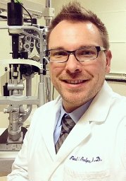 Paul O. Phelps合作论文数Department of Ophthalmology, Hospital of Cook County3Habib Larissa合作论文数Institute of Ophthalmology and Visual Science, Rutgers New Jersey Medical School3
 Jacob S Heng合作论文数Faculty of Medicine, Imperial College3




