Our previous work showed that a Sca-1(+) cell-based FGF2 therapy was capable of promoting robust increases in trabecular bone formation and connectivity on the endosteum of long bones. Past work reported that administration of FGF2 protein promoted bone formation in red marrow but not in yellow marrow. The issue as to whether the Sca-1(+) cell-based FGF2 therapy is effective in yellow marrow is highly relevant to its clinical potential for osteoporosis, as most red marrows in a person of an advanced age are converted to yellow marrows. Accordingly, this study sought to compare the osteogenic effects of this stem cell-based FGF2 therapy on red marrow-filled lumbar vertebrae with those on yellow marrow-filled caudal vertebrae of young adult W(41)/W(41) mice. The Sca-1(+) cell-based FGF2 therapy drastically increased trabecular bone formation in lumbar vertebrae, but the therapy not only did not promote bone formation but instead caused substantial loss of trabecular bone in caudal vertebrae. The lack of an osteogenic response was not due to insufficient engraftment of FGF2-expressing Sca-1(+) cells or inadequate FGF2 expression in caudal vertebrae. Previous studies have demonstrated that recipient mice of this stem cell-based FGF2 therapy developed secondary hyperparathyroidism and increased bone resorption. Thus, the loss of bone mass in caudal vertebrae might in part be due to an increase in resorption without a corresponding increase in bone formation. In conclusion, the Sca-1(+) cell-based FGF2 therapy is osteogenic in red marrow but not in yellow marrow.
It is generally well accepted that the pubertal surge in estrogen is responsible for the rapid bone accretion that occurs during puberty and that this effect is mediated by an estrogen-induced increase in GH/IGF action. To test the cause and effect relationship between estrogen and GH/IGF, we evaluated the consequence of ovariectomy (OVX) in prepubertal mice (C57BL/6J mice at 3 weeks of age) on skeletal changes and GH/IGF axis during puberty. Contrary to our expectations, OVX increased body weight (12 to 18%), bone mineral content (11%), bone length (4%), bone size (3%), and serum, liver and bone IGF-I (30 to 50%) and decreased total body fat (18%) at 3 weeks post-surgery. To determine if estrogen is the key ovarian factor responsible for these changes, we performed a second experiment in which OVX mice were treated with placebo or estrogen (E) implants. In addition to observing similar results compared with our first experiment, estrogen treatment partially rescued the increased body weight and bone size and completely rescued body fat and IGF-I levels. The increased bone accretion in OVX mice was due to increased bone formation rate (as determined by bone histomorphometry) and increased serum procollagen peptide. In conclusion, contrary to the known estrogen effect as an initiator of GH/IGF surge and thereby pubertal growth spurt, our findings demonstrate that loss of estrogen and/or other hormones during the pre-pubertal growth period effect leads to an increase in IGF-I production and bone accretion in mice.
Phenotype-driven mutagenesis approach in the mouse holds much promise as a method for revealing gene function. Earlier, we have described an N-ethyl-N-nitrosourea (ENU) mutagenesis screen to create genome-wide dominant mutations in the mouse model. Using this approach, we describe identification of two high bone density mutants in C57BL/6J (136) background. The mutants, named as 12184 and 12137, have been bred more than five generations with wild-type 136 mice, each producing >200 backcross progeny. The average total body areal bone mineral density (aBMD) was 13-17% higher in backcrossed progeny from both mutant lines between 6 and 10 weeks of age, as compared to wild-type (WT) 136 mice (n=60-107). At 3 weeks of age the aBMD of mutant progeny was not significantly affected as compared to WT 136 mice. Data from 10- and 16-week old progeny show that increased aBMD was mainly related to a 14-20% higher bone mineral content, whereas bone size was marginally increased. In addition, the average volumetric BMD (vBMD) was 5-15% higher at the midshaft tibia or femur, as compared to WT mice. Histomorphometric analysis revealed that bone resorption was 23-34% reduced in both mutant mice. Consistent with histomorphometry data, the mRNA expression of genes that regulate osteoclast differentiation and survival were altered in the 12137 mutant mice. To determine the chromosomal location of the ENU mutation, we intercrossed both mutant lines with C3H/HeJ (C3H) mice to generate B6C3H F2 mice (n = 164 for line 12137 and n= 137 F2 for line 12184). Interval mapping using 60 microsatellite markers and aBMD phenotype revealed only one significant or suggestive linkage on chromosome 4. Since body weight was significantly higher in mutant lines, we also used body weight as additive and interactive covariate for interval mapping; both analyses showed higher LOD scores for both 12137 and 12184 mutants without affecting the chromosomal location. The large phenotype in the mutant mice compared to generally observed QTL effects (<5%) would increase the probability of identifying the mutant gene. (c) 2007 Elsevier Inc. All rights reserved.
The findings that sex-specific effects on femoral structure and peak bone mineral density (BMD) are linked to quantitative trait loci (QTL) provide evidence for the involvement of specific genes that contribute to gender variation in skeletal phenotype. Based on previous findings that the BMD QTL in chromosome 1 (Chr 1) exerts a sex-specific effect on femoral structure, we predicted that congenic sublines of mice that carry one or more of the Chr 1 BMD loci would exhibit gender difference in the volumetric BMD (vBMD) phenotype. To test this hypothesis, we compared skeletal parameters of male and female of five C57BL/6J (B6).CAST/EiJ (CAST)-1 congenic sublines of mice that carry overlapping CAST chromosomal segments from the vBMD loci in Chr 1. Femur vBMD measurements were performed by the peripheral quantitative computed tomography in male and female mice at 16 weeks of age. The skeletal phenotype of the C175-185 and C178-185 congenic sublines of mice provided evidence for the presence of the BMD1-4 locus at 178-180 Mb from the centromere. This QTL affects femur vBMD only in female mice. In contrast, CAST chromosomal region carrying BMD1-1 locus increased femur vBMD both in male and female mice. Further-more, a gender specific effect on BMD of femur mid-shaft region (mid-BMD) was identified at 168-176 Mb in Chr 1 (F= 16.49, P=0.0002), while no significant effect was found on total femur BMD (F= 2.67, P=0.11). Moreover, this study allowed us to locate a body weight QTL at 168172 Mb of Chr 1, the effect of this locus was altered in female mice that carry CAST chromosomal segment 168-176 Mb of Chr 1. Based on this study, we conclude that Chr 1 carries at least two vBMD gender-dependent loci; one genetic locus at 178-180 Mb (BMD1-4 locus) which affects both mid-shaft and total femur vBMD in female mice only, and another gender-dependent locus at 168-176 Mb (BMD1-2 locus) which affects femur mid-shaft vBMD in female but not male mice. (C) 2007 Elsevier Inc. All rights reserved.
Most previous studies to identify loci involved in bone mineral density (BMD) regulation have used inbred strains with high and low BMD in generating F2 mice. However, differences in BMD may not be a requirement in selecting parental strains for BMD quantitative trait loci (QTL) studies. In this study, we intended to identify novel QTL using a cross of two strains, MRL/MpJ (MRL) and CAST/EiJ (CAST), both of which exhibit relatively high BMD when compared to previously used strains. In addition, CAST was genetically distinct. We generated 328 MRL × CAST F2 mice of both sexes and measured femur BMD and periosteal circumference (PC) using peripheral quantitative computed tomography. Whole-genome genotyping was performed with 86 microsatellite markers. A new BMD QTL on chromosome 10 and another suggestive one on chromosome 15 were identified. A significant femur PC QTL identified on chromosome 9 and a suggestive one on chromosome 2 were similar to those detected in MRL × SJL. QTL were also identified for other femur and forearm bone density and bone size phenotypes, some of which were colocalized within the same chromosomal positions as those for femur BMD and femur PC. This study demonstrates the utility of crosses involving inbred strains of mice which exhibit a similar phenotype in QTL identification.
Microarray analysis of gene expression was performed in the healing femur fractures of 13-week-old male rats during the inflammatory stage of repair, at 3 days post-fracture, and the endochondral bone formation stage of repair, at 11 days post-fracture. Multiple replicate pairs of fracture tissues paired with unfractured tissues, and unfractured control bones that had the stabilizing K-wire were introduced. This approach normalized the marrow contributions to the RNA repertoire. We identified 6555 genes with significant changes in expression in fracture tissues at 3 days and 11 days healing. The repertoire of growth factor genes expressed was also surprisingly restricted at both post-fracture intervals. The large number of Expressed Sequence Tags (ESTs) expressed at both post-fracture times indicates that several molecular pathways yet to be identified regulate fracture repair. The number of genes expressed during immune responses and inflammatory processes was restricted with higher expression largely during the early post-fracture analysis. Several of the genes identified in this study have been associated with regulation of cell and extracellular matrix interactions during scarless healing of fetal skin wounds. These observations suggest that these genes might also regulate the scarless healing characteristic of bone regeneration by similar mechanisms.
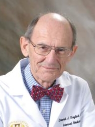 David J. Baylink合作论文数Regenerative Medicine Division, School of Medicine, Loma Linda University;School of Dentistry, Loma Linda University83
 S Mohan合作论文数56Wesley G Beamer合作论文数19KHW Lau合作论文数17MHC Sheng合作论文数12John R. Farley合作论文数9AK Srivastava合作论文数8ST Chen合作论文数8
 K-H William Lau合作论文数Dept Med, Loma Linda Univ8N Miyakoshi合作论文数8Yuji Kasukawa合作论文数8Susan L. Hall合作论文数7LR Donahue合作论文数7Bouchra Edderkaoui合作论文数7Hongrun Yu合作论文数7RB Chadwick合作论文数6Cj Rosen合作论文数6Thomas a Linkhart合作论文数6
 Cheryl L. Ackert-Bicknell合作论文数The Jackson Laboratory6CH Rundle合作论文数6
 Cesar Libanati合作论文数UCB Pharma6DD STRONG合作论文数5EE SCHULZ合作论文数5Kristen E Govoni合作论文数VA Medical Center5Chandrasekhar Kesavan合作论文数5
 Subburaman Mohan合作论文数Musculoskeletal Disease Center, VA Loma Linda Healthcare System;Biochemistry Division, School of Medicine, Loma Linda University4A Srivastava合作论文数3Mw Lundy合作论文数3KL Shultz合作论文数3SM FARLEY合作论文数3JR TALBOT合作论文数3CR LIBANATI合作论文数3R Gysin合作论文数3R Guo合作论文数3Wesley G. Beamer合作论文数3T. Ohta合作论文数3David J Baylink合作论文数3Godfred Masinde合作论文数VA Medical Center, Loma Linda University;Musculoskeletal Disease Center;Molecular Genetics Division3M. H.-C. Sheng合作论文数3Roderick T. Bronson合作论文数Harvard Medical School;Dana-Farber / Harvard Cancer Center3Patricia Ward-Bailey合作论文数3Clarita V. Odvina合作论文数3Y Amaar合作论文数3Toshikatsu Matsuyama合作论文数2KE Govoni合作论文数2AK TAYLOR合作论文数2AL BEACHLER合作论文数2K Johnson合作论文数2Y Kodama合作论文数2




