Cardiac transthyretin amyloidosis (ATTR-CA) is caused by myocardial deposition of misfolded transthyretin, leading to progressive heart failure. Disease pathology, however, extends beyond passive amyloid deposition and also involves active processes such as extracellular matrix (ECM) remodeling and immune activation. Mass spectrometry (MS) is the gold standard for amyloid typing in diagnostics. Here, we applied quantitative MS-driven proteomics on formalin-fixed paraffin-embedded whole cardiac tissue sections from six ATTR-CA cases, ten unaffected controls and four AL-CA controls to investigate protein expression changes. In addition to transthyretin, over 500 proteins were upregulated in ATTR-CA biopsies, including complement and coagulation factors as well as extracellular matrix (ECM) remodeling proteins. Among these, members of the A Disintegrin and Metalloproteinase with Thrombospondin Motifs (ADAMTS) family, metalloproteinases (MMPs), and Tissue Inhibitor of Metalloproteinase-3 (TIMP3) showed significant upregulation. These proteins are key regulators of ECM turnover and structural integrity. Immunohistochemistry confirmed ADAMTS4 enrichment in amyloid deposits, while TIMP3 showed strong expression in cardiomyocytes and weaker staining within amyloid deposits. Together, these findings indicate that ECM remodeling, alongside complement and coagulation activation, represents a reproducible feature of cardiac ATTR amyloidosis. Whole-tissue proteomics provides biological insights that extend beyond amyloid typing, with potential implications for biomarker discovery and therapeutic targeting in ATTR-CA.
Tauopathy, including Alzheimer's disease, is the most prevalent neurodegenerative disorder, yet current therapies fail to halt disease progression, partly because of limited understanding of early tau pathological structures. Cryo-EM has clarified distinct Tau structures tied to disease phenotypes but only in their mature forms and at low throughput. Hence, molecular probes that can recognize early Tau conformations in high throughput and in situ hold the potential to transform our understanding of Tau aggregation. Tau undergoes sequential changes, including hyperphosphorylation and misfolding, with the aggregation-prone paired helical filament 6 region in R3 shown to drive its self-assembly and fibrillar core formation. In this study, we used existing and novel monoclonal antibodies to map the Tau protein in Alzheimer's disease and other tauopathies. The conformation-specific antibody, 16B12, targeting the R1-R3 regions, showed high sensitivity in detecting early Tau structures and inhibited seed-induced aggregation in vitro. Another antibody, 9H6F2, detected P1-P2 interactions and functioned as a panbiomarker across tauopathies. Our findings underscore the critical role of early P1-P2 and R1-R3 interactions in Tau fibril maturation, and our monoclonal antibodies show promise as early diagnostic markers for Tau-related neurodegeneration.
The continuous risk of antibiotic resistance development underscores the demand for new agents with mechanisms distinct from existing antibacterial drugs. Here, we investigated HSI#6, a small-molecule antibacterial previously identified as a SecA activator, using integrated omics and functional assays. HSI#6 exhibits a rapid, broad-spectrum bacteriostatic activity, and induces a distinct cell envelope-homeostasis stress signature accompanied by global stress reprogramming. Time-resolved transcriptomics and proteomics revealed early activation of envelope stress regulons and oxidative stress pathways, followed by suppression of ribosome biogenesis and central metabolism. Comparative analysis and biomarker-based principal component analysis (PCA) positioned HSI#6 within the envelope stress mechanistic space, closely aligned with membrane-active antibiotics yet displaying a distinct signature. Adaptive laboratory evolution (ALE) combined with whole-genome sequencing (WGS) revealed compensatory mutations in topoisomerase 1A gene (topA) and transcriptional regulators, without adaptive resistance emerged even under prolonged selection pressure. These findings establish HSI#6 as a mechanistically unique antibacterial agent with low resistance potential.
The prevailing view frames microglia and macrophages as guardians against amyloid beta (Aβ) accumulation in Alzheimer’s disease (AD). Here, we overturn this paradigm by demonstrating that human phagocytic cells, including differentiated THP-1 macrophages and hESC-derived microglia, are not merely passive responders but active producers of extracellular, seeding-competent Aβ42 fibrils, the amyloid species most strongly linked to parenchymal plaque formation and neurodegeneration. These cell-generated aggregates differ structurally and functionally from synthetic fibrils, displaying enhanced seeding and tau cross-seeding activity in biosensor models. Notably, Aβ42 fibril formation in this system requires active cellular processes and is exacerbated by loss of Triggering Receptor Expressed on Myeloid Cells 2 (TREM2), a major AD risk gene. Transcriptomic profiling reveals an early inflammatory response resembling microglial states observed in human AD models. Together, these findings support emerging evidence from in vivo studies that macrophages and microglia can influence amyloid seeding and introduce a human-relevant in vitro platform to explore how Aβ aggregation intersects with innate immune function and genetic risk. Our results reinforce the concept that microglia may play a dual role in AD, acting both as responders and inadvertent facilitators of amyloid assembly, with implications for early therapeutic intervention.
Co-translational protein folding is shaped by the vectorial nature of translation, which causes residues to emerge sequentially from the ribosome. As a result, residues whose native interaction partners lie downstream in sequence cannot immediately form their native contacts and remain transiently unsatisfied until those partners are synthesized. These unsatisfied residues are vulnerable to non-native interactions and often require the engagement of co-translational chaperones. We previously developed the Native Fold Delay (NFD) metric to quantify the time lag between the synthesis of a residue and the point at which it can form all its native contacts. Here, we present the FoldDelay web server, a freely accessible platform that extends the NFD concept into a more comprehensive framework for analyzing native residue-residue contact formation during translation. Starting from user-submitted AlphaFold or PDB structures, the site identifies all N- to C-terminal residue-residue contacts, estimates their earliest possible formation times, and integrates domain annotations to distinguish between intra- and inter-domain contacts. The server provides a suite of linked interactive visualizations that allows users to explore native contact formation dynamics and detect transiently unsatisfied regions. The FoldDelay web server is freely accessible at https://folddelay.switchlab.org.
Designing antibodies is complex and resource intensive. While deep learning and generative approaches have shown promise in the design of protein binders, achieving high affinity and stability remains challenging. We introduce EvolveX, a structure-based antibody design pipeline leveraging the empirical force field FoldX to design complementarity-determining regions (CDRs) of single-domain antibodies (VHHs). We demonstrate the ability of EvolveX to redesign a VHH targeting mouse Vsig4 (mVsig4) to address two challenges: enhancing stability and affinity for mVsig4 and redesigning it for high affinity to the human ortholog. Notably, EvolveX improved the binding affinity of VHHs to human Vsig4 by over 1,000-fold. Structural analyses by X-ray crystallography confirmed design accuracy. Next-generation sequencing (NGS) analysis further demonstrated the efficiency of FoldX-based design pipeline. Collectively, our study highlights EvolveX's potential to overcome current limitations in antibody design, offering a powerful tool for the development of therapeutics with enhanced specificity, stability, and efficacy.
This Perspective explores recent methods and prospective ideas for developing hybrid AI-physics-based pipelines for protein and antibody de novo design. We argue that the highest-confidence candidates emerge where deep learning and first-principles models agree, a "sweet spot" that balances generative flexibility with thermodynamic realism. For example, although interface confidence scores such as ipTM, pDockQ2, or ipSAE are widely used to rank generated designs, we show that they are not well suited to rank similar sequences, which suggests the need to combine them with physics-based methods to improve design filtering and ranking. Furthermore, we describe a generalizable framework for implementing antibody design pipelines that combine AI with physics-based modeling and scoring methods and also showcase MadraX, a differentiable and AI-compatible implementation of the FoldX force field. In addition, we classify three tiers of AI-physics integration, from post hoc filtering to full embedding of differentiable physics inside deep learning models. Finally, we discuss the future of the protein design community and underline the need to support current initiatives for community wide blind assessments of the growing number of de novo design pipelines.
Amyloid assembly in vivo occurs in complex environments where multiple aggregation-prone species coexist. Aβ and medin are prevalent amyloids in ageing humans that co-localize in cerebral amyloid angiopathy (CAA), yet their structural interactions remain poorly understood. Here, using cryo-electron microscopy, we determine high-resolution fibril structures from in vitro mixtures of Aβ40 and medin. From the same reaction, we resolve three distinct fibril populations: (i) a previously characterized Aβ40 polymorph that also forms in isolation, (ii) a Aβ40 polymorph with Aβ42-like features, including ordered N- and C-terminal regions, and (iii) the atomic structure of full-length medin fibrils. Biochemical and immunogold analyses demonstrate Aβ-medin association within mixed assemblies, though medin is not resolved within the ordered Aβ core. These findings support two non-exclusive mechanisms: transient heterotypic interactions redirecting Aβ folding, or partial medin incorporation into fibril architecture. Our data reveal how coexisting amyloids remodel each other's polymorphic landscapes.
Drug-resistant bacteria pose an urgent global health threat, necessitating the development of antibacterial compounds with novel modes of action. Protein biosynthesis accounts for up to half of the energy expenditure of bacterial cells, and consequently inhibiting the efficiency or fidelity of the bacterial ribosome is a major target of existing antibiotics. Here, we describe an alternative mode of action that affects the same process: allowing translation to proceed but causing co-translational aggregation of the nascent peptidic chain. We show that treatment with an aggregation-prone peptide induces formation of polar inclusion bodies and activates the SsrA ribosome rescue pathway in bacteria. The inclusion bodies contain ribosomal proteins and ribosome hibernation factors, as well as mRNAs and cognate nascent chains of many proteins in amyloid-like structures, with a bias for membrane proteins with a fold rich in long-range beta-sheet interactions. The peptide is bactericidal against a wide range of pathogenic bacteria in planktonic growth and in biofilms, and reduces bacterial loads in mouse models of Escherichia coli and Acinetobacter baumannii infections. Our results indicate that disrupting protein homeostasis via co-translational aggregation constitutes a promising strategy for development of broad-spectrum antibacterials.
In Alzheimer's disease (AD), amyloid β (Aβ) aggregates, ranging from soluble Aβ oligomers to insoluble Aβ plaques. During Aβ aggregation, in vitro and in silico screening has shown that aggregation-prone regions (APRs) appear as an optimal binding site for protein-protein interactions. Interacting partners that define disease pathways provide potential insight into the cellular environment and protein interactions present during pathological Aβ aggregation. Using mass spectrometry, several proteins beyond Aβ have been discovered in AD pathogenesis. However, there is currently limited knowledge about proteins interacting with soluble Aβ in human brain tissue. This study aimed to systematically assess which proteins interact with soluble Aβ in human cortex across the AD continuum (i.e. pathologically-definite symptomatic AD, asymptomatic AD and non-AD controls) through affinity purification-mass spectrometry with a Aβ-specific binding condition using two Aβ antibodies (4G8: Aβ 17-24 and 6E10: Aβ 1-17 ) and a non-specific binding condition (magnetic beads-bound proteins). For differential expression, Paired T tests were performed to detect proteins significantly more abundant in Aβ 17-24 and Aβ 1-17 binding conditions respectively, considered as the ‘’Aβ interactome’’. Co-expression network analysis was performed to assess which proteins correlated with neuropathological staging. Proteins derived were then assessed for diagnostic group differences to detect early mediators of AD pathogenesis. The combined Aβ 17-24 and Aβ 1-17 interactome consisted out of 129 proteins, including APOE and the glial fibrillary acidic protein and vimentin, which were significantly more abundant in Aβ-IPs versus beads. Co-expression analysis revealed a protein module which was significantly negatively correlated with Aβ phase severity. Diagnostic group comparison indicated that proteins involved in cytoskeletal modelling (septins), clathrin-mediated endocytosis, chaperone function, and Rho GTPase activity were already reduced in the interactome of early stage AD cases. In silico and in vitro validation confirmed the existence of C-terminal Aβ APR homology in 40% of the mass-spectrometry derived Aβ interactome candidates (including PCSK1, HS105 and FN3K). Interactions of Aβ with proteins involved in septins, clathrin-mediated endocytosis, and chaperone functions are diminished in AD, even in asymptomatic individuals. This observation suggests that the impaired physiological protein-binding function of soluble Aβ might represent an early pathogenic event in the development of AD.
Tauopathies are neurodegenerative diseases characterized by the pathological aggregation of tau, which forms distinct polymorphic structures in a disease-specific manner. A recent study presents macrocyclic tau-mimicking peptides that structurally and functionally resemble these disease-associated folds, providing new tools for studying tau aggregation and potential therapeutic strategies.
Cytoplasmic aggregation and nuclear depletion of TAR DNA-binding protein 43 (TDP-43) are hallmarks of several neurodegenerative disorders. Yet, recapitulating both features in cellular systems has been challenging. Here, we produced amyloid-like fibrils from recombinant TDP-43 low-complexity domain and demonstrate that sonicated fibrils trigger TDP-43 pathology in human cells, including induced pluripotent stem cell (iPSC)-derived neurons. Fibril-induced cytoplasmic TDP-43 inclusions acquire distinct biophysical properties, recapitulate pathological hallmarks such as phosphorylation, ubiquitin, and p62 accumulation, and recruit nuclear endogenous TDP-43, leading to its loss of function. A transcriptomic signature linked to both aggregation and nuclear loss of TDP-43, including disease-specific cryptic splicing, is identified. Cytoplasmic TDP-43 aggregates exhibit time-dependent heterogeneous morphologies as observed in patients-including compacted, filamentous, or fragmented-which involve upregulation/recruitment of protein clearance pathways. Ultimately, cell-specific progressive toxicity is provoked by seeded TDP-43 pathology in human neurons. These findings identify TDP-43-templated aggregation as a key mechanism driving both cytoplasmic gain of function and nuclear loss of function, offering a valuable approach to identify modifiers of sporadic TDP-43 proteinopathies.
Amyloid fibrils underpin both functional and pathological protein assemblies and exhibit extensive structural polymorphism. More than 600 high-resolution fibril structures are now available across over 50 protein sequences, yet comparative analysis has been hindered by fragmented resources and inconsistent annotation. Here we present Amyloid Explorer, an open-access platform that integrates the complete fibril archive into a standardized, quality-controlled, and thermodynamically annotated framework. Each entry includes residue- and segment-level stability maps, structural quality metrics, and interactive visualization tools. Global analysis across the dataset reveals conserved rules of amyloid assembly, including cooperative cores, framework polymorphism through reuse of conserved segments in distinct folds, polymorph-specific energetic anchors, and short frustration zones. By combining scale with accessibility, Amyloid Explorer transforms a static archive into a discovery platform for mechanistic insight, classification, and the design of aggregation modulators. ### Competing Interest Statement The authors have declared no competing interest. VIB, C0401 FWO, G0A6724N SAO-FRA, 2023/0005 The University of Texas Southwestern Medical Center, https://ror.org/05byvp690, Thomas O. Hicks Endowed Scholars Award
Amyloid fibrils adopt diverse structural polymorphs linked to disease-specific phenotypes, but the thermodynamic principles guiding their formation and maturation remain unclear. Here, we apply energetic profiling to structural time series from cryo-EM datasets of IAPP, tau, and α-synuclein to decode the principles governing fibril maturation and polymorphic divergence. By mapping residue-level free energy contributions across experimentally resolved assembly pathways, we reconstruct their maturation trajectories and find that amyloid assembly is anchored by aggregation-prone regions that serve as sequence-encoded stabilizing motifs. As assembly progresses, these motifs are reorganized and expanded, while additional regions introduce structural frustration that enables conformational flexibility. Environmental cofactors such as metal ions or polyanions are observed in association with regions of structural remodeling, where they may act to compensate for otherwise energetically strained conformations. This framework offers mechanistic insight into how distinct polymorphs arise from a common sequence and mature through both intrinsic and extrinsic thermodynamic influences.
Medin, a 50-amino acid fragment derived from the protein MFG-E8 (lactadherin), is the most prevalent amyloid found in humans, present in the vasculature of nearly all individuals over the age of 50. Its biological relevance is highlighted by its co-localization with amyloid-β (Aβ) deposits in both Alzheimer's disease patients and transgenic mice models. Notably, Medin promotes amyloid-β aggregation, forming mixed fibrils with Aβ and enhancing its deposition in blood vessels. Here we report a new and efficient strategy to chemically access this compound. Our approach employs a solubilizing linker that not only ensures high solubility but also suppresses aggregation, allowing efficient purification of the product. The linker can be removed without a trace, after which the product behaves identically to wild-type Medin and forms amyloid fibrils. The synthesis route allows opening up a new chemical space, including nonnatural modifications like biotinylation. Together with the control over the aggregation properties, this is a powerful tool for amyloid protein studies.
The prevailing view frames microglia and macrophages as guardians against amyloid beta (Aβ) accumulation in Alzheimer's disease (AD). Here, we overturn this paradigm by demonstrating that human phagocytic cells-including differentiated THP-1 macrophages and iPSC-derived microglia-are not merely passive responders but active producers of extracellular, seeding-competent Aβ42 fibrils, the amyloid species most strongly linked to parenchymal plaque formation and neurodegeneration. These cell-generated aggregates differ structurally and functionally from synthetic fibrils, exhibiting heightened seeding activity and the ability to cross-seed tau aggregation, a key driver of AD progression. Notably, Aβ42 fibril formation in this system requires active cellular processes and is exacerbated by loss of TREM2, a major AD risk gene. Transcriptomic profiling reveals an early inflammatory response resembling microglial states observed in human AD models, positioning this system as a tractable, human-relevant platform to dissect the interplay between Aβ aggregation, innate immunity, and genetic susceptibility. Our findings suggest that macrophages and microglia play a dual role in AD, acting both as responders and inadvertent catalysts of pathogenic amyloid formation, with implications for early therapeutic intervention.
Synthetic oligo pools offer a cost-effective way for generating hundreds of thousands of antibody sequences but pose technical challenges, particularly in constructing full-length, complex antibody libraries and minimizing chimera formation during polymerase chain reaction (PCR) amplification. In this study, we developed a versatile combinatorial library cloning method capable of constructing single-domain antibody (VHH) and antigen-binding fragment (Fab) libraries with diversities reaching tens of thousands and lengths extending up to four complementarity-determining regions (CDRs). Our protocol allows to largely avoid chimera formation or, if desired, to induce full CDR recombination, starting from the same synthetic oligo pool. This method achieved a full-length rate exceeding 90% for both VHH and Fab libraries and a perfect construct error-free rate above 80% for VHH libraries. We also investigated various PCR conditions and validated that lowering template concentration during amplification effectively reduces chimera formation. Additionally, we established a PCR-free next-generation sequencing (NGS) platform to assess the quality of assembled VHH libraries. Overall, these advancements provide a more efficient approach for constructing high-diversity multi-CDR combinatorial libraries with high coverage and accuracy, along with a reliable NGS preparation method for quality control and downstream analysis, aiding the rapid discovery and development of novel therapeutic biologics.
Recalcitrant bacterial infections can be caused by various types of dormant bacteria, including persisters and viable but nonculturable (VBNC) cells. Despite their clinical importance, we know fairly little about bacterial dormancy development and recovery. Previously, we established a correlation between protein aggregation and dormancy in Escherichia coli. Here, we present further support for a direct relationship between both. Our experiments demonstrate that aggregates progressively sequester proteins involved in energy production, thereby likely causing ATP depletion and dormancy. Furthermore, we demonstrate that structural features of protein aggregates determine the cell’s ability to exit dormancy and resume growth. Proteins were shown to first assemble in liquid-like condensates that solidify over time. This liquid-to-solid phase transition impedes aggregate dissolution, thereby preventing growth resumption. Our data support a model in which aggregate structure, rather than cellular activity, marks the transition from the persister to the VBNC state. Recalcitrant infections are often caused by dormant bacteria, including persisters and viable but nonculturable cells. Here, the authors explore how composition and structure of protein aggregates affect dormancy and exit from this state.
 Frederic Rousseau合作论文数Switch Laboratory, Flanders Interuniversity Institute of Biotechnology, Vrije Universiteit Brussel Pleinlaan 2, Brussels, Belgium244
 Nikolaos Louros合作论文数Faculty of Biology, University of Athens43Rodrigo Gallardo合作论文数40
 Rob Van der Kant合作论文数28De Baets Greet合作论文数27Ramakers Meine合作论文数VIB Switch Laboratory, VIB;Department of Cellular and Molecular Medicine, KU Leuven26
 Luis Serrano Pubul合作论文数Centre for Genomic Regulation26Van Durme Joost合作论文数24
 Bert Houben合作论文数KULeuven, VIB, Ctr Brain & Dis Res, Switch Lab, B-3000 Leuven, Belgium23
 Bart De Strooper合作论文数Department of Neurosciences, KU Leuven;Institute of Neurology, University College London;UK Dementia Research Institute, University College London22
 Ladan Khodaparast合作论文数20
 Laleh Khodaparast合作论文数20
 Emiel Michiels合作论文数VIB Ctr Brain & Dis Res, Leuven, Belgium18Joke Reumers合作论文数18Matthias De Vleeschouwer合作论文数VIB Ctr Brain & Dis Res, Switch Lab, Herestr 49, B-3000 Leuven, Belgium17
 Maurer-Stroh Sebastian合作论文数ASTAR, Bioinformat Inst BII16
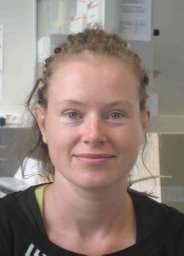 Kerensa Broersen合作论文数Switch Laboratory, Flanders Institute for Biotechnology (VIB) and Vrije Universiteit Brussel (VUB), Pleinlaan 2, Brussels, 1050 Belgium16
 Grigoria Tsaka合作论文数14
 Tom Lenaerts合作论文数Artificial Intelligence lab, Vrije Universiteit Brussel;Interuniversity Institute of Bioinformatics in Brussels, Universite Libre de Bruxelles-Vrije Universiteit Brussel;Machine Learning Group, Universite Libre de Bruxelles14
 Frederik De Smet合作论文数Center for Transgene Technology and Gene Therapy;Flanders Interuniversity Institute for Biotechnology;University of Leuven;Flanders Interuniversity Institute for Biotechnology, University of Leuven14Hannah Wilkinson合作论文数14
 Katerina Konstantoulea合作论文数Switch Laboratory, Department of Cellular and Molecular Medicine, KU Leuven, Leuven, Belgium.13
 Iryna Benilova合作论文数MRC Prion Unit and Institute of Prion diseases, Faculty of Brain Sciences, University College London12Vandersteen Annelies合作论文数12Gabriele Orlando合作论文数12
 Christine Van Broeckhoven合作论文数Center for Molecular Neurology, University of Antwerp;Institute Born-Bunge, University of Antwerp12
 Jan Delcour合作论文数Faculty of Bioscience Engineering, Katholieke Universiteit Leuven;Laboratory of Food Chemistry and Biochemistry, Katholieke Universiteit Leuven12Ramon Duran-Romaña合作论文数11Margarita Monge-Morera合作论文数11
 Rik Vandenberghe合作论文数Departement Neurowetenschappen, KU Leuven;Laboratorium voor Cognitieve Neurologie, KU Leuven;Leuven Brain Institute, KU Leuven11
 Guy Bormans合作论文数Department of Pharmaceutical and Pharmacological Sciences, KU Leuven;Faculty of Pharmaceutical Sciences, KU Leuven11Francois Stricher合作论文数11
 Kristel Sleegers合作论文数Department of Molecular Genetics, University of Antwerp;VIB Center for Molecular Neurology, University of Antwerp11Wim Jonckheere合作论文数10
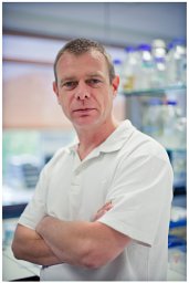 Marc Cruts合作论文数Laboratory of Neurogenetics, University of Antwerp (UIA),10
 Kris Gevaert合作论文数Department of Biomolecular Medicine, Faculty of Medicine and Health Sciences, Ghent University;VIB-UGent Center for Medical Biotechnology10
 Filip Claes合作论文数10
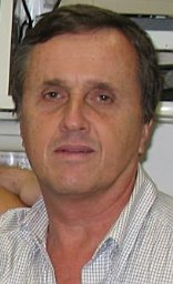 Jose R Couceiro合作论文数10
 Julie van der Zee合作论文数Biobank Expertise Unit, Center for Molecular Neurology, University of Antwerp;VIB-Department of Molecular Genetics, University of Antwerp10
 Sebastiaan Engelborghs合作论文数Center for Neurosciences, Vrije Universiteit Brussel;University Hospital Brussels;Department of Biomedical sciences, University of Antwerp9
 P.P. (Peter) De Deyn合作论文数Faculty of Medical Sciences, University of Groningen;University of Antwerp9Ludo Van Den Bosch合作论文数9
 Marlies A. Lambrecht合作论文数8Baatsen Pieter合作论文数KU Leuven, Katholieke Univ Leuven8
 Nathalie Brouwers合作论文数8
 Christine A. F. Von Arnim合作论文数Department of Neurology, University of Ulm8
 Jesper Ferkinghoff-Borg合作论文数Novo Nordisk A/S8Peter Vanhee合作论文数Vrije Universiteit Brussel8




