Aims:Synthetic electrocardiograms (ECGs) for inherited cardiac diseases may overcome the issue related to data scarcity for artificial intelligence (AI)-based algorithms. This study aimed to evaluate experienced cardiologists' ability to differentiate synthetic and real Brugada ECGs. Methods and results:A total of 2244 ECG instances (50% synthetic generated by a generative adversarial network, 50% real Brugada patients' ECGs) were evaluated by 7 cardiologists, each with >15 years of experience. All ECGs were standard 12-lead recordings acquired with identical settings (paper speed 25 mm/s, amplitude 10 mm/mV) and randomly assigned without identifying markers. The examination was blinded and conducted in 2 rounds with at least 2 h gap between rounds to assess potential learning effects and intra-rater reliability. Each physician classified the recordings as 'real' or 'synthetic' without having any additional information. Performance metrics, including accuracy, sensitivity, specificity, and intra-rater reliability (Cohen's Kappa), were analyzed. Brugada syndrome (BrS) specialists' repeated evaluations were characterized by low accuracy (first round 40%, second round 42%), specificity (first round 22%, second round 26%) and sensitivity (first round 58%, second round 58%). Intra-rater reliability varied widely (Cohen's Kappa: -0.12 to 0.80). Conclusion:Synthetic Brugada ECGs cannot be adequately distinguished from real patients' ECGs by BrS specialists.
Familial cardiopathies (FC) are predominantly inherited in an autosomal dominant manner. However, multiple cases with ≥1 pathogenic or likely pathogenic (P/LP) variants in associated genes have been reported, particularly in children presenting with severe phenotypes. To characterize the genetic features of severe FC cases in the pediatric population. A single-center, retrospective descriptive study of severe FC in pediatric patients. Probands with severe FC, aged ≤18 years, and undergoing genetic analysis of 116 FC-associated genes were included. Severe FC was defined as FC with early onset and/or the occurrence of ≥1 cardiac event (CE). Patients with syndromic and/or autosomal recessive FC were excluded. CEs were defined as cardiogenic syncope, ventricular tachycardia/fibrillation (VT/VF), appropriate ICD shocks, sudden cardiac death (SCD), and sudden cardiac arrest (SCA). Demographic, clinical, and genetic data were extracted from electronic medical records. Between 2014 and 2024, 381 children were evaluated for FC onset, of whom 85 (22.3%; 41% female; mean age at onset 9±6.1 years) presented with severe FC. Diagnoses included 54 cardiomyopathies [63.5%; 29 hypertrophic cardiomyopathy (HCM), 10 dilated cardiomyopathy (DCM), 9 arrhythmogenic cardiomyopathy (ACM), 4 noncompaction cardiomyopathy (NCCM), and 2 restrictive cardiomyopathy (RCM)] and 31 channelopathies [36.5%; 14 Brugada syndrome (BrS), 10 long QT syndrome (LQTS), and 7 catecholaminergic polymorphic ventricular tachycardia (CPVT)]. Genetic testing yielded positive results in 73 cases (86%; 46 cardiomyopathies and 27 channelopathies). A single associated variant was identified in 34 cases [46.6%; 30 P/LP -5 de novo- and 4 variants of uncertain significance (VUS)], while >1 associated variant was detected in 39 cases (53%; 29 P/LP -2 de novo- and 10 VUS). Among these, 7 cases had a second P/LP variant, and 22 had an additional VUS. Pediatric FC predominantly exhibit a single causal genetic variant. However, severe FC frequently involves additional variants (digenic or composite), which may act as modulators of worse outcomes. Comprehensive genetic analysis is recommended in cases of severe FC in children to identify these contributory factors.
Little is known about the distribution and clinical course of patients with inherited arrhythmia syndrome (IAS) and concomitant atrial arrhythmias (AAs). 1) To characterize the distribution of AAs in patients with IAS and 2) evaluate the long-term clinical course of these patients. An international multicenter study was performed and involved 28 centers in 16 countries. Inclusion criteria were: 1) IAS and 2) ECG documentation of AAs. The primary endpoint was a composite of sudden cardiac death, sustained VAs or appropriate ICD interventions. Strokes, inappropriate ICD shocks due to AAs, and the occurrence of sinus node dysfunction were assessed. A total of 522 patients with IAS and AAs were included. Most patients were diagnosed with Brugada syndrome (n=355, 68%) and long-QT syndrome (n=93, 18%). The remaining patients (n=71, 14%) presented with short-QT syndrome, early repolarization syndrome (ERS), catecholaminergic polymorphic ventricular tachycardia (CPVT), progressive cardiac conduction diseases, or idiopathic ventricular fibrillation. Atrial fibrillation (AF) was the most prevalent AA (82%), followed by atrial flutter (9%) and atrial tachycardia (9%). AA was the first clinical manifestation of IAS in 52% of patients. More than one type of AAs was documented in 23% of patients. The incidence of the primary endpoint was 1.4% per year, with a twofold increase observed in patients who experienced their first AA before the age of 20 (OR 2.2, p=0.043). This was consistent across the different forms of IAS. Inappropriate ICD shock due to AAs were reported in 2.8% of patients, strokes in 4.4% and sinus node dysfunction in 9.6%. Among patients with IAS and AAs, AA is the first clinical manifestation in about half of the cases, with more than one form of AAs present in one-fourth of the patients. The occurrence of AA earlier in life is associated with a higher risk of ventricular arrhythmias. The occurrence of stroke and sinus node dysfunction is not-infrequently in this cohort.Figure 1 Figure 2
BACKGROUND Brugada syndrome (BrS) is a genetic disorder that increases the risk of ventricular tachyarrhythmias and sudden cardiac death (SCD). Certain drugs (propofol, local anaesthetics), fever, bradycardia, increased vagal tone and electrolyte imbalances can trigger or worsen BrS arrhythmias. OBJECTIVE To evaluate the incidence of malignant ventricular arrhythmias during the perioperative period in patients with BrS, hypothesising that common anaesthetic drugs may be safe to use during daily clinical practice. DESIGN The BRUGANAES study was an observational, retrospective project including BrS patients who underwent various types of anaesthesia. SETTING BrS patients undergoing any type of anaesthesia intervention from 1 January 2006, to 31 December 2023, from a tertiary hospital in Barcelona. MAIN OUTCOME MEASURES The primary outcome was the occurrence of malignant ventricular arrhythmias and/or SCD during and up to 30 days postanaesthesia. Secondary outcomes included adverse events during hospitalisation, 30-day readmission rates and 30-day mortality rates. RESULTS Among 652 BrS patients registered in the hospital, 111 patients and 189 procedures were analysed. General anaesthesia was administered in 51.3% of cases, sedation in 36% and regional/neuraxial anaesthesia exclusively in 12.7%. Overall, nonrecommended drugs (propofol, ketamine and local anaesthetics) were used in 129 (68.3%) procedures, either bolus and/or continuous infusion. Epidural blocks were performed in 34% of regional anaesthesia cases, mostly in obstetrics, and subarachnoid blocks in 31.8%. The primary outcome occurred in two patients intraoperatively (1% of procedures): one with bradycardia-induced ventricular fibrillation after a nonrecommended drug and one with transient ventricular tachycardia after a drug not listed as potentially harmful. CONCLUSION To date, this is one of the largest cohorts describing the perioperative approach for BrS patients, including a wide range of anaesthesia procedures and drugs. Most of the patients undergoing anaesthesia for an interventional procedure received an anaesthetic drug classified as not recommended.
Abstract Background Fibrosis is a hallmark of atrial fibrillation (AF) arrhythmogenic substrate, and the extent of atrial fibrosis, as determined by late gadolinium enhancement (LGE)-MRI, predicts AF recurrence after ablation. However, ablation fundamentally alters the individual composition of fibrotic tissue, and inhomogeneous ablation lesions may constitute an arrhythmogenic substrate themselves. From ventricular arrhythmias (VA) we know that it is not the extent of scar tissue that determines arrhythmogenicity, but rather the scar-pervading channels of surviving tissue with residual conduction. Purpose To investigate post-ablation arrhythmogenic substrate in terms of atrial scar channels using LGE-MRI. Methods Patients with PVI-only AF ablation were included. All patients received a systematic 12-months follow-up and an LGE-MRI 3 months post-ablation. Scar channels were defined as corridors of borderzone tissue (residual conduction), protected by dense scar. Channels were automatically identified using a software algorithm that has been extensively validated in the ventricle and was now adapted for the atrium. To differentiate healthy tissue, borderzone and dense scar, LGE was quantified based on the signal intensity ratios of each voxel relative to the blood pool. Signal intensity ratio thresholds defining dense scar and borderzone, were then empirically tested to enhance the predictive value of the identified channels. The algorithm was then applied to an independent validation cohort using the empirically determined thresholds. Results 150 patients were included. 10 different combinations of signal intensity ratio thresholds defining borderzone tissue and dense scar were empirically tested regarding the prediction of potentially arrhythmogenic channels in 50 patients (derivation cohort). For each of the threshold combinations, the number of predicted channels was linked to 12-months arrhythmia-free survival using logistic regression. The computed channels best predicted 12-months arrhythmia recurrence, when based on signal intensity ratios of 1.1-1.32 (borderzone) and >1.32 (dense scar). This threshold combination was then validated in an independent cohort of 100 patients. Patients with scar channels had significantly lower arrhythmia-free survival than those without (55% vs. 88%, p=0.016, Fig. 1). The number of detected scar channels was predictive of 12-months arrythmia recurrence (OR 4.9; p=0.012) – independent of other factors like LA diameter, AF type or the extent of native atrial fibrosis (pre-ablation LGE-MRI). Of note, the number of gaps in the PV-encircling lesions was not predictive of AF recurrence. Conclusions LGE-MRI-detected post-ablation scar channels predict recurrent AF after PVI and may be a surrogate of iatrogenic substrate. In light of large-area ablations with PFA and other single-shot devices fueling a trend towards more and more extensive lesions, the concept of iatrogenic arrhythmogenic substrate may have to be reconsidered.
Brugada syndrome (BrS) is a cardiac channelopathy associated with an elevated risk of arrhythmias and sudden cardiac death compared with the general population. Since its initial description in 1992 by Pedro and Josep Brugada, there has been tremendous progress in our understanding and management of BrS. The condition is characterized by ‘coved’ ST segment elevations in the anterior precordial electrocardiogram leads, which occasionally requires additional pharmacological provocation for diagnosis. Substantial geographical variation in the prevalence, genetic characteristics and clinical behaviour of BrS exists. Improvements in the understanding of the genetic and molecular mechanisms of the condition have been made over the past 30 years, opening avenues for the discovery of diagnostic and management opportunities. In this Primer, we discuss the evolving epidemiology of BrS, the emerging genetic understanding of the condition, as well as its diagnosis and management. We summarize the major societal guideline recommendations pertaining to BrS and highlight the potential for technological advancements, such as digital health and machine learning, to improve patient care. Brugada syndrome is a rare heart condition that results in abnormal cardiac electrophysiology. In this Primer, Narasimhan and colleagues discuss the epidemiology, mechanisms, diagnosis and management of Brugada syndrome, and outline how the syndrome affects patient quality of life.
deceleration zones (DZs) evidenced by isochronal late activation mapping (ILAM) have shown a very good sensitivity and specificity for detection of ventricular tachycardia (VT) isthmus. A correlation between these zones and cardiac magnetic resonance imaging (MRI) findings has been demonstrated. Nevertheless, the usefulness of cardiac computed tomography (CCT) in identifying DZs and the role of lipomatous metaplasia (LM) are not well established. the objective of this study is to assess the correlation of CCT with DZs evidenced by ILAM and the added value of lipomatous metaplasia to increase the diagnostic accuracy. This is an observational study of consecutive patients with structural heart disease who were indicated for ventricular tachycardia ablation from January 2019 to July 2024. All patients underwent a pre-procedural CCT. CCT channels were automatically detected by specific software as well as the presence of lipomatous metaplasia. ILAM was performed via endocardial or epicardial approaches, using a high-density mapping catheter and an electroanatomic navigation system. The correlation between CCT and DZs identified by ILAM was assessed anatomically according to the AHA classification. a total of 39 patients were included in the study (90% male, 70% with ischemic cardiomyopathy, average LVEF of 33 ± 9.8%). Overall, ILAM maps showed 82 deceleration zones. Sensitivity of CCT to detect these DZ areas was 70% but the positive predictive value was low (PPV 34 %) that increased slightly when considering only CCT channels associated with lipomatous metaplasia (PPV 50%). When analyzing the results by type of cardiomyopathy, there is an improvement in the detection of DZs by CCT in patients with ischemic cardiomyopathy (ICM) compared to other types of cardiomyopathies (PPV 40% vs 20%, respectively). Conclusión: CCT shows a moderate sensitivity to detect DZs, with improved effectiveness of this imaging technique in patients with ICM and when channels are in contact with LM.
Severe transient episodes of bradycardia followed by syncope are common in children and generally have a benign prognosis. However, some patients may experience prolonged episodes of asystole due to significant sinus pauses (SP) or paroxysmal atrioventricular block (AVB). Cardioneuroablation (CNA) is an intervention based on denervation of the epicardial ganglionic plexuses (GP) by radiofrequency ablation, interrupting the vagal parasympathetic input to the sinus node and the atrioventricular node. To describe the methodology and role of CNA in the treatment of pediatric patients with functional AVB or SP. Case series study in a single center. Patients aged 18 years or younger with SP or AVB, undergoing CNA between 2023 and 2024, were included. CNA was performed by cardiac navigator with minimal fluoroscopy and guided by anatomy and fractionated components. 10 patients were included (mean age: 13.8 years, min 11- max 17 years; 83.3% female). Four patients had prolonged PS, two with paroxysmal AVB, and four with both SP and AVB. Eight patients had a history of syncope. There were no complications during the procedure. The longest pause pre-CNA was 20 s (range: 4.9-120s) with a total of 12 documented pauses (range: 2-112) in the 6 months prior to CNA. Post-CNA, the mean longest pause was 1.1 s (range: 0.8-2.1 s) at 3-month follow-up. At 6 months, the mean longest pause was 1.1 s (range: 0.8-1.3 s) with no documented pauses. No patient developed post-ANC syncope or other bradycardia-related symptoms during follow-up. CNA may be an effective alternative to pacemaker implantation in pediatric patients with syncope or significant symptoms secondary to PS or functional AVB.20sec sinus pause and CNA zones
Incessant neonatal supraventricular tachycardias (SVT) are associated with high morbidity and mortality rates. In cases refractory to pharmacological treatment, cardiac ablation (CA) has emerged as a viable therapeutic option. To evaluate the safety, effectiveness, and complications of CA in neonates with refractory SVT, as well as long-term survival outcomes. A retrospective observational study was conducted on 38 neonates (mean weight 3,250g [± 760g]; mean age at procedure 21.8 days [± 10.77d]) who underwent CA between 1998 and 2024. Acute success rates, recurrences, intra- and post-procedural complications, and long-term survival, recurrence, and ischemic signs were analyzed. Follow-up included electrocardiograms, echocardiograms, and stress tests. Radiofrequency ablation was used in all cases. The acute success rate reached 97.3%. Eight patients experienced SVT recurrence; of these, three required a second ablation, and five were successfully managed with pharmacological treatment. One major complication (mechanical perforation in a patient with cardiac malformation) was reported, with no cases of permanent atrioventricular block (AVB) or neurological damage. Minor complications included transient AVB in two patients and moderate to severe valvular insufficiency in four, all managed without significant consequences. The recurrence-free survival rate was 93.8% at 10 days, increasing to 97% from the seventh month onward. No ischemic abnormalities have been detected in long-term follow-up. The mean follow-up duration was 10.17 years, demonstrating the durability of the treatment. Cardiac ablation is a safe and effective intervention for neonates with pharmacologically refractory SVT, particularly in specialized centers. The results suggest low complication rates and excellent long-term recurrence-free survival, supporting its consideration as a rescue treatment in this vulnerable population.
BACKGROUND:Atrial fibrillation (AF) occurs in up to 20% of patients with Brugada syndrome (BrS), yet its risk factors and prognostic implications remain uncertain. OBJECTIVES:This study sought to identify risk factors for AF in patients with non-high-risk BrS and to evaluate the impact of AF on ventricular arrhythmias (VAs), sick sinus syndrome (SSS), and stroke in non-high-risk BrS. METHODS:This was a multicenter, retrospective study conducted across 20 international centers. Non-high-risk BrS patients were stratified based on the presence or absence of AF. The primary endpoint was the occurrence of VAs, defined as sustained ventricular tachycardia, ventricular fibrillation, or arrhythmic sudden cardiac death. RESULTS:A total of 686 BrS patients were analyzed (39.3 years of age, 33.1% female, 31.8% spontaneous type 1 electrocardiogram, 36.0% pathogenic/likely pathogenic SCN5A variant), including 280 with AF (40.8%). Proband status and older age were associated with AF at Cox regression analysis. Over a median follow-up of 48.8 months, the incidence of VAs was 0.26% per year, with no significant difference between patients with and without AF (HR: 0.67; P = 0.58). Early-onset AF (<20 years) was associated with significantly higher risk of VAs (P < 0.001). SSS was twice as prevalent in BrS patients with AF (10.0% vs 6.2%; P = 0.047), and stroke occurred exclusively in the AF group (2.5%), despite low CHA2DS2-VA (mean 0.5). CONCLUSIONS:The presence of AF in non-high-risk BrS does not identify patients with higher risk of VAs. However, early-onset AF (<20 years) defines a distinct subgroup with elevated risk. Patients with AF and BrS have a significantly higher risk of SSS and stroke.
Conduction physiological pacing (CPP), stimulating the bundle of His or the left bundle branch (LBBP), allows physiological activation of both ventricles. Studies in adults have shown improvement in electrical and mechanical dyssynchrony, ejection fraction and reversal of pacemaker-induced cardiomyopathy. Studies in pediatric patients are scarce compared to adults. To describe the characteristics of patients who have undergone CPP and compare them with those with traditional RV stimulation. ambispective study, in a single center. Patients who underwent transvenous PM implantation with an age less than or equal to 18 years were included. Generator replacements without implantation of new leads were excluded. From 2019 to 2024, 36 transvenous PM implants were included, 13 of which were CPP (2 His and 11 LBBP). The youngest age in CPP was 2 years old. Compared to the non-CPP group, in the CPP cases a longer fluoroscopy time was required (18 min 44 sec vs 12 min 14 sec, p=0.03), QRS shortening was observed (QRS differential -10.2 ms vs 30 ms, p <0.01) as well as a narrower stimulated QRS (110.9 ms vs 137.2 ms, p < 0.01). There were no differences in complications during or after the procedure between groups. CPP in Pediatrics allows obtaining narrower paced QRS, without longer duration of the procedure or complications compared to traditional RV stimulation, but with longer fluoroscopy time.
Novel concepts for pulmonary vein isolation (PVI) like pulsed-field ablation (PFA) or high-power short-duration ablation (HPSD) promise favorable profiles of safety and efficacy. However, clinical comparisons of those novel concepts with conventional ablation approaches regarding ablation lesions are lacking. To systematically investigate lesion characteristics of novel ablation concepts, we performed a prospective head-to-head comparison using late gadolinium enhancement (LGE)-CMR. This study included patients undergoing first-time PVI-only atrial fibrillation ablation—either by ablation index–guided radiofrequency ablation (RF), cryoballoon ablation (CRYO), HPSD (90W, 4 s), or PFA (Farapulse). All patients received an LGE-CMR 3 months post-ablation to assess ablation lesions. Post-ablation LGE-CMRs from 138 patients were analyzed (43 RF, 40 CRYO, 25 PFA, 30 HPSD). PFA resulted in the least continuous LGE lesion with the lowest proportion of complete PV-encircling LGE lesions (PFA 12
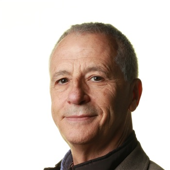 Lluís Mont合作论文数Atrial Fibrillation Unit, Clínic Barcelona;University of Barcelona471
 Elena Arbelo Lainez合作论文数Hospital Clínic de Barcelona;Universitat de Barcelona;Institut d’Investigacions Biomèdiques August Pi i Sunyer230
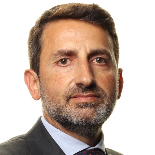 Antonio Berruezo合作论文数Department of Arrhythmias and Electrophysiology, Teknon Medical Center;Heart Institute, Teknon Medical Center218
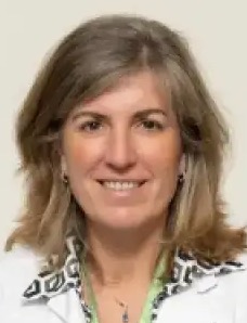 Marta Sitges合作论文数Cardiovascular Institute, Hospital Clinic of Barcelona;Universitat de Barcelona200
 Ramon Brugada合作论文数185
 Pedro Brugada合作论文数181
 José M Tolosana Viu合作论文数Institut d'investigacions Biomèdiques August Pi i Sunyer, Hospital Clinic de Barcelona;Arrhythmia Section, Cardiovascular Clinical Institute, Hospital Clínic of Barcelona173
 Oscar Campuzano合作论文数School of Medicine, and Institute of Neurosciences, Autonomous University Barcelona127
 Georgia Sarquella Brugada合作论文数Arrhythmias Unit, Hospital Sant Joan de Déu Barcelona;Sant Cugat High Performance Centre, University of Barcelona;Faculty of Medicine, University of Girona124
 Eduard Guasch合作论文数110Barbara Vidal合作论文数Hospital Clinic, Cardiovascular Institute94Borràs Roger合作论文数88David Tamborero合作论文数Hospital Clinic, Thorax Institute86R Brugada合作论文数75
 David Andreu合作论文数Department of Medicine and Life Sciences, Universitat Pompeu Fabra61
 Ivo Roca-Luque合作论文数50
 Anna Iglesias合作论文数50
 Juan Fernandez-Armenta合作论文数47
 Rosario Jesús Perea合作论文数Hospital Clinic de Barcelona;Division of Respiratory, Cardiovascular and Renal Pathobiology and Bioengineering, Consorci Institut D'Investigacions Biomediques August Pi I Sunyer;University of Barcelona;Institut D’investigacions Biomèdiques August Pi I Sunyer47
 Sergi Cesar合作论文数46
 Adelina Doltra合作论文数Hospital Clínic, Universitat de Barcelona43Etelvino Silva合作论文数40
 Charles Antzelevitch合作论文数Medicine and Pharmacology, Sidney Kimmel Medical College of Thomas Jefferson University/Lankenau Heart Institute38Manel Azqueta合作论文数37
 Paola Berne合作论文数36Coll Monica合作论文数35
 Bart Bijnens合作论文数Department of Cardiology, University Hospital Gasthuisberg35M Matas合作论文数32
 Victoria Delgado合作论文数Leiden University Medical Center;Department of Cardiovascular Imaging, University Hospital Germans Trias I Pujol30
 Ernesto Diaz-Infante合作论文数29Diego Penela合作论文数28Alexandra Pérez-Serra合作论文数Catalan Institute of Health28
 Andreu Porta Sánchez合作论文数Arrhythmia Section, Hospital Clinic de Barcelona;Facultad de Medicina, Universitat de Barcelona28
 Maurits A. Allessie合作论文数Biomedical Center, University ofLimburg27Maria Matiello合作论文数27Paz Garre合作论文数26
 Silvia G Priori合作论文数Department of Molecular Medicine, University of Pavia;Hospital ICS Maugeri26
 Angelo Auricchio合作论文数Faculty of Biomedical Sciences, Università della Svizzera;University Hospital in Magdeburg;Center for Computational Medicine in Cardiology, Università Della Svizzera Italiana in Lugano;Division of Cardiology, Istituto Cardiocentro Ticino25
 Bisbal Felipe合作论文数Hospital Clínic, Universitat de Barcelona24Begona Benito合作论文数24
 Josef Kautzner合作论文数Institute for Clinical and Experimental Medicine;Lékařské Fakultě Univerzity Karlovy v Praze23Aguinaga Luis合作论文数22Castel Angeles合作论文数22Vincent Probst合作论文数Hopital Nord, Institut du thorax22
 M. Angeles Castel合作论文数Unit for Heart Failure and Cardiac Transplantation, Hospital Clinic Barcelona22Victoria Fiol合作论文数21Anna Fernandez-Falgueras合作论文数21
 Jesus Mates合作论文数21
 Jeffrey Towbin合作论文数University of Tennessee Health Science Center;Le Bonheur Children's Hospital;St. Jude Children's Research Hospital21




