From its inception, the International Society of Oxygen Transport to Tissue (ISOTT) included several core members who were intrigued by the possibility of using light to probe the metabolic state of the cerebral cortex. This 50th anniversary review will focus on optical methods (UV, Visible, NIR) used to study brain oxygen metabolism in the intact brain. Chance, Lübbers, and Jöbsis did fundamental work in the 1970s and 1980s. These investigators spearheaded the use of fluorescence of pyridine nucleotides (e.g., NADH) and flavoprotein, and visible and near-infrared spectroscopy of haemoglobin amount and saturation, as well as the mitochondrial cytochrome chain. Two of our society awards are named for these pioneers and founders. By the new millennium, their many scientific descendants (although still too few for the task at hand) have been productive and moved the field forward. Technical advances in materials, detectors, devices, and computer power have made great advances possible. Physiological theory and understanding of cell and molecular mechanisms still lag, providing fertile territory for future goals. There are at least two major directions currently that will certainly continue to illuminate brain metabolism (obvious-and well worn-pun intended). These are the burgeoning NIR instrumentation variations, and second, the tremendous potential of two-photon phosphorescence lifetime microscopy. These advances have been introduced through the years and continue to be advanced at our annual ISOTT symposia.
People resuscitated after sudden cardiac arrest remain at high risk for mortality, with treatment for survivors varying from monitoring to life support. With respect to assessing survivability post cardiac arrest and resuscitation (CAR), we previously demonstrated the potential of the hypoxic ventilatory response (HVR) as a reliable indicator for discerning between survivors and non-survivors in the early stages of recovery following CAR in rats. Since HVR describes the increase in ventilation in response to hypoxia, we hypothesize that damage to cardiorespiratory regulatory centers in the brainstem underlie the loss of HVR observed post resuscitation in nonsurvivors. Wistar rats underwent cardiac arrest (12-min) and resuscitation. At 1 day post-resuscitation, rats were perfused transcardially and the brains were harvested and processed for immunohistostaining of caspase-3, a marker of apoptosis. Positive caspase-3 staining was observed in brainstem regions such as the rostral ventral lateral medulla (RVLM); Co-localization of caspase-3 and NeuN was observed in the RVLM as well, suggesting that apoptosis most likely occurs in neurons. Our results showed positive markers for neuronal apoptosis present in pathways of the brainstem involved in respiratory and cerebrovascular regulation, suggesting brain stem damage underlies changes in HVR following CAR.
Hypoxia-inducible factor 1 (HIF-1) is a major player in the oxygen sensor system as well as a transcription factor. HIF-1 is also associated in the pathogenesis of many brain diseases including Alzheimer's disease (AD), epilepsy and stroke. HIF-1 regulates the expression of many genes such as those involved in glycolysis, erythropoiesis, angiogenesis and proliferation in hypoxic condition. Despite several studies, the mechanism through which HIF-1 confers neuroprotection remains unclear, one of them is modulating metabolic profiles and inflammatory pathways. Characterization of the neuroprotective role of HIF-1 may be through its stabilization and the regulation of target genes that aid in the early adaptation to the oxidative stressors. It is interesting to note that mounting data from recent years point to an additional crucial regulatory role for hypoxia-inducible factors (HIFs) in inflammation. HIFs in immune cells regulate the production of glycolytic energy as well as innate immunity, pro-inflammatory gene expression, and mediates activation of pro-survival pathways. The present review highlights the contribution of HIF-1 to neuroprotection where inflammation is the crucial factor in the pathogenesis contributing to neural death. The potential mechanisms that contribute to neuroprotection as a result of the downstream targets of HIF-1α are discussed. Such mechanisms include those mediated through IL-10, an anti-inflammatory molecule involved in activating pro-survival signaling mechanisms via AKT/ERK and JAK/STAT pathways.
Perinatal hypoxia leads to changes in cerebral angiogenesis and persistent structural and functional changes in the adult brain. It may also result in greater vulnerability to subsequent challenges. We investigated the effect of postnatal day 2 (P2) hypoxic preconditioning on adult brain capillary density and brain vascular endothelial growth factor (VEGF) expression in mice. P2 mice were exposed to hypoxia (5% O2) in a normobaric chamber for 2 h then returned to normoxia while their littermates remained in normoxia (P2 control). After 2-6 months, they were euthanised and their brains were removed for capillary density determination. Another set of animals (P2 hypoxic mice and P2 controls) were euthanised at 2, 10, 23, and 60 days after birth and brain VEGF expression was assessed by western blot. Adult brain capillary density was significantly increased in the P2 hypoxic mice when compared to the P2 control mice. Additionally, VEGF expression appeared to be elevated in the P2-hypoxia mice when compared to the P2-control mice at all time points, and VEGF levels in P2-hypoxia mice declined with age similarly to P2-control mice. These data demonstrate that transient early-postnatal hypoxic stress leads to an increase in capillary density that persists in the adult, possibly due to increased VEGF expression. These results might be explained by epigenetic factors in the VEGF gene.
Supplementation of oxygen at concentrations significantly above environmental level for prolonged periods may lead to hyperoxia and tissue toxicity. The mammalian brain undergoes structural and functional changes during adaptation to hypoxia and hyperoxia. In this study we investigated the effect of prolonged hyperoxic exposure on cognitive and motor performance in mice. Two-month-old male mice were placed in either hyperoxic (50% O2) or normoxic conditions for 3 weeks. Cognitive function was measured using the Y-maze test. High alteration rate between the three arms of the maze is indicative of sustained memory and cognitive function. Motor function was measured using the grip strength and rotarod tests. In the rotarod test high speed and long latency are indicative of coordination and resistance. After 3 weeks of exposure, hematocrit levels were significantly decreased in the hyperoxia group compared to normoxic control littermates (%, mean ± SD, 37.8 ± 1.3, n = 15 vs. 49.9 ± 5.1, n = 15, p < 0.05). In the Y-maze test, chronic hyperoxic exposure resulted in a statistically significant decrease in alteration rate compared to normoxic control (%, mean ± SD, 53.4 ± 9.9, n = 30 vs. 61.2 ± 9.5, n = 15, p < 0.05). The rotarod and grip strength tests did not show statistically significant changes between the two groups. Our data suggest that chronic hyperoxia may lead to decreased cognitive performance in adult mice, which may be secondary to structural and functional changes in the brain.
Aims: Brief episodes of sublethal hypoxia reprogram brain response to face possible subsequent lethal stimuli by triggering adaptive and prosurvival events-a phenomenon denominated hypoxic preconditioning (HP). To date, the potential therapeutic implications of HP to forestall sporadic Alzheimer's disease (sAD) pathology remain unexplored. Using a well-established protocol of HP and focusing on hippocampus as a first brain region affected in AD, this study was undertaken to investigate the potential protective effects of HP in a sAD rat model induced by the intracerebroventricular (icv) administration of streptozotocin (STZ) and to uncover the mitochondrial adaptations underlying this nonpharmacological strategy.Results: HP prevented the memory and learning deficits as well as tau pathology in the icvSTZ rat model. HP also attenuated icvSTZ-related reactive astrogliosis, as noted by increased glial fibrillary acidic protein immunoreactivity and myo-inositol levels. Notably, HP abrogated the icvSTZ-related impaired energy metabolism and oxidative damage. Particularly, HP averted increased lactate, glutamate, and succinate levels, and decreased mitochondrial respiratory chain function and mitochondrial DNA content. Concerning mitochondrial adaptations underlying HP-triggered tolerance to icvSTZ, preconditioned hippocampal mitochondria displayed an enhanced complex II-energized mitochondrial respiration, which resulted from a coordinated interaction between mitochondrial biogenesis and fusion-fission. Mitochondrial biogenesis was stimulated immediately after HP, whereas in a latter phase mitochondrial fusion-fission events are modulated favoring the generation of elongated mitochondria.Innovation and Conclusion: Overall, these results demonstrate for the first time that HP prevents the sAD-like phenotype, in part, by targeting mitochondria emerging as a preventive strategy in the context of AD.
HypoxiaHypoxia inducible factor alpha (HIF1α) is associated with neuroprotectionNeuroprotection conferred by diet-induced ketosis but the underlying mechanism remains unclear. In this study we use a ketogenic dietKetogenic diets in rodents to induce a metabolic state of chronic ketosis, as measured by elevated blood ketone bodies. Chronic ketosis correlates with neuroprotectionNeuroprotection in both aged and following focal cerebral ischaemia and reperfusion (via middle cerebral artery occlusion, MCAO) in mouse and rat models. Ketone bodies are known to be used efficiently by the brain and metabolism of ketone bodies is associated with increased cytosolic succinateSuccinate levels that inhibits prolyl hydroxylases allowing HIF1α to accumulate. Ketosis also regulates inflammatory pathways, and HIF1α is reported to be essential for gene expression of interleukin10 (IL10Interleukin 10 (IL10)). Therefore we hypothesised that ketosis-stabilised HIF1α modulates the expression of inflammatory cytokines orchestrating neuroprotectionNeuroprotection. To test changes in cytokine levels in rodent brain, eight-week-old rats were fed either the standard chow diet (SD) or the ketogenic (KG) diet for 4 weeks before ischaemia experiments (MCAO) were performed and the brain tissues were collected. Consistent with our hypothesis, immunoblotting analysis shows IL10Interleukin 10 (IL10) levels were significantly higher in KG diet rat brain compared to SD, whereas the TNFα and IL6 levels were significantly lower in the brains of KG diet fed group.
Hypoxia-inducible factors (HIFs) are transcriptional regulators that mediate in mice for HIF-1 and HIF-2. The objective of this study was to investigate the effect of neuronal deletion of HIF-1 and HIF-2 in hypoxic adaptation by using the neuron-specific knockout (KO) mice. The floxed control and KO mice were used. Hypoxic mice were kept in a hypobaric chamber at a pressure of 300 torr (0.4 ATM, which was equivalent to 8% oxygen under normobaric condition) for 3 weeks. The littermate, normoxic control mice were housed in the same room next to the chamber to match ambient conditions. Body weights were monitored throughout the 3-week course. Cognitive function was measured using a Y-maze test; motor functions were measured using the rotarod test and the grip strength test. The hematocrit increased significantly at the end of 3-week hypoxic exposure in both control and KO mice. In the Y-maze test, the alternation rate (indicative of sustained cognition) trended lower in the KO mice compared to the controls following hypoxia (%, 51.3 +/- 13.1, n = 6 vs. 63.2 +/- 12.0, n = 8). In the rotarod test, the latency (seconds) in the KO mice was significantly lower compared to the controls (50.4 +/- 5.7 vs. 77.1 +/- 5.0, n = 3 each before hypoxia and 66.4 +/- 3.4, n = 6 vs. 98.1 +/- 15.4 after hypoxia, n = 3). The grip strength in the KO mice was similar compared to the control mice before hypoxia, but the strength of KO mice trended higher after hypoxic exposure. Our data suggest that deficiency of neuronal HIF-1 and HIF-2 may result in changes in behavioral performance and other adaptative responses to hypoxia.
The mammalian brain modulates its microvascular network to accommodate tissue energy demand in a process referred to as angioplasticity. There is an aging effect on cognitive function and adaptive responses to hypoxia. Hypoxia-induced angiogenesis is delayed in the aging mouse brain. Additionally, it has been shown that environmental enrichment provides an environment that fosters increased physical activity and sensory stimulation for mice as compared to standard housing; this stimulation increases neuronal activity and consequently brain oxygen demand. In this study, we investigated the effect of environmental enrichment and chronic hypoxia on cognitive performance in the young (2-4 months old) and the aged mice (17-21 months old). Mice were placed in a non-enriched or an enriched environment for 4 weeks under normoxia followed by 3 weeks of hypobaric hypoxia (similar to 0.4 atm, equivalent to 8% normobaric oxygen at sea level). Cognitive function was evaluated using the Y-maze and the novel object recognition tests in the enriched or non-enriched mice under normoxic or hypoxic conditions. In Y-maze, a high alternation rate is indicative of sustained cognition as the animals must remember which arm was entered last, so as not to re-enter it. Novel object recognition is based on the natural tendency of rodents to investigate a novel object instead of a familiar one; a higher novel object exploration rate is indicative of better cognitive function. The young mice showed a significantly higher alternation rate (%, 63 +/- 7 vs. 48 +/- 10, n = 8 and 10, respectively) in the Y-Maze test as compared to the aged mice. Under normoxia, the enriched mice showed an improved alternation rate (%, 63 +/- 10, n = 10) in Y-Maze test and a higher novel object exploration rate (%, 68 +/- 10 vs. 52 +/- 10) compared to the non-enriched controls. Similar results were observed for both young and aged mice following hypoxic exposure. Our data suggests that environmental enrichment improved the cognitive performance in the young and aged mice under normoxic and hypoxic conditions.
We have previously reported that in a rat model of chronic hypoxia, HIF-1 alpha and its target genes have significantly accumulated by 3 days of exposure, whereas no significant increase in capillary density has occurred; there is a significant increase in capillary density at 21 days of chronic hypoxic exposure. In this study we hypothesize that by utilizing 3 days and 21 days of hypoxic preconditioning, we would distinguish between the relative neuroprotective contributions of the accumulation of HIF-1 alpha and its target genes and angiogenic adaptation in a rat middle cerebral artery occlusion (MCAO) model. Rats were randomly assigned to either hypoxic precondition groups (3-day and 21-day hypoxia) or normoxic control group. Hypoxic animals were kept in a hypobaric chamber at a constant pressure of 0.5 atmosphere (380 mmHg, equivalent to 10% normobaric oxygen at sea level) for either 3 or 21 days. Normoxic controls were housed in the same room next to the hypobaric chamber. Erythropoietin (EPO) was measured at 3 and 21 days of hypoxia using Western blotting analysis. Infarct volumes were measured following 24 hours of permanent MCAO. We found that EPO is upregulated at 3 days of hypoxia and returns to baseline by 21 days of hypoxia. The infarct volumes following 24-hour MCAO were significantly reduced with 3-day hypoxic preconditioning when compared to normoxic controls (%, 31.8 +/- 5, n = 9 vs. 50.1 +/- 10.9, n = 7). No significant differences in infarct volume were seen between the normoxic controls and 21-day hypoxic preconditioned rats. We have shown that a 3-day hypoxic preconditioning, but not 21-day hypoxic preconditioning, provides significant neuroprotection against focal ischemia in rats, supporting a larger role for the accumulations of HIF-1 alpha and upregulation of its target genes in the neuroprotection against focal ischemia.
Key points Extreme aviation is accompanied by ever‐present risks of hypobaric hypoxia and decompression sickness. Neuroprotection against those hazards is conferred through fractional inspired oxygen () concentrations of 60–100% (hyperoxia). Hyperoxia reduces global cerebral perfusion (gCBF), increases reactive oxygen species within the brain and leads to cell death within the hippocampus. However, an understanding of hyperoxia's effect on cortical activity and concomitant levels of cognitive performance is lacking. This limits our understanding of whether hyperoxia could lower the brain's threshold of tolerance to physiological stressors inherent to extreme aviation, such as high gravitational forces. This study aimed to quantify the impact of hyperoxia upon global cerebral perfusion (gCBF), cognitive performance and cortical electroencephalography (EEG). Hyperoxia evoked a rapid reduction in gCBF, yet cognitive performance and vigilance were enhanced. EEG measurements revealed enhanced alpha power, suggesting less desynchrony, within the cortical temporal regions. Collectively, this work suggests hyperoxia‐induced brain hypoperfusion is accompanied by enhanced cognitive processing and cortical arousal. AbstractExtreme aviators continually inspire hyperoxic gas to mitigate risk of hypoxia and decompression injury. This neuroprotection carries a physiological cost: reduced cerebral perfusion (CBF). As reduced CBF may increase vulnerability to ever‐present physiological challenges during extreme aviation, we defined the magnitude and duration of hyperoxia‐induced changes in CBF, cortical electrical activity and cognition in 30 healthy males and females. Magnetic resonance imaging with pulsed arterial spin labelling provided serial measurements of global CBF (gCBF), first during exposure to 21% inspired oxygen () followed by a 30‐min exposure to 100% . High‐density EEG facilitated characterization of cortical activity during assessment of cognitive performance, also measured during exposure to 21% and 100% . Acid‐base physiology was measured with arterial blood gases. We found that exposure to 100% reduced gCBF to 63% of baseline values across all participants. Cognitive performance testing at 21% was accompanied by increased theta and beta power with decreased alpha power across multiple cortical areas. During cognitive testing at 100% , alpha activity was less desynchronized within the temporal regions than at 21% . The collective hyperoxia‐induced changes in gCBF, cognitive performance and EEG were similar across observed partial pressures of arterial oxygen (), which ranged between 276–548 mmHg, and partial pressures of arterial carbon dioxide (), which ranged between 34–50 mmHg. Sex did not influence gCBF response to 100% . Our findings suggest hyperoxia‐induced reductions in gCBF evoke enhanced levels of cortical arousal and cognitive processing, similar to those occurring during a perceived threat.
Cerebral edema in ischemic stroke can lead to increased intracranial pressure, reduced cerebral blood flow and neuronal death. Unfortunately, current therapies for cerebral edema are either ineffective or highly invasive. During the development of cytotoxic and subsequent ionic cerebral edema water enters the brain by moving across an intact blood brain barrier and through aquaporin-4 (AQP4) at astrocyte endfeet. Using AQP4-expressing cells, we screened small molecule libraries for inhibitors that reduce AQP4-mediated water permeability. Additional functional assays were used to validate AQP4 inhibition and identified a promising structural series for medicinal chemistry. These efforts improved potency and revealed a compound we designated AER-270, N-[3,5-bis (trifluoromethyl)phenyl]-5-chloro-2-hydroxybenzamide. AER-270 and a prodrug with enhanced solubility, AER-271 2-{[3,5-Bis(trifluoromethyl) phenyl]carbamoyl}-4-chlorophenyl dihydrogen phosphate, improved neurological outcome and reduced swelling in two models of CNS injury complicated by cerebral edema: water intoxication and ischemic stroke modeled by middle cerebral artery occlusion.
Aquaporin-4 (AQP4) plays an important role in regulating water exchange across the blood-brain barrier (BBB) and brain-cerebrospinal fluid interface. Studies on AQP-4 knockout mice (AQP4-KO) have reported considerable protection from brain edema induced by acute water intoxication and ischemic stroke, identifying AQP4 as a potential target for therapeutic interventions. However, the long-term effects of chronic AQP4 suppression are yet to be elucidated. In the current study, we evaluated the physiological and structural changes in adult AQP4-KO mice using magnetic resonance imaging (MRI) and immunohistochemical analysis. Water exchange across BBB was assessed by tracking an intravenous bolus injection of oxygen-17 (17O) water (H217O) using 17O-MRI. Cerebral blood flow (CBF) was quantified using arterial spin-labeling (ASL) MRI. Capillary density was determined by immunohistochemical staining for glucose transporter-1 (GLUT1). Compared to wildtype control mice, AQP4-KO mice showed a significant reduction in peak and steady-state H217O uptake despite unaltered CBF. Interestingly, a 22% increase in cortical capillary density was observed in AQP4-KO mice. These results suggest that increased cerebral vascularization may be an adaptive response to chronic reduction in water exchange across BBB in AQP4-KO mice.
 Kui Xu合作论文数59Myron Rosenthal合作论文数42
 Wesley David Lust合作论文数Laboratory of Neuropathology and Neuroanatomical Sciences, National Institute of Neurological Diseases and Stroke National Institutes of Health36
 Michelle Puchowicz合作论文数The Department of Physiology and Biophysics, School of Medicine, Case Western Reserve University32S I Harik合作论文数29
 George Perry合作论文数College of Sciences, The University of Texas at San Antonio23
 Gjumrakch Aliev合作论文数GALLY International Biomedical Research;Department of Pharmacology, Institute of Pharmacy- FSAEI HE, I.M. Sechenov First Moscow State Medical University;Institute of Physiologically Active Compounds, Russian Academy of Sciences23K Xu合作论文数18Xiaoyan Sun合作论文数Case Western Reserve Univ, Sch Med, Dept Physiol & Biophys, Cleveland, OH 44106 USA18
 Justin Sick合作论文数17
 Constantinos Tsipis合作论文数16Juan C. Chavez合作论文数14
 Mark A. Smith合作论文数Institute of Pathology, Case Western Reserve University, Cleveland, Ohio 4410613
 Robert P. Friedland合作论文数Department of Neurology, School of Medicine, University of Louisville13FF JOBSIS合作论文数12Warren R. Selman合作论文数Departments of Neurology12
 Mark E Obrenovich合作论文数School of Medicine, Case Western Reserve University12P Pichiule合作论文数11Kreisman N R合作论文数10Ts Whittingham合作论文数9Ralph C. Crumrine合作论文数9M A Smith合作论文数Division of Neuropathology, Institute of Pathology, Case Western Reserve University, 2085 Adelbert Road, Cleveland, OH 44106-4901, USA Tel.: 1-216-368-2488/216-844-1808; Fax: 1-216-368-8964/216-844-1810; E-mail: gxp7@po.cwru.edu US9Benderro Girriso F合作论文数9
 Walter F. Boron合作论文数Case Western Reserve University9Fisher Elaine M合作论文数8
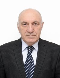 Eldar Gasimov合作论文数Department of Cytology, Histology and Embryology, Azerbaijan Medical University8Youzhi Kuang合作论文数7Bernadette O Erokwu合作论文数7Sandra Siedlak合作论文数Department of Pathology, Case Western Reserve University7
 Bruce N. Ames合作论文数Graduate School, University of California, Berkeley7Svetlana Pundik合作论文数7
 Maxwell Lewis Neal合作论文数Department of Biomedical & Health Informatics, University of Washington7Ma Haxhiu合作论文数6Ma Hritz合作论文数6Ra Ratcheson合作论文数6
 Nicole L Ward合作论文数School of Medicine, Case Western Reserve University6
 Chris Flask合作论文数Dept Radiol, Case Western Reserve Univ6
 Chii-Wann Lin合作论文数Institute of Electrical Engineering, National Taiwan University6
 Bruce T. Lamb合作论文数Department of Genetics, Case Western Reserve University6
 Justin C Shenk合作论文数University of Texas Health Science Center at San Antonio6Ma Puchowicz合作论文数5D Seyidova合作论文数5Kutina-Nelson K L合作论文数5Duckrow R B合作论文数5Lin Wen-Chuan合作论文数5Paola Pichiule合作论文数5Andrew I. Light合作论文数5
 Gemma Casadesus合作论文数Institute of Pathology, Case Western Reserve University5




