Primary bone malignancies - including chordoma, chondrosarcoma, osteosarcoma, and Ewing sarcoma - originate from bone or cartilage cells and often develop in anatomically complex or surgically challenging regions. While surgical resection remains the standard of care for most localized tumors, radiation therapy (RT) has become an increasingly integral component of multidisciplinary management, particularly when complete surgical excision is not feasible, margins are close or positive, or the tumor is adjacent to critical structures. Historically, conventional photon-based RT has shown limited efficacy in many of these tumors due to factors such as relative radioresistance and proximity to radiosensitive normal tissues. However, advances in conformal photon techniques such as intensity-modulated radiation therapy (IMRT) and stereotactic body radiation therapy (SBRT), along with hadron-based approaches like proton beam therapy (PBT) and carbon ion radiation therapy (CIRT), have expanded the therapeutic potential of RT in bone sarcomas. This review highlights the evolving role of RT in the management of primary bone malignancies, with a focus on technological advances, clinical outcomes, ongoing trials, and future directions in the field.
Abstract Background: Molecular therapies for chondrosarcomas remain limited, in part due to the rarity of these cancers, which hampers efforts at broad genomic characterization. The establishment of large, standardized, multicenter databases, such as the AACR Project GENIE, along with subsequent individual studies, has begun to assemble an extensive mutational profile of this disease. This abstract provides a preliminary analysis of the mutational data from these studies. Methods: The GENIE Cohort v18.0, MSK Nature Communications Sarcoma 2022, and UCLA Cell 2024 sarcoma datasets, accessed via cBioPortal, were collated and analyzed. These datasets included patient information for conventional chondrosarcoma (CS), dedifferentiated chondrosarcoma (DDCS), and mesenchymal chondrosarcoma (MCS). Available data included patient age, sex, race, mutational profiles, and, for CS and DDCS, survival outcomes. Results: In total, the datasets encompassed 518 patients with chondrosarcoma: 428 CS, 38 DDCS, and 52 MCS. The average age at biopsy was 52 for CS patients, 63 for DDCS patients, and 33 for MCS patients. Women comprised 40.4%, 54.1%, and 55.8% of CS, DDCS, and MCS patients, respectively. Among patients with recorded race, the racial distribution was 74% White, 5% Black, 10% Asian, and 12% Other for CS; 89% White, 5% Black, 3% Asian, and 3% Other for DDCS; and 83% White, 13% Black, and 4% Other for MCS. The average mutation count per patient sample was 10.1 for CS, 5.4 for DDCS, and 2.6 for MCS. The top five most frequently mutated genes for each disease were as follows - CS:TP53 (124/439), IDH1 (113/437), IDH2 (35/423), KMT2D (15/186), and TERT (14/179); DDCS:TP53 (24/40), IDH1 (18/40), TERT (13/38), IDH2 (12/40), FLT4 (8/40); MCS: MAP3K13 (5/18), INSR (4/17), SDHA (5/23), KMT2D (4/24), and KMT5A (2/13). Median overall survival was 58 months for CS patients versus 25 months for DDCS patients. Notably, many of the most frequently mutated genes beyond TP53, IDH1, and IDH2 are involved in chromatin regulation and genomic stability (KMT2A, KMT2D, TERT, etc.), and these mutations are more common in DDCS. Conclusion: This multicenter mutational analysis confirms that, after TP53, the most frequently mutated genes in CS and DDCS are IDH1 and IDH2. Mutations in genes involved in chromatin regulation and genomic stability are more prevalent in DDCS, which may contribute to its increased aggressiveness. These findings support ongoing efforts to target these pathways in chondrosarcoma. Citation Format: Luyuan Li, Wensi Tao, Robert L. Walker, Manish KC, Darshan Gundala, Josiane E. Eid, Zhenfeng Duan, Jonathan C. Trent. Mutational profiling of chondrosarcoma across multicenter cohorts [abstract]. In: Proceedings of the American Association for Cancer Research Annual Meeting 2026; Part 1 (Regular Abstracts); 2026 Apr 17-22; San Diego, CA. Philadelphia (PA): AACR; Cancer Res 2026;86(7 Suppl):Abstract nr 14.
Chondrosarcoma is a heterogeneous group of malignant bone tumors characterized by the production of neoplastic hyaline cartilage. These tumors are often indolent and typically resistant to conventional chemotherapy and radiotherapy, with surgical resection remaining the cornerstone of treatment. However, patients with unresectable or metastatic disease face poor outcomes due to the lack of effective systemic therapies. While advancements in systemic treatments - such as immunotherapy and anti-angiogenic agents - and novel radiation modalities like proton beam therapy and carbon ion therapy have shown some benefit in select cases, their overall efficacy remains limited, underscoring the urgent need for more effective treatment options. Emerging novel therapies are beginning to illuminate potential new treatment pathways. Current efforts are increasingly focused on targeting recurrent genetic alterations and dysregulated signaling pathways, including IDH mutations, DR5-mediated apoptotic pathway, hedgehog signaling, Src kinase pathway, the PI3K-Akt-mTOR axis, histone deacetylation, and angiogenesis. Although these targeted strategies are still under investigation, they show promise in overcoming therapeutic resistance and advancing personalized treatment. This review provides an overview of the clinical characteristics of major chondrosarcoma subtypes, key genetic alterations implicated in tumor pathogenesis, and recent advances in treatment strategies, including surgery, chemotherapy, radiotherapy, immunotherapy, and emerging targeted therapies.
Supplementary Figure S8 - PDF file 1084K, Figure S8: Effects of SSTC-104 on Wnt-regulated tissues
Transduction with lentiviral vectors is a useful approach to study the molecular function of specific genes in mammalian cells. Here, we present a calcium phosphate-based transfection protocol that guarantees highly efficient production and delivery of lentiviral vectors in adherent cultured cells. We also describe in detail a direct lysis technique to measure protein expression, an optimized sulforhodamine B proliferation assay, and a step-by-step chromatin immunoprecipitation procedure to verify the binding of ETV5 to E2F1 first intron in SYO-1 sarcoma cells. For complete details on the use and execution of this protocol, please refer to Kingston et al. (2003),1 Ireton et al. (2002),2 Brown et al. (2009),3 DeSalvo et al. (2021),4 Vichai and Kirtikara (2006),5 and Boyer et al. (2005).6
Supplementary Figure S10 4882K, Figure S10a and S10b: Nuclear beta-catenin accumulation in human synovial sarcoma tumors
Supplementary Methods and Legends - PDF file 130K, Methods:Additional information on experimental techniques Legends: describing supplementary figures
Supplementary Figure S9 - PDF file 867K,Figure S9: The SYT-SSX2 domain SSXRD executes -catenin activation and myogenic differentiation
Supplementary Figures S2 and S3 - PDF file 2332K, Figure S2:SG3 tumors are positive for beta-catenin and Myf5 Figure S3:Normal development of Myf5 myoblasts in beta-catenin knockout mice
Supplementary Figure S1 - PDF file 554K, Breeding strategy used to generate beta-catenin-null SYT-SSX2 transgenic mice
Supplementary Figure S11 and Tables S1-S3 - PDF file 460K, Figure S11: Regulation of an embryonic Wnt-interactive network by SYT-SSX2 and in human synovial sarcomas Table S1: Differentially regulated Wnt components in mesenchymal precursor cells expressing SYT-SSX2 and in synovial sarcoma tumors Table S2: Differentially regulated Wnt targets in mesenchymal precursor cells expressing SYT-SSX2 and in synovial sarcoma tumors Table S3: The embryonic Wnt interactive network in mesenchymal precursor cells expressing SYTSSX2 and in synovial sarcoma tumors
Supplementary Figures S4, S5, S6, S7 - PDF file 1832K, Figure S4:Pyrvinium effect on beta-catenin in SYT-SSX2 expressing C2C12 and synovial sarcoma cells Figure S5:Phosphorylated -catenin and full-length APC in HS-SY-II and SYO-1 SS cells Figure S6:SSTC-104 effects on growth and beta-catenin in HS-SY-II synovial sarcoma cells Figure S7: LRP6 depletion in HS-SY-II synovial sarcoma cells induces -catenin nuclear exit
Chondrosarcoma is a group of primary bone cancers that arise from transformed cells of chondrocytic lineage. Tumor recurrence and metastasis are devastating for patients with chondrosarcoma since there are no effective treatment options. IDH mutations occur in over 50% of tumors from patients with conventional or dedifferentiated chondrosarcomas and represent an attractive target for therapy. However, their role in the pathogenesis of chondrosarcoma remains largely unknown. In this study, we sought to determine the association of IDH mutation and HIF-1α in chondrosarcoma. We used the chondrosarcoma JJ012 cell line and its derived CRISPR/Cas9 mutant IDH1 (IDH1mut) knockout (KO) cells. RNA-Seq data analysis revealed downregulation of several HIF-1α target genes upon loss of IDH1mut. This was associated with reduced HIF-1α levels in the IDH1mut KO cells and tumors. Loss of IDH1mut also attenuated the expression of angiogenic markers in tumor tissues and abrogated the angiogenic capacity of JJ012 cells. Moreover, we observed that exogenous expression of HIF-1α significantly promoted anchorage-independent colony-formation by IDH1mut KO cells. These results suggest IDH1 mutation confers angiogenic and tumorigenic properties of JJ012 cells by inducing HIF-1α. Thus, the HIF pathway represents a promising candidate for combinatorial regimens to target IDH1 mutated chondrosarcomas.
Synovial sarcoma is an aggressive malignancy with no effective treatments for patients with metastasis. The synovial sarcoma fusion SS18-SSX, which recruits the SWI/SNF-BAF chromatin remodeling and polycomb repressive complexes, results in epigenetic activation of FGF receptor (FGFR) signaling. In genetic FGFR-knockout models, culture, and xenograft synovial sarcoma models treated with the FGFR inhibitor BGJ398, we show that FGFR1, FGFR2, and FGFR3 were crucial for tumor growth. Transcriptome analyses of BGJ398-treated cells and histological and expression analyses of mouse and human synovial sarcoma tumors revealed prevalent expression of two ETS factors and FGFR targets, ETV4 and ETV5. We further demonstrate that ETV4 and ETV5 acted as drivers of synovial sarcoma growth, most likely through control of the cell cycle. Upon ETV4 and ETV5 knockdown, we observed a striking upregulation of DUX4 and its transcriptional targets that activate the zygotic genome and drive the atrophy program in facioscapulohumeral dystrophy patients. In addition to demonstrating the importance of inhibiting all three FGFRs, the current findings reveal potential nodes of attack for the cancer with the discovery of ETV4 and ETV5 as appropriate biomarkers and molecular targets, and activation of the embryonic DUX4 pathway as a promising approach to block synovial sarcoma tumors.
Chondrosarcomas are a heterogeneous group of malignant bone tumors that produce hyaline cartilaginous matrix. Mutations in isocitrate dehydrogenase enzymes (IDH1/2) were recently described in several cancers, including conventional and dedifferentiated chondrosarcomas. These mutations lead to the inability of IDH to convert isocitrate into α-ketoglutarate (α-KG). Instead, α-KG is reduced into D-2-hydroxyglutarate (D-2HG), an oncometabolite. IDH mutations and D-2HG are thought to contribute to tumorigenesis due to the role of D-2HG as a competitive inhibitor of α-KG-dependent dioxygenases. However, the function of IDH mutations in chondrosarcomas has not been clearly defined. In this study, we knocked out mutant IDH1 (IDH1mut) in two chondrosarcoma cell lines using the CRISPR/Cas9 system. We observed that D-2HG production, anchorage-independent growth, and cell migration were significantly suppressed in the IDH1mut knockout cells. Loss of IDH1mut also led to a marked attenuation of chondrosarcoma formation and D-2HG production in a xenograft model. In addition, RNA-Seq analysis of IDH1mut knockout cells revealed downregulation of several integrin genes, including those of integrin alpha 5 (ITGA5) and integrin beta 5 (ITGB5). We further demonstrated that deregulation of integrin-mediated processes contributed to the tumorigenicity of IDH1-mutant chondrosarcoma cells. Our findings showed that IDH1mut knockout abrogates chondrosarcoma genesis through modulation of integrins. This suggests that integrin molecules are appealing candidates for combinatorial regimens with IDH1mut inhibitors for chondrosarcomas that harbor this mutation.
Chondrosarcomas are malignant bone tumors that produce cartilaginous matrix. Mutations in isocitrate dehydrogenase enzymes (IDH1/2) were recently described in several cancers, including chondrosarcomas. IDH mutations detected in human cancers invariably are heterozygous missense substitutions. These mutations lead to the inability of IDH to convert isocitrate into α-ketoglutarate (α-KG). Instead, α-KG is reduced into 2-hydroxyglutarate (D-2HG), an oncometabolite. Due to the structural similarity between D-2HG and α-KG, it has been reported that high levels of D-2HG competitively inhibit α-KG-dependent dioxygenases such as TET, JHDM and PHD enzymes, thus contributing to tumorigenesis. We sought to determine the role of IDH1 mutations in the tumorigenesis of human chondrosarcomas by inactivating mutant IDH1 using pharmacological and genetic approaches. In our study, we employed two human chondrosarcoma cell lines, JJ012 and HT1080, that carry endogenous IDH1 mutations. IDH mutation analysis was performed by PCR-based DNA sequencing, and D-2HG levels were measured by tandem mass spectrometry. Mutant IDH1 was knocked down via siRNA and knocked out via CRISPR/Cas9. We analyzed the effect of mutant IDH1 on chondrosarcoma growth in murine xenograft models. We found that knockdown of mutant IDH1 via siRNA significantly reduced D-2HG production in chondrosarcoma cells. In addition, mutant IDH1 knockdown dramatically inhibited colony formation in the sarcoma cells. Consistently, genetic knockout of mutant IDH1 almost completely depleted D-2HG production and significantly inhibited colony formation in the chondrosarcoma cells. To assess the significance of these results in vivo, we implanted the mutant IDH1- knockout cells in nude mice and studied their capacity for tumor initiation and growth. In these models, we observed that loss of mutant IDH1 led to a marked attenuation of chondrosarcoma formation. Our findings clearly demonstrate that mutant IDH1 plays a vital role in chondrosarcoma tumor formation. By investigating the role of IDH mutations in the pathogenesis of chondrosarcomas, we aim to uncover the potential therapeutic targets against this aggressive cancer. Citation Format: Luyuan Li, Xiaoyu Hu, Josiane E. Eid, Joanna DeSalvo, Jonathan C. Trent. Mutant IDH1 is essential for chondrosarcoma growth [abstract]. In: Proceedings of the American Association for Cancer Research Annual Meeting 2019; 2019 Mar 29-Apr 3; Atlanta, GA. Philadelphia (PA): AACR; Cancer Res 2019;79(13 Suppl):Abstract nr 864.
Cellular metabolism reprogramming is an emerging hallmark of cancer, which provides tumor cells with not only necessary energy but also crucial materials to support growth. Exploiting the unique features of cancer metabolism is promising in cancer therapies. The growing interest in this field has led to numerous inhibitors being developed against key molecules in metabolic pathways, though most of them are still in preclinical development. Potential targeted cancer cell metabolic pathways under investigation include glycolysis, tricarboxylic acid (TCA) cycle, oxidative phosphorylation (OXPHOS), glutaminolysis, pentose phosphate pathway (PPP), lipid synthesis, amino acid and nucleotide metabolism. Sarcoma is a type of cancer that arises from transformed cells of mesenchymal origin, in contrast to carcinoma which originates from epithelial cells. Compared with carcinoma, progress towards harnessing the therapeutic potential of targeting sarcoma cell metabolism has been relatively slow. Recently however, with the discovery of cancer-specific mutations in metabolic enzymes such as isocitrate dehydrogenase (IDH) and succinate dehydrogenase (SDH) in certain sarcoma types, cancer cellular metabolism has been considered more as a source of new targets for treating sarcoma. In this article, we review metabolic enzymes currently tested for cancer therapies and describe the therapeutic potential of targeting IDH mutations and SDH deficiency in sarcomas.
Mesenchymal stem cells (MSCs) originate from embryonic mesoderm and give rise to the multiple lineages of connective tissues. Transformed MSCs develop into aggressive sarcomas, some of which are initiated by specific chromosomal translocations that generate fusion proteins with potent oncogenic properties. The sarcoma oncogenes typically prime MSCs through aberrant reprogramming. They dictate commitment to a specific lineage but prevent mature differentiation, thus locking the cells in a state of proliferative precursors. Deregulated expression of lineage-specific transcription factors and controllers of chromatin structure play a central role in MSC reprogramming and sarcoma pathogenesis. This suggests that reversing the epigenetic aberrancies created by the sarcoma oncogenes with differentiation-related reagents holds great promise as a beneficial addition to sarcoma therapies.
 Christina B Garcia合作论文数15
 Frump Andrea L合作论文数12
 Ethan Lee合作论文数12
 Mario R. Capecchi合作论文数Department of Human Genetics, School of Medicine, University of Utah;Eccles Institute of Human Genetics, School of Medicine, University of Utah;Howard Hughes Medical Institute10
 Michael N Vansaun合作论文数10Gleaves Linda A合作论文数9Orton Darren合作论文数9
 Barbara M. Fingleton合作论文数Department of Pharmacology, Vanderbilt University;Department of Surgery, Vanderbilt University Medical Center9
 Timothy S. Blackwell合作论文数Department of Internal Medicine, The University of Michigan9
 Kenyi Saito-Diaz合作论文数Department of Cell and Developmental Biology and Program in Developmental Biology, Vanderbilt University Medical Center9Taylor P. Sherrill合作论文数9
 Fiona Elizabeth Yull合作论文数9
 Whitney Barham合作论文数9
 Jonathan C Trent合作论文数Sylvester Comprehensive Cancer Center, University of Miami;Division of Hematology, Department of Medicine, Miller School of Medicine, University of Miami8Luyuan Li (李路远)合作论文数6
 Xiaoyu Hu合作论文数3
 Pampee Young合作论文数Dept Pathol, Vanderbilt Univ3DeSalvo Joanna合作论文数3Yuguang Ban合作论文数Department of Public Health Sciences, University of Miami2Xiaodian Sun合作论文数2Roy Barco合作论文数2
 Jinbo Yue (岳金波)合作论文数The Second Affiliated Hospital of Shandong First Medical University;Cancer Hospital of Shandong First Medical University2Jerome Jourquin合作论文数1C. M. Shaffer合作论文数1Robert L. Caldwell合作论文数1a paz合作论文数1
 Mahsa Khanlari合作论文数Department of Pathology, St. Jude Children's Research Hospital1Christian M Shaffer合作论文数Department of Biomedical Informatics, Vanderbilt University Medical Center, Nashville, TN, USA.1
 Robert H Carnahan合作论文数Vanderbilt Vaccine Center, and Department of Pediatrics, Vanderbilt University Medical Center1
 Curtis Thorne合作论文数1Kerr Darcy A合作论文数1Meyers Kelly C合作论文数1
 David M. Livingston合作论文数Department of Cancer Biology, Dana-Farber Cancer Institute1Jiang Zhijie合作论文数1
 Breelyn Wilky合作论文数Department of Medicine-Medical Oncology, School of Medicine, University of Colorado Anschutz Medical Campus;University of Colorado Hospital1
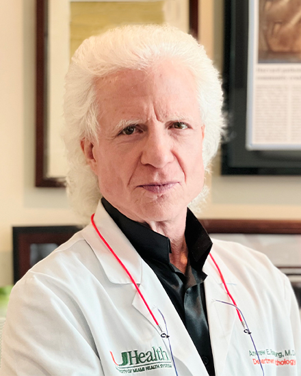 Andrew Eric Rosenberg合作论文数Division of Anatomic Pathology, Department of Pathology and Laboratory Medicine, Miller School of Medicine, University of Miami1Scully Ralph合作论文数1
 Shuhua Zheng合作论文数Nova Southeastern University, College of Osteopathic Medicine, Fort Lauderdale, FL 33134, USA1
 Yuejun Kang (康跃军)合作论文数Institute for Clean Energy and Advanced Materials, School of Materials & Energy, Southwest University1Lobdell Nichole A合作论文数1
 Zhongming Zhao合作论文数Department of Bioinformatics and Systems Medicine, D.Bradley McWilliams School of Biomedical Informatics, The University of Texas Health Science Center at Houston;Center for Precision Health, The University of Texas Health Science Center at Houston1Juha Partanen合作论文数1
 Tadashi Kondo (近藤格)合作论文数Division of Rare Cancer, National Cancer Center Research Institute;Department of Proteomics, National Cancer Center Research Institute1
 Dongqing Li (李冬青)合作论文数Department of Mechanical and Mechatronics Engineering, University of Waterloo1
 Lin Chen (陈林)合作论文数The Second Affiliated Hospital of Amu1
 Andrew Kung合作论文数Department of Pediatrics, Memorial Sloan Kettering Cancer Center1
 David M. Ornitz合作论文数Department of Developmental Biology, School of Medicine, Washington University in St. Louis1
 Marzenna Blonska合作论文数Department of Molecular and Cellular Oncology, University of Texas M.D. Anderson Cancer Center1
 Chao Liu合作论文数1




