Artificial intelligence (AI) is transforming morphological assessment in hematologic malignancies by enabling automated cell classification, detection of disease-specific features, and prediction of genetic alterations from blood and bone marrow smear images. Deep learning models have achieved expert-level performance in identifying malignant patterns, uncovering morphologic-molecular associations, and providing patient-level diagnoses through multiple instance learning approaches. This review summarizes recent advances in AI-driven morphological analysis across myeloid and lymphoid neoplasms, emphasizing their impact on diagnosis and disease classification. We further examine emerging evidence linking image-derived features, such as nuclear texture, cytoplasmic granularity, and spatial cell organization, to molecular and prognostic variables. In addition, we discuss enabling technologies, including virtual staining and interpretability methods, that support clinical integration. Finally, we address key challenges, including data heterogeneity, the need for prospective validation, and interdisciplinary collaboration. AI-powered, explainable morphological analysis has the potential to advance precision hematology and personalized patient management.
Detecting BK Virus (BKV) is crucial for managing post-transplant outcomes in kidney patients. While BKV is typically identified using SV40 immunohistochemistry (IHC), this method is time-consuming, limited by tissue availability and resource-intensive, especially in low-resource settings. Recent advances in computational pathology have shown potential for automating disease detection from Hematoxylin and Eosin (H E)-stained images, though BKV detection remains understudied due to its low prevalence and limited data. We hypothesize that BKV-positive cells exhibit unique morphological patterns in H E-stained tissue, detectable via computational methods. To address this, we developed BKVision, a weakly-supervised deep learning model for BKV detection in H E whole-slide images (WSIs). Trained on 3,734 WSIs, BKVision achieves an F1-score of 0.984 ± 0.008 on a test cohort of 936 slides. Additionally, we conducted a morphological analysis on 774 H E image patches, extracting 37 human interpretable features and validating them against IHC with pathologist guidance. This identified 11 cell attributes, such as nuclear enlargement and chromatin texture changes, that distinguish BKV-positive from negative cases. These findings highlight the potential to enhance BKV diagnostic criteria by integrating these identified morphological features. BKVision demonstrates the potential of computational methods to provide accurate, accessible, and interpretable BKV detection without the need for IHC, offering a cost-effective alternative in low-resource settings while revealing key morphological features of BKV infection.
Breast‐conserving surgery is typically performed with the patient in a supine position, whereas preoperative diagnostic MRI breast images are obtained with the patient in a prone position. The change in patient positioning causes significant large deformations, requiring preoperative localization of the detected lesions. Developing an individual‐specific breast biomechanical model capable of simulating these deformations remains challenging yet highly desirable. This study presents a novel approach that combines finite element analysis with the optimization of mechanical properties of breast tissues, using only surface information to construct a personalized deformation model of the breast. A visco‐hyperelastic model is employed to characterize the stress–strain relationship of breast tissue. The proposed method has been tested on 15 cases of breast cancer and achieves a tumor localization error of 8.12 ± 4.15 mm. The results show that this approach provides an accurate and realistic estimation of large breast tissue deformations and yields smaller tumor localization errors compared to previously reported methods.
In this work we propose and compare different deep learning algorithms for the segmentation of stromal regions in pancreatic histopathological image using three consecutive tissue sections, each uniquely stained with Hematoxylin and Eosin (H&E), Masson's Trichrome, and Alcian Blue. After a non-rigid registration process, variations in tissue distribution between consecutive slides still persist, which leads to distinct desired segmentations of tissues for each stain, thus underscoring the need for a specific segmentation and co-segmentation approaches to achieve higher accuracy. We compare single stain models, with respect to multi-stain techniques that either consider the multiple stains all at once in training or are based on multi-branch siamese and co-segmentation techniques. We demonstrate superior performance in identifying stromal regions with the multi-stain approaches in comparison to the segmentation techniques applied to individual stains, by effectively utilizing the complementary information each staining technique provides. This advancement is poised to enhance the further evaluation of tumor microenvironment and stromal characteristics in patients with pancreatic cancer.
The incorporation of digital pathology in clinical practice will require the training of pathologists in digital skills. Our study aimed to assess the reliability among pathologists in determining tumor percentage in whole slide images (WSI) of non-small cell lung cancer (NSCLC) using digital image analysis, and study how the results correlate with the molecular findings. Pathologists from nine centers were trained to quantify epithelial tumor cells, tumor-associated stromal cells, and non-neoplastic cells from NSCLC WSI using QuPath. Then, we conducted two consecutive ring trials. In the first trial, analyzing four WSI, reliability between pathologists in the assessment of tumor cell percentage was poor (intraclass correlation coefficient (ICC) 0.09). After performing the first ring trial pathologists received feedback. The second trial, comprising 10 WSI with paired next-generation sequencing results, also showed poor reliability (ICC 0.24). Cases near the recommended 20% visual threshold for molecular techniques exhibited higher values with digital analysis. In the second ring trial reliability slightly improved and human errors were reduced from 5.6% to 1.25%. Most discrepancies arose from subjective tasks, such as the annotation process, suggesting potential improvement with future artificial intelligence solutions.
Magnetic resonance imaging of whole fetal body and placenta is limited by different sources of motion affecting the womb. Usual scanning techniques employ single-shot multi-slice sequences where anatomical information in different slices may be subject to different deformations, contrast variations or artifacts. Volumetric reconstruction formulations have been proposed to correct for these factors, but they must accommodate a non-homogeneous and non-isotropic sampling, so regularization becomes necessary. Thus, in this paper we propose a deep generative prior for robust volumetric reconstructions integrated with a diffeomorphic volume to slice registration method. Experiments are performed to validate our contributions and compare with ifdefined tmiformat R2.5a state of the art method methods in the literature in a cohort of 72 fetal datasets in the range of 20-36 weeks gestational age. Results suggest improved image resolution Quantitative as well as radiological assessment suggest improved image quality and more accurate prediction of gestational age at scan is obtained when comparing to a state of the art reconstruction method methods. In addition, gestational age prediction results from our volumetric reconstructions compare favourably are competitive with existing brain-based approaches, with boosted accuracy when integrating information of organs other than the brain. Namely, a mean absolute error of 0.618 weeks ( R2=0.958 ) is achieved when combining fetal brain and trunk information.
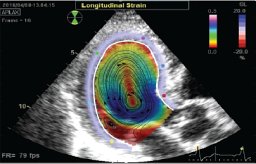
Background Estimates of regional left ventricular (LV) strains provide additional information to global function parameters such as ejection fraction (EF) and global longitudinal strain (GLS) and are more sensitive in detecting abnormal regional cardiac function. The accurate and reproducible assessment of regional cardiac function has implications in the management of various cardiac diseases such as heart failure, myocardial ischemia, and dyssynchrony. Purpose To develop a method that yields highly reproducible, high-resolution estimates of regional endocardial strains from 4DCT images. Methods A method for estimating regional LV endocardial circumferential (epsilon cc)$( {{\epsilon }_{cc}} )$ and longitudinal (epsilon ll${\epsilon }_{ll}$) strains from 4DCT was developed. Point clouds representing the LV endocardial surface were extracted for each time frame of the cardiac cycle from 4DCT images. 3D deformation fields across the cardiac cycle were obtained by registering the end diastolic point cloud to each subsequent point cloud in time across the cardiac cycle using a 3D point-set registration technique. From these deformation fields, epsilon ccand epsilon ll${\epsilon }_{cc}\ {\rm{and\ }}{\epsilon }_{ll}$ were estimated over the entire LV endocardial surface by fitting an affine transformation with maximum likelihood estimation. The 4DCT-derived strains were compared with strains estimated in the same subjects by cardiac magnetic resonance (CMR); twenty-four subjects had CMR scans followed by 4DCT scans acquired within a few hours. Regional LV circumferential and longitudinal strains were estimated from the CMR images using a commercially available feature tracking software (cvi42). Global circumferential strain (GCS) and global longitudinal strain (GLS) were calculated as the mean of the regional strains across the entire LV for both modalities. Pearson correlation coefficients and Bland-Altman analyses were used for comparisons. Intraclass correlation coefficients (ICC) were used to assess the inter- and intraobserver reproducibility of the 4DCT-derived strains. Results The 4DCT-derived regional strains correlated well with the CMR-derived regional strains (epsilon cc${\epsilon }_{cc}$: r = 0.76, p epsilon ll${\epsilon }_{ll}$: r = 0.64, p < 0.001). A very strong correlation was found between 4DCT-derived GCS and 4DCT-derived EF (r = -0.96; p < 0.001). The 4DCT-derived strains were also highly reproducible, with very low inter- and intraobserver variability (intraclass correlation coefficients in the range of [0.92, 0.99]). Conclusions We have developed a novel method to estimate high-resolution regional LV endocardial circumferential and longitudinal strains from 4DCT images. Except for the definition of the mitral valve and LV outflow tract planes, the method is completely user independent, thus yielding highly reproducible estimates of endocardial strain. The 4DCT-derived strains correlated well with those estimated using a commercial CMR feature tracking software. The promising results reported in this study highlight the potential utility of 4DCT in the precise assessment of regional cardiac function for the management of cardiac disease.
Breast cancer is the most common cancer in women worldwide. Screening programs and imaging improvements have increased the detection of clinically occult non-palpable lesions requiring preoperative localization. Wire guided localization (WGL) is the current standard of care for the excision of non-palpable carcinomas during breast conserving surgery. Due to the current limitations of intraoperative tumor localization approaches, the integration of multimodal imaging information may be especially relevant in surgical planning. This research proposes a novel method for performing preoperative image-to-surgical surface data alignment to determine the position of the tumor at the time of surgery and aid preoperative planning. First, the volume of the breast in the surgical position is reconstructed and a set of surface correspondences is defined. Then, the preoperative (prone) and intraoperative (supine) volumes are co-registered using landmark driven non-rigid registration methods. We compared the performances of diffeomorphic and Bspline based registration methods. Finally, our method was validated using clinical data from 67 patients considering as target registration error (TRE) the distance between the estimated tumor position and the reference surgical position. The proposed method achieved a TRE of 16.21 ± 8.18 mm and it could potentially assist the surgery planning and guidance of breast cancer treatment in the clinical practice.
Stroke affects primarily aged and co-morbid people, aspects not properly considered to date. Since angiogenesis/vasculogenesis are key processes for stroke recovery, we purposed to determine how different co-morbidities affect the outcome and angiogenesis/vasculogenesis, using a rodent model of metabolic syndrome, and by dynamic enhanced-contrast imaging (DCE-MRI) to assess its non-invasive potential to determine these processes. Twenty/twenty-two month-old corpulent (JCR:LA-Cp/Cp), a model of metabolic syndrome and lean rats were used. After inducing the experimental ischemia by transient MCAO, angiogenesis was analyzed by histology, vasculogenesis by determination of endothelial progenitor cells in peripheral blood by flow cytometry and evaluating their pro-angiogenic properties in culture and the vascular function by DCE-MRI at 3, 7 and 28 days after tMCAO. Our results show an increased infarct volume, BBB damage and an impaired outcome in corpulent rats compared with their lean counterparts. Corpulent rats also displayed worse post-stroke angiogenesis/vasculogenesis, outcome that translated in an impaired vascular function determined by DCE-MRI. These data confirm that outcome and angiogenesis/vasculogenesis induced by stroke in old rats are negatively affected by the co-morbidities present in the corpulent genotype and also that DCE-MRI might be a technique useful for the non-invasive evaluation of vascular function and angiogenesis processes.
Microscopy plays a crucial role in the diagnosis of numerous diseases. However, the need for trained microscopists and pathologists, the complexity of pathology, and the accessibility and affordability of the technology can hinder the provision of rapid and high-quality diagnoses and healthcare. In this work, we present an affordable, 3D-printed, portable, robotic, mobile-based slide scanning microscope. The proposed device is composed of electronic, mechanical, and optical modules operated via smartphone with a control app. The device is connected and fully integrated with a telemedicine web platform, where digitized microscopy images can be remotely visualized and analyzed. The robotic scanner, which has approximately 1-µm resolution, has been evaluated in two clinical scenarios with histology and stool samples. The results showed sufficient image quality for performing a proper diagnosis in all cases under study.
A new workflow is proposed to update the intraoperative electron radiotherapy (IOERT) planning refreshing the position and orientation (pose) of a virtual applicator with respect to the preoperative computed tomography (CT) with the actual pose during surgery. The workflow proposed relies on a robust registration of the preoperative CT and intraoperative projection radiographs acquired with a C-arm system. The workflow initially performs a geometric calibration of the C-arm using fiducials placed on the applicator. In the next step, a point-based 2D-3D registration based on fiducials positioned on the patient's skin is performed, followed by an intensity-based registration that refines the point-based registration result. The performance of the workflow has been evaluated using a realistic physical phantom consisting of a pig lower limb and its corresponding CT and 7 C-arm projections at different poses. The accuracy has been measured with respect to the applicator origin and axis before and after the registration refinement process. A feasibility study with human data is also included. Error analysis revealed angular accuracy of 0.9 ± 0.7 degrees and translational accuracy of 1.9 ± 1 mm. Our experiments demonstrated that the proposed workflow can achieve subdegree angular accuracy in locating the applicator with respect to the preoperative CT to update and supervise the IOERT planning right before radiation delivery. The proposed workflow could be easily implementable in a routine, corresponding to a significant improvement in quality assurance during IOERT procedures.
Left ventricular (LV) torsion is an important prognostic parameter ([1][1]). We introduce here a new algorithm for measuring endocardial LV torsion from standard four-dimensional computed tomography (4DCT) imaging validated with cardiac magnetic resonance (CMR) tagging. We analyzed 16 consecutive
This work is framed within the general objective of helping to reduce the cost of telepathology in developing countries and rural areas with no access to automated whole slide imaging (WSI) scanners. We present an automated software pipeline to the problem of mosaicing images acquired with a smartphone, attached to a portable, low-cost, robotic microscopic scanner fabricated using 3D printing technology. To achieve this goal, we propose a robust and automatic workflow, which solves all necessary steps to obtain a stitched image, covering the area of interest, from a set of initial 2D grid of overlapping images, including vignetting correction, lens distortion correction, registration and blending. Optimized solutions, like Voronoi cells and Laplacian blending strategies, are adapted to the low-cost optics and scanner device, and solve imperfections caused using smartphone camera optics. The presented solution can obtain histopathological virtual slides with diagnostic value using a low-cost portable device.
Introduction: We developed a method that uses 4DCT to measure endocardial LV torsion. 4DCT provides high spatial resolution images which allows for visualization of submillimeter features on the endocardial surface with the use of a contrast bolus and blood pool segmentation. These features can serve as fiducial markers which enable motion-tracking by point-cloud registration of the segmented endocardial surface across time. In this study, we compare estimates of torsion in human subjects derived from 4DCT to those obtained from CMR tagging in the same subjects on the same day.
The location of the mitral and aortic valves in dynamic cardiac imaging is useful for extracting functional derived parameters such as ejection fraction, valve excursions, and global longitudinal strain, and when performing anatomical structures tracking using slice following or valve intervention's planning. Completely automatic segmentation methods are still challenging tasks because of their fast movements and the different positions that prevent good visibility of the leaflets along the full cardiac cycle. In this article, we propose a processing pipeline to track the displacement of the aortic and mitral valve annuli from high-resolution cardiac four-dimensional computed tomographic angiography (4D-CTA). The proposed method is based on the dynamic separation of left ventricle, left atrium and aorta using statistical shape modeling and an energy minimization algorithm based on graph-cuts and has been evaluated on a set of 15 electrocardiography-gated 4D-CTAs. We report a mean agreement distance between manual annotations and our proposed method of 2.52 +/- 1.06 mm for the mitral annulus and 2.00 +/- 0.69 mm for the aortic valve annulus based on valve locations detected from manual anatomical landmarks. In addition, we show the effect of detecting the valvular planes on derived functional parameters (ejection fraction, global longitudinal strain, and excursions of the mitral and aortic valves). (c) 2020 The Authors. Published by Elsevier B.V.
Breast cancer is the most frequent cancer in women worldwide. Screening programs and imaging improvements have increased the detection of clinically occult non-palpable lesions requiring preoperative localization. Image-guided wire localization (WGL) is the current standard of care for the excision of non-palpable carcinomas during breast conserving surgery (BCS). Due to the current limitations of intraoperative tumor localization approaches, the integration of the information from multimodal imaging may be especially relevant in surgical planning. This work presents a workflow to perform a prone image-to-surgical physical data alignment in order to determine the correspondence between the tumor identified in the preoperative image and the final position of the tumor in the surgical position. The evaluation of the methodology has been carried out in 18 cases achieving an average localization error of 10.40 mm and 9.84 mm in 11 small lesion cases (less than 1 cm in diameter).
Breast cancer is the most common invasive cancer in women worldwide. Many women have their tumors detected before the lesions become clinically palpable. Occult lesions must be marked for the surgeon to ensure that they can be effectively resected. Image-guided wire localization (WGL) is the current standard of care for the excision of non-palpable carcinomas during breast conserving surgery (BCS). The integration of the information from multimodal imaging may be especially relevant in surgical planning as a complement or an alternative to WGL. The combination of information from images in different positions is especially difficult due to large breast deformation. This work presents a system to localize the target lesion in the operative supine position, starting from a prone Magnetic Resonance Imaging (MRI) study and performing a surface based registration. The evaluation of the methodology has been carried out in 13 cases achieving an average localization error of 6.7 mm.
Optical Coherence Tomography (OCT) has shown a great potential as a complementary imaging tool in the diagnosis of skin diseases. Speckle noise is the most prominent artifact present in OCT images and could limit the interpretation and detection capabilities. In this work we propose a new speckle reduction process and compare it with various denoising filters with high edge-preserving potential, using several sets of dermatological OCT B-scans. To validate the performance we used a custom-designed spectral domain OCT and two different data set groups. The first group consisted in five datasets of a single B-scan captured N times (with N<20), the second were five 3D volumes of 25 B-scans. As quality metrics we used signal to noise (SNR), contrast to noise (CNR) and equivalent number of looks (ENL) ratios. Our results show that a process based on a combination of a 2D enhanced sigma digital filter and a wavelet compounding method achieves the best results in terms of the improvement of the quality metrics. In the first group of individual B-scans we achieved improvements in SNR, CNR and ENL of 16.87 dB, 2.19 and 328 respectively; for the 3D volume datasets the improvements were 15.65 dB, 3.44 and 1148. Our results suggest that the proposed enhancement process may significantly reduce speckle, increasing SNR, CNR and ENL and reducing the number of extra acquisitions of the same frame.
Optical Coherence Tomography (OCT) has shown a great potential as a complementary imaging tool in the diagnosis of skin diseases. Speckle noise is the most prominent artifact present in OCT images and could limit the interpretation and detection capabilities. In this work we evaluate various denoising filters with high edge-preserving potential for the reduction of speckle noise in 256 dermatological OCT B-scans. Our results show that the Enhanced Sigma Filter and the Block Matching 3-D (BM3D) as 2D denoising filters and the Wavelet Multiframe algorithm considering adjacent B-scans achieved the best results in terms of the enhancement quality metrics used. Our results suggest that a combination of 2D filtering followed by a wavelet based compounding algorithm may significantly reduce speckle, increasing signal-to-noise and contrast-to-noise ratios, without the need of extra acquisitions of the same frame.
 Andrés Santos合作论文数technical university of madrid40
 Mª Jesús Ledesma Carbayo合作论文数Departamento de Ingeniería Electrónica, Universidad Politécnica de Madrid35
 Pedro Guerra合作论文数University of Granada19
 George Kontaxakis合作论文数ETS de Telecomunicacion, Universidad Politecnica de Madrid17
 Manuel Desco合作论文数12
 Juan Jose Vaquero Lopez合作论文数Departamento de Bioingenieria, Universidad Carlos III de Madrid;Biomedical Sciences and Engineering Laboratory, Universidad Carlos III de Madrid9
 Giancarlo Sportelli合作论文数Biomed. Image Technol. Group, Univ. Politec. de Madrid;c;Biomed. Image Technol. Group, Univ. Politec. de Madrid5
 Rubio, J.L.合作论文数5
 Javier Pascau Gonzalez Garzon合作论文数Department of Biengineering and Aerospace Engineering, Universidad Carlos III de Madrid5J. C. Antoranz合作论文数Dpto F ´ õsica Teorica;Universidad de La Habana;Dpto F ´ õsica Teorica, Universidad de La Habana5Daniel Rodríguez-Pérez合作论文数Department of Mathematical Physics and Fluids, Universidad Nacional de Educación a Distancia5Javier Bermejo合作论文数4Esther Pérez-David合作论文数Dept Cardiol, Hosp Gen Univ Gregorio Maranon4Lizarraga Santiago合作论文数4
 Francisco Fernández - Avilés合作论文数Centro de Investigación Biomedica en Red en Enfermedades Cardiovasculares;Cardiology Department, Hospital General Universitario Gregorio Marañon;Centro Cardiovascular Madrid4
 Elliot McVeigh合作论文数Department of Biomedical Engineering, Jacobs School of Engineering, University of California, San Diego4Angel Arenal合作论文数Department of Cardiology, Hospital General Universitario Gregorio Marañón4javier bermejo thomas合作论文数3
 Juan Jose Gomez-Valverde合作论文数3
 F. Alfano合作论文数3
 M. Herrero Conde合作论文数3candelas perez del villar moro合作论文数3
 Gabrielle M Colvert合作论文数Dept Bioengn, Univ Calif San Diego3
 Maria Linares合作论文数3Benito Yolanda合作论文数3
 Jose Luis Rubio Guivernau合作论文数3
 Raquel Yotti合作论文数3
 Wolfgang Drexler合作论文数Center for Medical Physics and Biomedical Engineering, Medical University Vienna;School of Optometry and Vision Sciences, Cardiff University3Jaime García-Villena合作论文数2María del Pilar Ortega合作论文数2
 Mónica García-Sevilla合作论文数Univ Carlos III Madrid, Dept Bioengn & Aerosp Engn, Madrid, Spain2Daniel Cuadrado合作论文数2
 Rui V Simoes合作论文数2
 Damián Sánchez-Quintana合作论文数Faculty of Medicine, University of Extremadura2
 Felipe Alonso-Atienza合作论文数Client Solut Adv Analyt, BBVA2Niccolo Camarlinghi合作论文数2
 Behrooz Zabihian合作论文数DEEI, Univ. of Algarve;c;DEEI, Univ. of Algarve2
 W. Patricia Ingkanisorn Bandettini合作论文数National Heart, Lung, and Blood Institute, National Institutes of Health2
 Pradillo Jesús M合作论文数Pharmacol, Univ Complutense Madrid2
 Pablo Martinez-Legazpi合作论文数Hospital General Universitario Gregorio Marañón, University of California San Diego2Straub K合作论文数2
 Stefano Ferretti合作论文数2
 Marcus Chen合作论文数Laboratory of Cardiovascular Intervention, National Heart, Lung, and Blood Institute, National Institutes of Health2
 Carles Arús合作论文数2
 Felipe Calvo合作论文数Department of Oncology, School of Medicine, University of Navarra;Proton Therapy Center, Cancer Center University of Navarra2A. Kraan合作论文数INFN Sezione di Pisa, Pisa, Italy2
 Ana Paula Candiota合作论文数Edifici, Universitat Autònoma de Barcelona2Jaime Elizaga合作论文数Complutense University;European University2
 Miguel Luengo-Oroz合作论文数Spotlab;Universidad Politécnica de Madrid2




